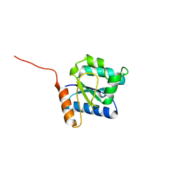
7VOP
 
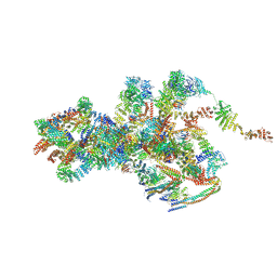 | | Cryo-EM structure of Xenopus laevis nuclear pore complex cytoplasmic ring subunit | | 分子名称: | GATOR complex protein SEC13, IL4I1 protein, MGC154553 protein, ... | | 著者 | Tai, L, Zhu, Y, Sun, F. | | 登録日 | 2021-10-14 | | 公開日 | 2022-02-02 | | 最終更新日 | 2024-06-19 | | 実験手法 | ELECTRON MICROSCOPY (8.7 Å) | | 主引用文献 | 8 angstrom structure of the outer rings of the Xenopus laevis nuclear pore complex obtained by cryo-EM and AI.
Protein Cell, 13, 2022
|
|
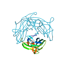
7VCI
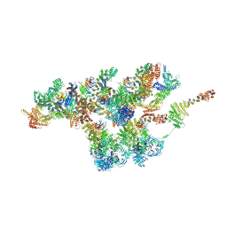 | | Structure of Xenopus laevis NPC nuclear ring asymmetric unit | | 分子名称: | GATOR complex protein SEC13, MGC154553 protein, MGC83295 protein, ... | | 著者 | Tai, L, Zhu, Y, Sun, F. | | 登録日 | 2021-09-03 | | 公開日 | 2022-02-02 | | 最終更新日 | 2024-06-19 | | 実験手法 | ELECTRON MICROSCOPY (8.1 Å) | | 主引用文献 | 8 angstrom structure of the outer rings of the Xenopus laevis nuclear pore complex obtained by cryo-EM and AI.
Protein Cell, 13, 2022
|
|
7WLY
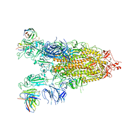 | | Cryo-EM structure of the Omicron S in complex with 35B5 Fab(1 down- and 2 up RBDs) | | 分子名称: | 2-acetamido-2-deoxy-beta-D-glucopyranose, 2-acetamido-2-deoxy-beta-D-glucopyranose-(1-4)-2-acetamido-2-deoxy-beta-D-glucopyranose, Heavy chain of 35B5 Fab, ... | | 著者 | Wang, X, Zhu, Y. | | 登録日 | 2022-01-14 | | 公開日 | 2022-05-25 | | 最終更新日 | 2022-06-22 | | 実験手法 | ELECTRON MICROSCOPY (3.4 Å) | | 主引用文献 | 35B5 antibody potently neutralizes SARS-CoV-2 Omicron by disrupting the N-glycan switch via a conserved spike epitope.
Cell Host Microbe, 30, 2022
|
|
7WLZ
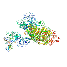 | |
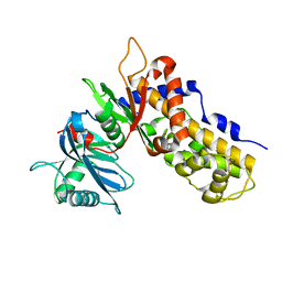
6AEG
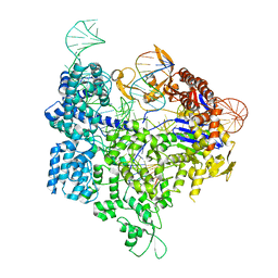 | | Crystal structure of xCas9 in complex with sgRNA and target DNA (GAT PAM) | | 分子名称: | DNA (25-MER), DNA (5'-D(*AP*AP*AP*GP*AP*TP*TP*AP*TP*TP*G)-3'), DNA nuclease, ... | | 著者 | Guo, M, Ren, K, Zhu, Y, Huang, Z. | | 登録日 | 2018-08-04 | | 公開日 | 2019-03-27 | | 最終更新日 | 2023-11-22 | | 実験手法 | X-RAY DIFFRACTION (2.701 Å) | | 主引用文献 | Structural insights into a high fidelity variant of SpCas9.
Cell Res., 29, 2019
|
|
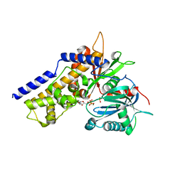
6AEB
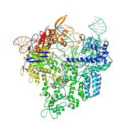 | | Crystal structure of xCas9 in complex with sgRNA and target DNA (AAG PAM) | | 分子名称: | DNA (25-MER), DNA (5'-D(*AP*AP*AP*AP*AP*GP*TP*AP*TP*TP*G)-3'), DNA Nuclease, ... | | 著者 | Guo, M, Ren, K, Zhu, Y, Huang, Z. | | 登録日 | 2018-08-04 | | 公開日 | 2019-03-27 | | 最終更新日 | 2023-11-22 | | 実験手法 | X-RAY DIFFRACTION (3.004 Å) | | 主引用文献 | Structural insights into a high fidelity variant of SpCas9.
Cell Res., 29, 2019
|
|
7ZBT
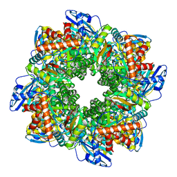 | | Subtomogram averaging of Rubisco from native Halothiobacillus carboxysomes | | 分子名称: | Ribulose bisphosphate carboxylase large chain, Ribulose bisphosphate carboxylase small subunit | | 著者 | Ni, T, Zhu, Y, Yu, X, Sun, Y, Liu, L, Zhang, P. | | 登録日 | 2022-03-24 | | 公開日 | 2022-07-20 | | 最終更新日 | 2024-07-24 | | 実験手法 | ELECTRON MICROSCOPY (3.3 Å) | | 主引用文献 | Structure and assembly of cargo Rubisco in two native alpha-carboxysomes.
Nat Commun, 13, 2022
|
|
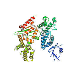
7Y04
 | | Hsp90-AhR-p23 complex | | 分子名称: | ADENOSINE-5'-DIPHOSPHATE, Aryl hydrocarbon receptor, BERYLLIUM TRIFLUORIDE ION, ... | | 著者 | Wen, Z.L, Zhai, Y.J, Zhu, Y, Sun, F. | | 登録日 | 2022-06-03 | | 公開日 | 2023-01-04 | | 最終更新日 | 2024-07-03 | | 実験手法 | ELECTRON MICROSCOPY (3.5 Å) | | 主引用文献 | Cryo-EM structure of the cytosolic AhR complex.
Structure, 31, 2023
|
|
5XBL
 | | Structure of nuclease in complex with associated protein | | 分子名称: | Associated protein, CRISPR-associated endonuclease Cas9/Csn1, RNA (98-MER) | | 著者 | Dong, D, Guo, M, Wang, S, Zhu, Y, Huang, Z. | | 登録日 | 2017-03-20 | | 公開日 | 2017-06-14 | | 最終更新日 | 2024-03-27 | | 実験手法 | X-RAY DIFFRACTION (3.052 Å) | | 主引用文献 | Structural basis of CRISPR-SpyCas9 inhibition by an anti-CRISPR protein
Nature, 546, 2017
|
|
5XLX
 | |
5XLY
 | | Crystal structure of CheR1 in complex with c-di-GMP-bound MapZ | | 分子名称: | 9,9'-[(2R,3R,3aS,5S,7aR,9R,10R,10aS,12S,14aR)-3,5,10,12-tetrahydroxy-5,12-dioxidooctahydro-2H,7H-difuro[3,2-d:3',2'-j][1,3,7,9,2,8]tetraoxadiphosphacyclododecine-2,9-diyl]bis(2-amino-1,9-dihydro-6H-purin-6-one), Chemotaxis protein methyltransferase 1, Cyclic diguanosine monophosphate-binding protein PA4608 | | 著者 | Yuan, Z, Zhu, Y, Gu, L. | | 登録日 | 2017-05-12 | | 公開日 | 2017-08-23 | | 最終更新日 | 2024-03-27 | | 実験手法 | X-RAY DIFFRACTION (1.763 Å) | | 主引用文献 | Structural basis for the regulation of chemotaxis by MapZ in the presence of c-di-GMP
Acta Crystallogr D Struct Biol, 73, 2017
|
|
5XN7
 | |
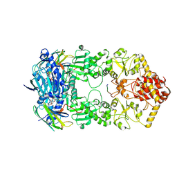
5WTI
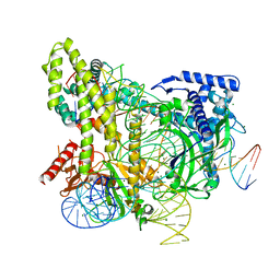 | | Crystal structure of the CRISPR-associated protein in complex with crRNA and DNA | | 分子名称: | CRISPR-associated protein, DNA (28-MER), DNA (5'-D(P*GP*TP*GP*TP*GP*GP*AP*TP*TP*CP*CP*G)-3'), ... | | 著者 | Wu, D, Guan, X, Zhu, Y, Huang, Z. | | 登録日 | 2016-12-13 | | 公開日 | 2017-11-01 | | 実験手法 | X-RAY DIFFRACTION (2.682 Å) | | 主引用文献 | Structural basis of stringent PAM recognition by CRISPR-C2c1 in complex with sgRNA
Cell Res., 27, 2017
|
|
5Y8E
 | |
5YHU
 | |
5Y8F
 | |
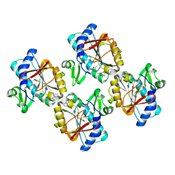
6JJ7
 | | Crystal structure of OsHXK6-Glc complex | | 分子名称: | Rice hexokinase 6, beta-D-glucopyranose | | 著者 | He, C, Wei, P, Chen, J, Wang, H, Wan, Y, Zhou, J, Zhu, Y, Huang, W, Yin, L. | | 登録日 | 2019-02-25 | | 公開日 | 2019-07-03 | | 最終更新日 | 2023-11-22 | | 実験手法 | X-RAY DIFFRACTION (2.9 Å) | | 主引用文献 | Crystal structure of OsHXK6-Glc complex
To Be Published
|
|
6JJ4
 | | Crystal structure of OsHXK6-apo form | | 分子名称: | Hexokinase-6 | | 著者 | He, C, Wei, P, Chen, J, Wang, H, Wan, Y, Zhou, J, Zhu, Y, Huang, W, Yin, L. | | 登録日 | 2019-02-25 | | 公開日 | 2019-07-03 | | 最終更新日 | 2023-11-22 | | 実験手法 | X-RAY DIFFRACTION (2.6 Å) | | 主引用文献 | Crystal structure of OsHXK6-apo
To Be Published
|
|
6JJ9
 | | Crystal structure of OsHXK6-Glc-ATP-Mg2+ complex | | 分子名称: | ADENOSINE-5'-DIPHOSPHATE, Hexokinase-6, MAGNESIUM ION, ... | | 著者 | He, C, Wei, P, Chen, J, Wang, H, Wan, Y, Zhou, J, Zhu, Y, Huang, W, Yin, L. | | 登録日 | 2019-02-25 | | 公開日 | 2019-07-03 | | 最終更新日 | 2023-11-22 | | 実験手法 | X-RAY DIFFRACTION (3 Å) | | 主引用文献 | Crystal structure of OsHXK6-Glc-ATP-Mg2+ complex
To Be Published
|
|
6JXR
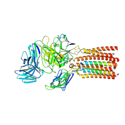 | | Structure of human T cell receptor-CD3 complex | | 分子名称: | T cell receptor alpha variable 12-3,Possible J 11 gene segment,T cell receptor alpha constant, T cell receptor beta variable 6-5,M1-specific T cell receptor beta chain,T cell receptor beta constant 2, T-cell surface glycoprotein CD3 delta chain, ... | | 著者 | Dong, D, Zheng, L, Lin, J, Zhu, Y, Li, N, Zhang, B, Xie, S, Zheng, J, Wang, Y, Gao, N, Huang, Z. | | 登録日 | 2019-04-24 | | 公開日 | 2019-09-11 | | 最終更新日 | 2020-09-16 | | 実験手法 | ELECTRON MICROSCOPY (3.7 Å) | | 主引用文献 | Structural basis of assembly of the human T cell receptor-CD3 complex.
Nature, 573, 2019
|
|
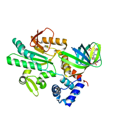
6K2U
 | | Crystal structure of Thr66 ADP-ribosylated ubiquitin | | 分子名称: | ADENOSINE-5-DIPHOSPHORIBOSE, MAGNESIUM ION, Polyubiquitin-C, ... | | 著者 | Wang, X, Zhou, Y, Zhu, Y. | | 登録日 | 2019-05-15 | | 公開日 | 2020-03-18 | | 最終更新日 | 2023-11-29 | | 実験手法 | X-RAY DIFFRACTION (2.554 Å) | | 主引用文献 | Threonine ADP-Ribosylation of Ubiquitin by a Bacterial Effector Family Blocks Host Ubiquitination.
Mol.Cell, 78, 2020
|
|
6A0A
 | |
6A0C
 | | Structure of a triple-helix region of human collagen type III | | 分子名称: | 1,2-ETHANEDIOL, GLYCEROL, collagen type III peptide | | 著者 | Yang, X, Zhu, Y, Ye, S, Zhang, R. | | 登録日 | 2018-06-05 | | 公開日 | 2018-12-26 | | 最終更新日 | 2023-11-22 | | 実験手法 | X-RAY DIFFRACTION (1.501 Å) | | 主引用文献 | Characterization by high-resolution crystal structure analysis of a triple-helix region of human collagen type III with potent cell adhesion activity.
Biochem. Biophys. Res. Commun., 508, 2019
|
|
5ZHU
 | |
6JJ8
 | | Crystal structure of OsHXK6-ATP-Mg2+ complex | | 分子名称: | ADENOSINE-5'-DIPHOSPHATE, MAGNESIUM ION, PHOSPHATE ION, ... | | 著者 | He, C, Wei, P, Chen, J, Wang, H, Wan, Y, Zhou, J, Zhu, Y, Huang, W, Yin, L. | | 登録日 | 2019-02-25 | | 公開日 | 2019-07-03 | | 最終更新日 | 2023-11-22 | | 実験手法 | X-RAY DIFFRACTION (2.8 Å) | | 主引用文献 | Crystal structure of OsHXK6-ATP-Mg2+ complex
To Be Published
|
|


