3NIV
 
 | |
3NND
 | |
3NPK
 | |
3MPO
 | |
3MSR
 | |
3LIC
 | |
3LIB
 | |
3LID
 | |
3NAS
 | |
3M2T
 | |
3OVG
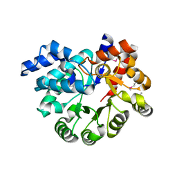
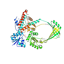
 | | The crystal structure of an amidohydrolase from Mycoplasma synoviae with Zn ion bound | | Descriptor: | PHOSPHATE ION, ZINC ION, amidohydrolase | | Authors: | Zhang, Z, Kumaran, D, Burley, S.K, Swaminathan, S, New York SGX Research Center for Structural Genomics (NYSGXRC) | | Deposit date: | 2010-09-16 | | Release date: | 2010-10-13 | | Last modified: | 2023-12-06 | | Method: | X-RAY DIFFRACTION (2.059 Å) | | Cite: | The crystal structure of an amidohydrolase from Mycoplasma synoviae with Zn ion bound
To be Published
|
|
8BDP
 | |
3PAN
 | |
3M2P
 | |
3OPN
 | |
3PAO
 | |
3PBK
 | |
3PBM
 | |
3OBZ
 | |
3OU8
 | |
3OTT
 | |
3OXN
 | |
3PWT
 | |
3Q34
 | |
3PX7
 | |


