Loading
PDBj
MenuPDBj@FacebookPDBj@TwitterPDBj@YouTubewwPDB FoundationwwPDB
RCSB PDBPDBeBMRBAdv. SearchSearch help
PDB: 545 results

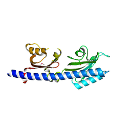
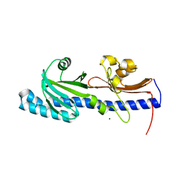
3LIF
DownloadVisualize
BU of 3lif by Molmil
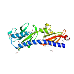
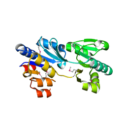
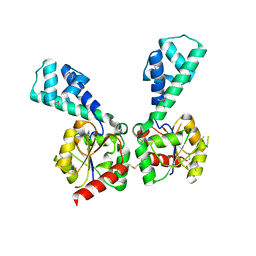
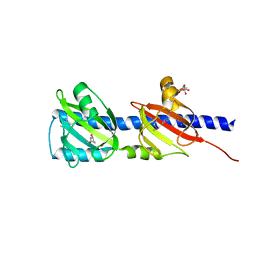
Crystal Structure of the extracellular domain of the putative histidine kinase rpHK1S-Z16
Descriptor: (4S)-2-METHYL-2,4-PENTANEDIOL, CITRIC ACID, Putative diguanylate cyclase (GGDEF) with PAS/PAC domain
Authors:Zhang, Z, Hendrickson, W.A.
Deposit date:2010-01-24
Release date:2010-05-05
Last modified:2017-11-01
Method:X-RAY DIFFRACTION (2.7 Å)
Cite:Structural characterization of the predominant family of histidine kinase sensor domains.
J.Mol.Biol., 400, 2010
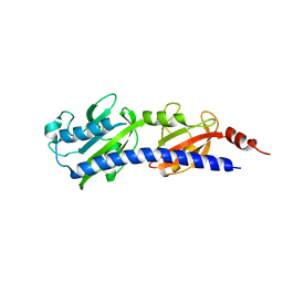
3LIE
DownloadVisualize
BU of 3lie by Molmil
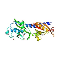
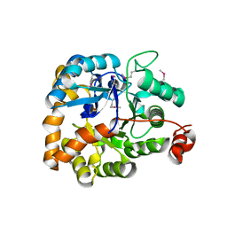
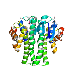
Crystal Structure of the extracellular domain of the putative histidine kinase vpHK1S-Z8
Descriptor: MAGNESIUM ION, Putative sensory box/GGDEF family protein
Authors:Zhang, Z, Hendrickson, W.A.
Deposit date:2010-01-24
Release date:2010-05-05
Last modified:2024-02-21
Method:X-RAY DIFFRACTION (2.59 Å)
Cite:Structural characterization of the predominant family of histidine kinase sensor domains.
J.Mol.Biol., 400, 2010
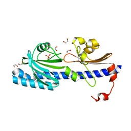
3LIC
DownloadVisualize
BU of 3lic by Molmil
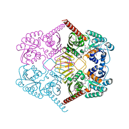
Crystal Structure of the extracellular domain of the putative histidine kinase soHK1S-Z6
Descriptor: 1,2-ETHANEDIOL, Sensor protein
Authors:Zhang, Z, Hendrickson, W.A.
Deposit date:2010-01-24
Release date:2010-05-05
Last modified:2017-11-01
Method:X-RAY DIFFRACTION (2.3 Å)
Cite:Structural characterization of the predominant family of histidine kinase sensor domains.
J.Mol.Biol., 400, 2010
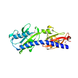
3LIB
DownloadVisualize
BU of 3lib by Molmil
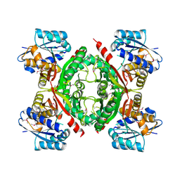
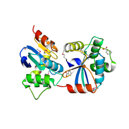
Crystal Structure of the extracellular domain of the putative histidine kinase mmHK1S-Z3
Descriptor: Hypothetical sensory transduction histidine kinase, POTASSIUM ION
Authors:Zhang, Z, Hendrickson, W.A.
Deposit date:2010-01-24
Release date:2010-05-05
Last modified:2024-02-21
Method:X-RAY DIFFRACTION (2.99 Å)
Cite:Structural characterization of the predominant family of histidine kinase sensor domains.
J.Mol.Biol., 400, 2010
3LID
DownloadVisualize
BU of 3lid by Molmil
Crystal Structure of the extracellular domain of the putative histidine kinase vpHK1S-Z8
Descriptor: 1,2-ETHANEDIOL, CHLORIDE ION, PHOSPHATE ION, ...
Authors:Zhang, Z, Hendrickson, W.A.
Deposit date:2010-01-24
Release date:2010-05-05
Last modified:2024-02-21
Method:X-RAY DIFFRACTION (1.76 Å)
Cite:Structural characterization of the predominant family of histidine kinase sensor domains.
J.Mol.Biol., 400, 2010
3LI9
DownloadVisualize
BU of 3li9 by Molmil
Crystal Structure of the extracellular domain of the putative histidine kinase mmHK1S-Z2
Descriptor: 2-[BIS-(2-HYDROXY-ETHYL)-AMINO]-2-HYDROXYMETHYL-PROPANE-1,3-DIOL, Hypothetical sensory transduction histidine kinase
Authors:Zhang, Z, Hendrickson, W.A.
Deposit date:2010-01-24
Release date:2010-05-05
Last modified:2017-11-01
Method:X-RAY DIFFRACTION (1.7 Å)
Cite:Structural characterization of the predominant family of histidine kinase sensor domains.
J.Mol.Biol., 400, 2010
3LNV
DownloadVisualize
BU of 3lnv by Molmil
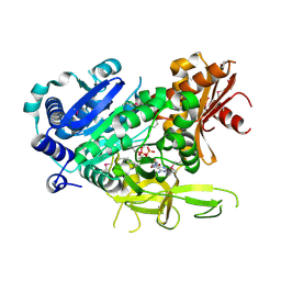
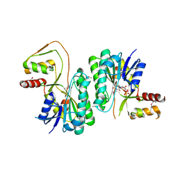
The crystal structure of fatty acyl-adenylate ligase from L. pneumophila in complex with acyl adenylate and pyrophosphate
Descriptor: 5'-O-[(S)-(dodecanoyloxy)(hydroxy)phosphoryl]adenosine, PYROPHOSPHATE 2-, Saframycin Mx1 synthetase B
Authors:Zhang, Z, Burley, S.K, Swaminathan, S, New York SGX Research Center for Structural Genomics (NYSGXRC)
Deposit date:2010-02-03
Release date:2010-04-07
Last modified:2023-11-22
Method:X-RAY DIFFRACTION (2 Å)
Cite:Structural and Functional Studies of Fatty Acyl Adenylate Ligases from E. coli and L. pneumophila.
J.Mol.Biol., 406, 2011
3LI8
DownloadVisualize
BU of 3li8 by Molmil
Crystal Structure of the extracellular domain of the putative histidine kinase mmHK1S-Z2
Descriptor: 1,2-ETHANEDIOL, Hypothetical sensory transduction histidine kinase, SULFATE ION
Authors:Zhang, Z, Hendrickson, W.A.
Deposit date:2010-01-24
Release date:2010-05-05
Last modified:2017-11-01
Method:X-RAY DIFFRACTION (1.75 Å)
Cite:Structural characterization of the predominant family of histidine kinase sensor domains.
J.Mol.Biol., 400, 2010
3LIA
DownloadVisualize
BU of 3lia by Molmil
Crystal Structure of the extracellular domain of the putative histidine kinase mmHK1S-Z2
Descriptor: 2-[BIS-(2-HYDROXY-ETHYL)-AMINO]-2-HYDROXYMETHYL-PROPANE-1,3-DIOL, Hypothetical sensory transduction histidine kinase, SULFATE ION
Authors:Zhang, Z, Hendrickson, W.A.
Deposit date:2010-01-24
Release date:2010-05-05
Last modified:2024-02-21
Method:X-RAY DIFFRACTION (1.99 Å)
Cite:Structural characterization of the predominant family of histidine kinase sensor domains.
J.Mol.Biol., 400, 2010
3LL3
DownloadVisualize
BU of 3ll3 by Molmil
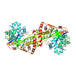
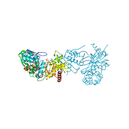
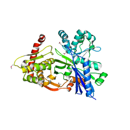
The crystal structure of ligand bound xylulose kinase from Lactobacillus acidophilus
Descriptor: 1-DEOXY-D-XYLULOSE-5-PHOSPHATE, ADENOSINE-5'-DIPHOSPHATE, ADENOSINE-5'-TRIPHOSPHATE, ...
Authors:Zhang, Z, Burley, S.K, Swaminathan, S, New York SGX Research Center for Structural Genomics (NYSGXRC)
Deposit date:2010-01-28
Release date:2010-03-23
Last modified:2023-11-22
Method:X-RAY DIFFRACTION (2.002 Å)
Cite:The crystal structure of xylulose kinase from Lactobacillus acidophilus
To be Published
3MPO
DownloadVisualize
BU of 3mpo by Molmil
The crystal structure of a hydrolase from Lactobacillus brevis
Descriptor: Predicted hydrolase of the HAD superfamily
Authors:Zhang, Z, Burley, S.K, Swaminathan, S, New York SGX Research Center for Structural Genomics (NYSGXRC)
Deposit date:2010-04-27
Release date:2010-05-12
Last modified:2021-02-10
Method:X-RAY DIFFRACTION (2.9 Å)
Cite:The crystal structure of a hydrolase from Lactobacillus brevis
To be Published
3MSR
DownloadVisualize
BU of 3msr by Molmil
The crystal structure of an amidohydrolase from Mycoplasma synoviae
Descriptor: GLYCEROL, PHOSPHATE ION, amidohydrolases
Authors:Zhang, Z, Burley, S.K, Swaminathan, S, New York SGX Research Center for Structural Genomics (NYSGXRC)
Deposit date:2010-04-29
Release date:2010-05-12
Last modified:2021-02-10
Method:X-RAY DIFFRACTION (2.162 Å)
Cite:The crystal structure of an amidohydrolase from Mycoplasma synoviae
To be Published
3KZB
DownloadVisualize
BU of 3kzb by Molmil
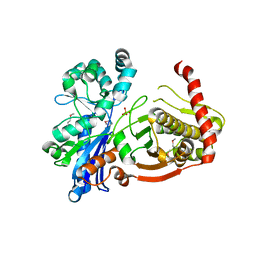
Crystal structure of xylulokinase from Chromobacterium violaceum
Descriptor: ADENOSINE-5'-DIPHOSPHATE, Xylulokinase
Authors:Zhang, Z, Burley, S.K, Swaminathan, S, New York SGX Research Center for Structural Genomics (NYSGXRC)
Deposit date:2009-12-08
Release date:2010-03-09
Last modified:2023-11-22
Method:X-RAY DIFFRACTION (2.705 Å)
Cite:Crystal structure of xylulokinase from Chromobacterium violaceum
To be Published
3M2P
DownloadVisualize
BU of 3m2p by Molmil
The crystal structure of UDP-N-acetylglucosamine 4-epimerase from Bacillus cereus
Descriptor: UDP-N-acetylglucosamine 4-epimerase, URIDINE-5'-DIPHOSPHATE
Authors:Zhang, Z, Burley, S.K, Swaminathan, S, New York SGX Research Center for Structural Genomics (NYSGXRC)
Deposit date:2010-03-08
Release date:2010-04-07
Last modified:2021-02-10
Method:X-RAY DIFFRACTION (2.95 Å)
Cite:The crystal structure of UDP-N-acetylglucosamine 4-epimerase from Bacillus cereus
To be Published
3NAS
DownloadVisualize
BU of 3nas by Molmil
The crystal structure of beta-phosphoglucomutase from Bacillus subtilis
Descriptor: beta-phosphoglucomutase
Authors:Zhang, Z, Burley, S.K, Swaminathan, S, New York SGX Research Center for Structural Genomics (NYSGXRC)
Deposit date:2010-06-02
Release date:2010-07-28
Last modified:2021-02-10
Method:X-RAY DIFFRACTION (3 Å)
Cite:The crystal structure of beta-phosphoglucomutase from Bacillus subtilis
To be Published
3NIV
DownloadVisualize
BU of 3niv by Molmil
The crystal structure of Glutathione S-transferase from Legionella pneumophila
Descriptor: Glutathione S-transferase
Authors:Zhang, Z, Burley, S.K, Swaminathan, S, New York SGX Research Center for Structural Genomics (NYSGXRC)
Deposit date:2010-06-16
Release date:2010-07-14
Last modified:2021-02-10
Method:X-RAY DIFFRACTION (2.3 Å)
Cite:The crystal structure of Glutathione S-transferase from Legionella pneumophila
To be Published
3M2T
DownloadVisualize
BU of 3m2t by Molmil
The crystal structure of dehydrogenase from Chromobacterium violaceum
Descriptor: NICOTINAMIDE-ADENINE-DINUCLEOTIDE, Probable dehydrogenase, SULFATE ION
Authors:Zhang, Z, Burley, S.K, Swaminathan, S, New York SGX Research Center for Structural Genomics (NYSGXRC)
Deposit date:2010-03-08
Release date:2010-04-07
Last modified:2024-10-16
Method:X-RAY DIFFRACTION (2.3 Å)
Cite:The crystal structure of dehydrogenase from Chromobacterium violaceum
To be Published
3NND
DownloadVisualize
BU of 3nnd by Molmil
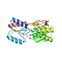
The crystal structure of ABC transporter from Rhodopseudomonas palustris
Descriptor: Possible substrate binding protein of ABC transporter system
Authors:Zhang, Z, Burley, S.K, Swaminathan, S, New York SGX Research Center for Structural Genomics (NYSGXRC)
Deposit date:2010-06-23
Release date:2010-10-13
Last modified:2024-10-16
Method:X-RAY DIFFRACTION (2.8 Å)
Cite:The crystal structure of ABC transporter from Rhodopseudomonas palustris
To be Published
3NPK
DownloadVisualize
BU of 3npk by Molmil
The crystal structure of geranyltranstransferase from Campylobacter jejuni
Descriptor: GLYCEROL, Geranyltranstransferase, PYROPHOSPHATE
Authors:Zhang, Z, Burley, S.K, Swaminathan, S, New York SGX Research Center for Structural Genomics (NYSGXRC)
Deposit date:2010-06-28
Release date:2010-07-14
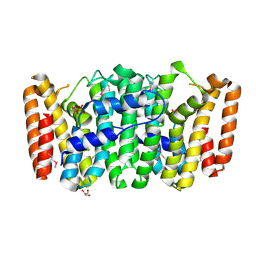
Last modified:2023-12-27
Method:X-RAY DIFFRACTION (1.5 Å)
Cite:The crystal structure of geranyltranstransferase from Campylobacter jejuni
To be Published
3GBT
DownloadVisualize
BU of 3gbt by Molmil
Crystal structure of gluconate kinase from Lactobacillus acidophilus
Descriptor: Gluconate kinase
Authors:Zhang, Z, Swaminathan, S, Burley, S.K, New York SGX Research Center for Structural Genomics (NYSGXRC)
Deposit date:2009-02-20
Release date:2009-03-10
Last modified:2021-02-10
Method:X-RAY DIFFRACTION (2.4 Å)
Cite:The crystal structure of gluconate kinase from Lactobacillus acidophilus
To be Published
3GV1
DownloadVisualize
BU of 3gv1 by Molmil
Crystal structure of disulfide interchange protein from Neisseria gonorrhoeae
Descriptor: BENZOIC ACID, Disulfide interchange protein
Authors:Zhang, Z, Burley, S.K, Swaminathan, S, New York SGX Research Center for Structural Genomics (NYSGXRC)
Deposit date:2009-03-30
Release date:2009-04-14
Last modified:2024-10-16
Method:X-RAY DIFFRACTION (2 Å)
Cite:The crystal structure of disulfide interchange protein from Neisseria gonorrhoeae
To be Published
3H5O
DownloadVisualize
BU of 3h5o by Molmil
The crystal structure of transcription regulator GntR from Chromobacterium violaceum
Descriptor: SULFATE ION, Transcriptional regulator GntR
Authors:Zhang, Z, Burley, S.K, Swaminathan, S, New York SGX Research Center for Structural Genomics (NYSGXRC)
Deposit date:2009-04-22
Release date:2009-05-05
Last modified:2021-02-10
Method:X-RAY DIFFRACTION (2.3 Å)
Cite:The crystal structure of transcription regulator GntR from Chromobacterium violaceum
To be Published
3G0B
DownloadVisualize
BU of 3g0b by Molmil
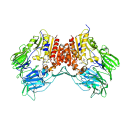
Crystal structure of dipeptidyl peptidase IV in complex with TAK-322
Descriptor: 2-({6-[(3R)-3-aminopiperidin-1-yl]-3-methyl-2,4-dioxo-3,4-dihydropyrimidin-1(2H)-yl}methyl)benzonitrile, 2-acetamido-2-deoxy-beta-D-glucopyranose, 2-acetamido-2-deoxy-beta-D-glucopyranose-(1-4)-2-acetamido-2-deoxy-beta-D-glucopyranose, ...
Authors:Zhang, Z, Wallace, M.B, Feng, J, Stafford, J.A, Kaldor, S.W, Shi, L, Skene, R.J, Aertgeerts, K, Lee, B, Jennings, A, Xu, R, Kassel, D, Webb, D.R, Gwaltney, S.L.
Deposit date:2009-01-27
Release date:2010-02-16
Last modified:2020-07-29
Method:X-RAY DIFFRACTION (2.25 Å)
Cite:Design and Synthesis of Pyrimidinone and Pyrimidinedione Inhibitors of Dipeptidyl Peptidase IV.
J.Med.Chem., 54, 2011
3G0C
DownloadVisualize
BU of 3g0c by Molmil
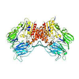
Crystal structure of dipeptidyl peptidase IV in complex with a pyrimidinedione inhibitor 1
Descriptor: 2-acetamido-2-deoxy-beta-D-glucopyranose, 2-acetamido-2-deoxy-beta-D-glucopyranose-(1-4)-2-acetamido-2-deoxy-beta-D-glucopyranose, 7-(2-chlorobenzyl)-1,3-dimethyl-8-piperazin-1-yl-3,7-dihydro-1H-purine-2,6-dione, ...
Authors:Zhang, Z, Wallace, M.B, Feng, J, Stafford, J.A, Kaldor, S.W, Shi, L, Skene, R.J, Aertgeerts, K, Lee, B, Jennings, A, Xu, R, Kassel, D, Webb, D.R, Gwaltney, S.L.
Deposit date:2009-01-27
Release date:2010-02-16
Last modified:2023-09-06
Method:X-RAY DIFFRACTION (2.69 Å)
Cite:Design and Synthesis of Pyrimidinone and Pyrimidinedione Inhibitors of Dipeptidyl Peptidase IV.
J.Med.Chem., 54, 2011
3IFR
DownloadVisualize
BU of 3ifr by Molmil
The crystal structure of xylulose kinase from Rhodospirillum rubrum
Descriptor: Carbohydrate kinase, FGGY, PHOSPHATE ION
Authors:Zhang, Z, Burley, S.K, Swaminathan, S, New York SGX Research Center for Structural Genomics (NYSGXRC)
Deposit date:2009-07-24
Release date:2009-08-25
Last modified:2024-10-09
Method:X-RAY DIFFRACTION (2.3 Å)
Cite:The crystal structure of xylulose kinase from Rhodospirillum rubrum
To be Published

226707

数据于2024-10-30公开中

PDB statisticsPDBj update infoContact PDBjnumon