Loading
PDBj
MenuPDBj@FacebookPDBj@TwitterPDBj@YouTubewwPDB FoundationwwPDB
RCSB PDBPDBeBMRBAdv. SearchSearch help
PDB: 744 results

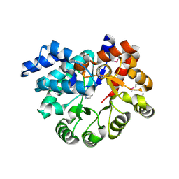
8WGY
DownloadVisualize
BU of 8wgy by Molmil
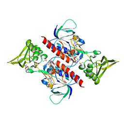
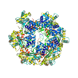
MPOX E5 hexamer AMP-PNP and ssDNA bound form with clear primase domain
Descriptor: DNA (5'-D(P*CP*CP*C)-3'), MAGNESIUM ION, PHOSPHOAMINOPHOSPHONIC ACID-ADENYLATE ESTER, ...
Authors:Zhang, Z, Dong, C.
Deposit date:2023-09-22
Release date:2024-10-16
Method:ELECTRON MICROSCOPY (2.91 Å)
Cite:Essential and multifunctional mpox virus E5 helicase-primase in double and single hexamer.
Sci Adv, 10, 2024
8WH0
DownloadVisualize
BU of 8wh0 by Molmil
MPOX E5 hexamer ssDNA and AMP-PNP bound conformation
Descriptor: DNA (5'-D(P*CP*CP*CP*CP*C)-3'), MAGNESIUM ION, PHOSPHOAMINOPHOSPHONIC ACID-ADENYLATE ESTER, ...
Authors:Zhang, Z, Dong, C.
Deposit date:2023-09-22
Release date:2024-10-09
Method:ELECTRON MICROSCOPY (2.51 Å)
Cite:Essential and multifunctional mpox virus E5 helicase-primase in double and single hexamer.
Sci Adv, 10, 2024
3D8X
DownloadVisualize
BU of 3d8x by Molmil
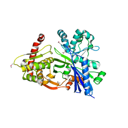
Crystal Structure of Saccharomyces cerevisiae NDPPH Dependent Thioredoxin Reductase 1
Descriptor: FLAVIN-ADENINE DINUCLEOTIDE, NADPH DIHYDRO-NICOTINAMIDE-ADENINE-DINUCLEOTIDE PHOSPHATE, Thioredoxin reductase 1
Authors:Zhang, Z.Y, Bao, R, Yu, J, Chen, Y.X, Zhou, C.-Z.
Deposit date:2008-05-26
Release date:2008-12-09
Last modified:2024-10-16
Method:X-RAY DIFFRACTION (2.8 Å)
Cite:Crystal structure of Saccharomyces cerevisiae cytoplasmic thioredoxin reductase Trr1 reveals the structural basis for species-specific recognition of thioredoxin
Biochim.Biophys.Acta, 1794, 2009
3GBT
DownloadVisualize
BU of 3gbt by Molmil
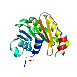
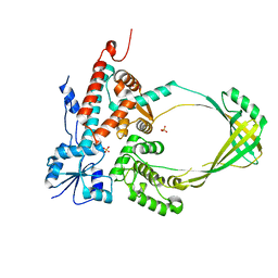
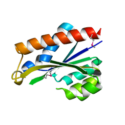
Crystal structure of gluconate kinase from Lactobacillus acidophilus
Descriptor: Gluconate kinase
Authors:Zhang, Z, Swaminathan, S, Burley, S.K, New York SGX Research Center for Structural Genomics (NYSGXRC)
Deposit date:2009-02-20
Release date:2009-03-10
Last modified:2021-02-10
Method:X-RAY DIFFRACTION (2.4 Å)
Cite:The crystal structure of gluconate kinase from Lactobacillus acidophilus
To be Published
4YK6
DownloadVisualize
BU of 4yk6 by Molmil
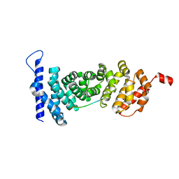
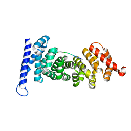
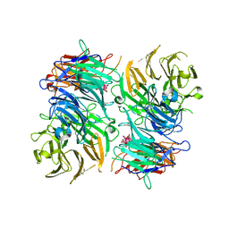
Crystal structure of APC-ARM in complexed with Amer1-A4
Descriptor: APC membrane recruitment protein 1, Adenomatous polyposis coli protein
Authors:Zhang, Z, Xiao, Y, Wu, G.
Deposit date:2015-03-04
Release date:2016-03-09
Last modified:2023-11-08
Method:X-RAY DIFFRACTION (1.7 Å)
Cite:Structures of the APC-ARM domain in complexes with discrete Amer1/WTX fragments reveal that it uses a consensus mode to recognize its binding partners
Cell Discov, 1, 2015
8UUZ
DownloadVisualize
BU of 8uuz by Molmil
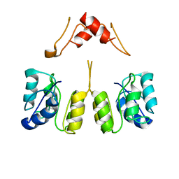
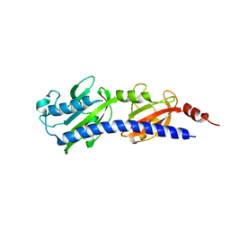
Campylobacter jejuni CosR apo form
Descriptor: DNA-binding response regulator
Authors:Zhang, Z.
Deposit date:2023-11-02
Release date:2024-01-31
Last modified:2024-04-03
Method:ELECTRON MICROSCOPY (3.77 Å)
Cite:Structural basis of DNA recognition of the Campylobacter jejuni CosR regulator.
Mbio, 15, 2024
3LIB
DownloadVisualize
BU of 3lib by Molmil
Crystal Structure of the extracellular domain of the putative histidine kinase mmHK1S-Z3
Descriptor: Hypothetical sensory transduction histidine kinase, POTASSIUM ION
Authors:Zhang, Z, Hendrickson, W.A.
Deposit date:2010-01-24
Release date:2010-05-05
Last modified:2024-02-21
Method:X-RAY DIFFRACTION (2.99 Å)
Cite:Structural characterization of the predominant family of histidine kinase sensor domains.
J.Mol.Biol., 400, 2010
4YJE
DownloadVisualize
BU of 4yje by Molmil
Crystal structure of APC-ARM in complexed with Amer1-A1
Descriptor: APC membrane recruitment protein 1, Adenomatous polyposis coli protein
Authors:Zhang, Z, Xiao, Y, Wu, G.
Deposit date:2015-03-03
Release date:2016-03-09
Last modified:2023-11-08
Method:X-RAY DIFFRACTION (1.9 Å)
Cite:Structures of the APC-ARM domain in complexes with discrete Amer1/WTX fragments reveal that it uses a consensus mode to recognize its binding partners
Cell Discov, 1, 2015
3IFR
DownloadVisualize
BU of 3ifr by Molmil
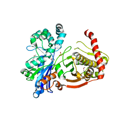
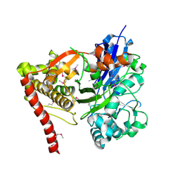
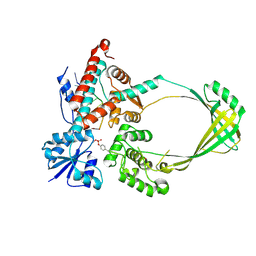
The crystal structure of xylulose kinase from Rhodospirillum rubrum
Descriptor: Carbohydrate kinase, FGGY, PHOSPHATE ION
Authors:Zhang, Z, Burley, S.K, Swaminathan, S, New York SGX Research Center for Structural Genomics (NYSGXRC)
Deposit date:2009-07-24
Release date:2009-08-25
Last modified:2024-10-09
Method:X-RAY DIFFRACTION (2.3 Å)
Cite:The crystal structure of xylulose kinase from Rhodospirillum rubrum
To be Published
8CX5
DownloadVisualize
BU of 8cx5 by Molmil
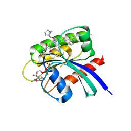
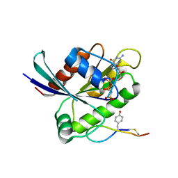
Crystal Structure of small molecule alpha,beta-ketoamide 4 covalently bound to K-Ras(G12R)
Descriptor: GUANOSINE-5'-DIPHOSPHATE, Isoform 2B of GTPase KRas, MAGNESIUM ION, ...
Authors:Zhang, Z, Morstein, J, Ecker, A, Guiley, K.Z, Shokat, K.M.
Deposit date:2022-05-19
Release date:2022-08-31
Last modified:2023-10-18
Method:X-RAY DIFFRACTION (1.72 Å)
Cite:Chemoselective Covalent Modification of K-Ras(G12R) with a Small Molecule Electrophile.
J.Am.Chem.Soc., 144, 2022
6WGN
DownloadVisualize
BU of 6wgn by Molmil
Crystal structure of K-Ras(G12D) GppNHp bound to cyclic peptide ligand KD2
Descriptor: Cyclic Peptide KD2, GTPase KRas, MAGNESIUM ION, ...
Authors:Zhang, Z, Gao, R, Hu, Q, Peacock, H, Peacock, D.M, Shokat, K.M, Suga, H.
Deposit date:2020-04-06
Release date:2020-10-14
Last modified:2023-10-18
Method:X-RAY DIFFRACTION (1.601 Å)
Cite:GTP-State-Selective Cyclic Peptide Ligands of K-Ras(G12D) Block Its Interaction with Raf.
Acs Cent.Sci., 6, 2020
3I8B
DownloadVisualize
BU of 3i8b by Molmil
The crystal structure of xylulose kinase from Bifidobacterium adolescentis
Descriptor: MAGNESIUM ION, Xylulose kinase
Authors:Zhang, Z, Burley, S.K, Swaminathan, S, New York SGX Research Center for Structural Genomics (NYSGXRC)
Deposit date:2009-07-09
Release date:2009-08-04
Last modified:2021-02-10
Method:X-RAY DIFFRACTION (2 Å)
Cite:The crystal structure of xylulose kinase from Bifidobacterium adolescentis
To be Published
8GUP
DownloadVisualize
BU of 8gup by Molmil
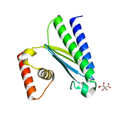
Crystal structure of EsaG from Staphylococcus aureus
Descriptor: CITRIC ACID, Type VII secretion system protein EsaG
Authors:Zhang, Z.M, Wang, Y.J.
Deposit date:2022-09-13
Release date:2022-11-09
Last modified:2024-05-29
Method:X-RAY DIFFRACTION (2.298725 Å)
Cite:A toxin-deformation dependent inhibition mechanism in the T7SS toxin-antitoxin system of Gram-positive bacteria.
Nat Commun, 13, 2022
3GG4
DownloadVisualize
BU of 3gg4 by Molmil
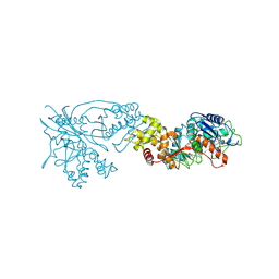
The crystal structure of glycerol kinase from Yersinia pseudotuberculosis
Descriptor: 2-[BIS-(2-HYDROXY-ETHYL)-AMINO]-2-HYDROXYMETHYL-PROPANE-1,3-DIOL, glycerol kinase
Authors:Zhang, Z, Swaminathan, S, Burley, S.K, New York SGX Research Center for Structural Genomics (NYSGXRC)
Deposit date:2009-02-27
Release date:2009-03-31
Last modified:2021-02-10
Method:X-RAY DIFFRACTION (2 Å)
Cite:The crystal structure of glycerol kinase from Yersinia pseudotuberculosis
To be Published
6L4K
DownloadVisualize
BU of 6l4k by Molmil
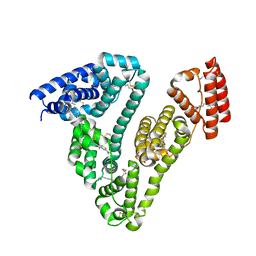
Human serum albumin-Palmitic acid-Cu compound
Descriptor: 2-bromanyl-9-ethyl-~{N},~{N},7-trimethyl-3-thia-1$l^{4},5,6$l^{4},10-tetraza-2$l^{4}-cupratricyclo[6.4.0.0^{2,6}]dodeca-1(8),4,6,9,11-pentaen-4-amine, PALMITIC ACID, Serum albumin
Authors:Zhang, Z.L.
Deposit date:2019-10-17
Release date:2020-10-21
Last modified:2024-10-30
Method:X-RAY DIFFRACTION (2.09 Å)
Cite:Novel Brain-Tumor-Inhibiting Copper(II) Compound Based on a Human Serum Albumin (HSA)-Cell Penetrating Peptide Conjugate.
J.Med.Chem., 62, 2019
3OU8
DownloadVisualize
BU of 3ou8 by Molmil
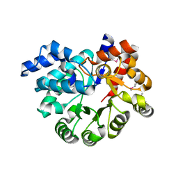
The crystal structure of adenosine deaminase from Pseudomonas aeruginosa
Descriptor: Adenosine deaminase, ZINC ION
Authors:Zhang, Z, Burley, S.K, Swaminathan, S, New York SGX Research Center for Structural Genomics (NYSGXRC)
Deposit date:2010-09-14
Release date:2010-10-13
Last modified:2021-02-10
Method:X-RAY DIFFRACTION (2.51 Å)
Cite:The crystal structure of adenosine deaminase from Pseudomonas aeruginosa
To be Published
3OTT
DownloadVisualize
BU of 3ott by Molmil
Crystal Structure of the extracellular domain of the putative one component system BT4673 from B. thetaiotaomicron
Descriptor: HEXATANTALUM DODECABROMIDE, Two-component system sensor histidine kinase
Authors:Zhang, Z, Liu, Q, Hendrickson, W.A.
Deposit date:2010-09-13
Release date:2011-08-10
Last modified:2024-02-21
Method:X-RAY DIFFRACTION (2.3 Å)
Cite:Crystal structures of apparent saccharide sensors from histidine kinase receptors prevalent in a human gut symbiont.
Febs J., 281, 2014
3OPN
DownloadVisualize
BU of 3opn by Molmil
The crystal structure of a putative hemolysin from Lactococcus lactis
Descriptor: GLYCEROL, SULFATE ION, putative hemolysin
Authors:Zhang, Z, Burley, S.K, Swaminathan, S, New York SGX Research Center for Structural Genomics (NYSGXRC)
Deposit date:2010-09-01
Release date:2010-10-13
Last modified:2024-10-30
Method:X-RAY DIFFRACTION (2.05 Å)
Cite:The crystal structure of a putative hemolysin from Lactococcus lactis
To be Published
3PAO
DownloadVisualize
BU of 3pao by Molmil
The crystal structure of adenosine deaminase with adenine bound from Pseudomonas aeruginosa
Descriptor: ADENINE, Adenosine deaminase, ZINC ION
Authors:Zhang, Z, Burley, S.K, Swaminathan, S, New York SGX Research Center for Structural Genomics (NYSGXRC)
Deposit date:2010-10-19
Release date:2010-12-22
Last modified:2024-10-30
Method:X-RAY DIFFRACTION (2.493 Å)
Cite:The crystal structure of adenosine deaminase with adenine bound from Pseudomonas aeruginosa
To be Published
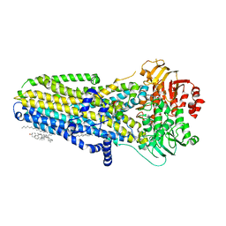
6MSM
DownloadVisualize
BU of 6msm by Molmil
Phosphorylated, ATP-bound human cystic fibrosis transmembrane conductance regulator (CFTR)
Descriptor: (2S)-3-(hexadecanoyloxy)-2-[(9Z)-octadec-9-enoyloxy]propyl 2-(trimethylammonio)ethyl phosphate, ADENOSINE-5'-TRIPHOSPHATE, CHOLESTEROL, ...
Authors:Zhang, Z, Liu, F, Chen, J.
Deposit date:2018-10-16
Release date:2018-11-21
Last modified:2024-03-13
Method:ELECTRON MICROSCOPY (3.2 Å)
Cite:Molecular structure of the ATP-bound, phosphorylated human CFTR.
Proc. Natl. Acad. Sci. U.S.A., 115, 2018
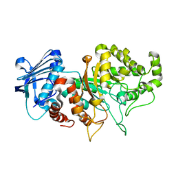
8BDP
DownloadVisualize
BU of 8bdp by Molmil
A GH20 family sulfoglycosidase Bt4394 in complex with NAG-thiazoline and sulfite
Descriptor: Beta-N-acetylhexosaminidase, CHLORIDE ION
Authors:Zhang, Z, He, Y, Jin, Y.
Deposit date:2022-10-19
Release date:2023-01-11
Last modified:2024-01-31
Method:X-RAY DIFFRACTION (1.47 Å)
Cite:Mechanistic and Structural Insights into the Specificity and Biological Functions of Bacterial Sulfoglycosidases
Acs Catalysis, 2022
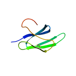
2LW6
DownloadVisualize
BU of 2lw6 by Molmil
Solution structure of an avirulence protein AvrPiz-t from pathogen Magnaportheoryzae
Descriptor: AvrPiz-t protein
Authors:Zhang, Z.-M, Zhang, X, Zhou, Z, Hu, H, Liu, M, Zhou, B, Zhou, J.
Deposit date:2012-07-23
Release date:2012-09-12
Last modified:2024-10-16
Method:SOLUTION NMR
Cite:Solution structure of the Magnaporthe oryzae avirulence protein AvrPiz-t.
J.Biomol.Nmr, 55, 2013
3PWT
DownloadVisualize
BU of 3pwt by Molmil
Crystal structure of mutant E.coli topoisomerase IA
Descriptor: DNA topoisomerase, SULFATE ION
Authors:Zhang, Z, Bokun, C, Tse-Dinh, Y.-C.
Deposit date:2010-12-08
Release date:2010-12-29
Last modified:2024-02-21
Method:X-RAY DIFFRACTION (1.9 Å)
Cite:Crystal structure of mutant E.coli topoisomerase IA
To be Published
3PU6
DownloadVisualize
BU of 3pu6 by Molmil
The crystal structure of an uncharacterized protein from Wolinella succinogenes
Descriptor: Uncharacterized protein
Authors:Zhang, Z, Burley, S.K, Swaminathan, S, New York SGX Research Center for Structural Genomics (NYSGXRC)
Deposit date:2010-12-03
Release date:2011-01-19
Last modified:2021-02-10
Method:X-RAY DIFFRACTION (2.6 Å)
Cite:The crystal structure of an uncharacterized protein from Wolinella succinogenes
TO BE PUBLISHED
3PX7
DownloadVisualize
BU of 3px7 by Molmil
Crystal Structure of covalent complex of topoisomerase 1A with substrate
Descriptor: DNA 5'-D(*A*AP*TP*GP*CP*GP*CP*T)-3', DNA 5'-D(*TP*TP*GP*GP*G)-3', DNA topoisomerase, ...
Authors:Zhang, Z, Tse-dinh, Y.C, Cheng, B.
Deposit date:2010-12-09
Release date:2011-04-20
Last modified:2024-10-30
Method:X-RAY DIFFRACTION (2.3 Å)
Cite:Structure of E. coli Topoisomerase I Trapped as Covalent Complex Intermediate with Cleaved DNA
To be Published

226707

数据于2024-10-30公开中

PDB statisticsPDBj update infoContact PDBjnumon