4Z96
 
 | |
1X24
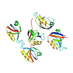
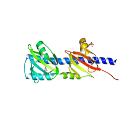
 | | Prl-1 (ptp4a) | | 分子名称: | protein tyrosine phosphatase 4a1 | | 著者 | Zhang, Z.Y, Sun, J.P, Liu, S, Wang, W.Q, Yang, H. | | 登録日 | 2005-04-20 | | 公開日 | 2005-10-11 | | 最終更新日 | 2011-07-13 | | 実験手法 | X-RAY DIFFRACTION (3.2 Å) | | 主引用文献 | Structure and Biochemical Properties of PRL-1, a Phosphatase Implicated in Cell Growth, Differentiation, and Tumor Invasion(,)
Biochemistry, 44, 2005
|
|
5WVW
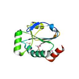
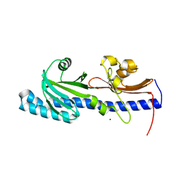
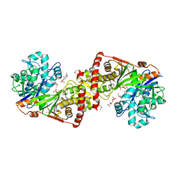
 | | The crystal structure of Cren7 mutant L28A in complex with dsDNA | | 分子名称: | Chromatin protein Cren7, DNA (5'-D(*GP*TP*GP*AP*TP*CP*AP*C)-3') | | 著者 | Zhang, Z.F, Zhao, M.H, Wang, L, Chen, Y.Y, Dong, Y.H, Gong, Y, Huang, L. | | 登録日 | 2016-12-29 | | 公開日 | 2017-04-26 | | 最終更新日 | 2023-11-22 | | 実験手法 | X-RAY DIFFRACTION (1.8 Å) | | 主引用文献 | Roles of Leu28 side chain intercalation in the interaction between Cren7 and DNA
Biochem. J., 474, 2017
|
|
3KZB
 | |
3HDC
 | |
3RL7
 | | Crystal structure of hDLG1-PDZ1 complexed with APC | | 分子名称: | 11-mer peptide from Adenomatous polyposis coli protein, Disks large homolog 1 | | 著者 | Zhang, Z, Li, H, Wu, G. | | 登録日 | 2011-04-19 | | 公開日 | 2011-12-14 | | 最終更新日 | 2023-11-01 | | 実験手法 | X-RAY DIFFRACTION (2.3 Å) | | 主引用文献 | Molecular basis for the recognition of adenomatous polyposis coli by the Discs Large 1 protein.
Plos One, 6, 2011
|
|
3RL8
 | | Crystal structure of hDLG1-PDZ2 complexed with APC | | 分子名称: | 11-mer peptide from Adenomatous polyposis coli protein, Disks large homolog 1 | | 著者 | Zhang, Z, Li, H, Wu, G. | | 登録日 | 2011-04-19 | | 公開日 | 2011-12-14 | | 最終更新日 | 2023-11-01 | | 実験手法 | X-RAY DIFFRACTION (2.2 Å) | | 主引用文献 | Molecular basis for the recognition of adenomatous polyposis coli by the Discs Large 1 protein.
Plos One, 6, 2011
|
|
3KCN
 | |
3KCM
 | |
3KXW
 | |
2OPW
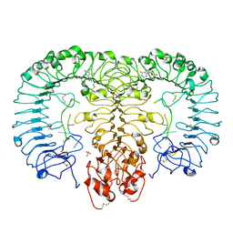
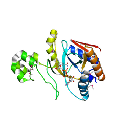
 | | Crystal structure of human phytanoyl-CoA dioxygenase PHYHD1 (apo) | | 分子名称: | PHYHD1 protein | | 著者 | Zhang, Z, Butler, D, McDonough, M.A, Kavanagh, K.L, Bray, J.E, Ng, S.S, von Delft, F, Arrowsmith, C.H, Weigelt, J, Edwards, A, Sundstrom, M, Schofield, C.J, Oppermann, U, Structural Genomics Consortium (SGC) | | 登録日 | 2007-01-30 | | 公開日 | 2007-03-06 | | 最終更新日 | 2024-04-03 | | 実験手法 | X-RAY DIFFRACTION (1.9 Å) | | 主引用文献 | Crystal structure of human phytanoyl-CoA dioxygenase PHYHD1 (apo)
To be Published
|
|
5GMG
 | | Crystal structure of monkey TLR7 in complex with loxoribine and polyU | | 分子名称: | 2-acetamido-2-deoxy-beta-D-glucopyranose, 2-acetamido-2-deoxy-beta-D-glucopyranose-(1-4)-2-acetamido-2-deoxy-beta-D-glucopyranose, 2-azanyl-9-[(2~{R},3~{R},4~{S},5~{R})-5-(hydroxymethyl)-3,4-bis(oxidanyl)oxolan-2-yl]-7-prop-2-enyl-1~{H}-purine-6,8-dione, ... | | 著者 | Zhang, Z, Ohto, U, Shimizu, T. | | 登録日 | 2016-07-14 | | 公開日 | 2016-11-02 | | 最終更新日 | 2023-11-08 | | 実験手法 | X-RAY DIFFRACTION (2.6 Å) | | 主引用文献 | Structural Analysis Reveals that Toll-like Receptor 7 Is a Dual Receptor for Guanosine and Single-Stranded RNA
Immunity, 45, 2016
|
|
3LIF
 | |
3LIE
 | |
3LID
 | |
3LNV
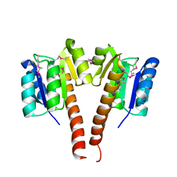
 | | The crystal structure of fatty acyl-adenylate ligase from L. pneumophila in complex with acyl adenylate and pyrophosphate | | 分子名称: | 5'-O-[(S)-(dodecanoyloxy)(hydroxy)phosphoryl]adenosine, PYROPHOSPHATE 2-, Saframycin Mx1 synthetase B | | 著者 | Zhang, Z, Burley, S.K, Swaminathan, S, New York SGX Research Center for Structural Genomics (NYSGXRC) | | 登録日 | 2010-02-03 | | 公開日 | 2010-04-07 | | 最終更新日 | 2023-11-22 | | 実験手法 | X-RAY DIFFRACTION (2 Å) | | 主引用文献 | Structural and Functional Studies of Fatty Acyl Adenylate Ligases from E. coli and L. pneumophila.
J.Mol.Biol., 406, 2011
|
|
3LIA
 | |
3LL3
 | |
8UX2
 | | Chromobacterium violaceum mono-ADP-ribosyltransferase CteC in complex with NAD+ | | 分子名称: | 1,2-ETHANEDIOL, CALCIUM ION, NAD(+)--protein-threonine ADP-ribosyltransferase, ... | | 著者 | Zhang, Z, Rondon, H, Das, C. | | 登録日 | 2023-11-08 | | 公開日 | 2024-01-17 | | 最終更新日 | 2024-02-07 | | 実験手法 | X-RAY DIFFRACTION (1.87 Å) | | 主引用文献 | Crystal structure of bacterial ubiquitin ADP-ribosyltransferase CteC reveals a substrate-recruiting insertion.
J.Biol.Chem., 300, 2023
|
|
3LWI
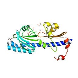
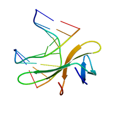 | | Crystal structure of Cren7-dsDNA complex | | 分子名称: | Chromatin protein Cren7, DNA (5'-D(*GP*CP*GP*AP*TP*CP*GP*C)-3') | | 著者 | Zhang, Z.F, Gong, Y, Guo, L, Jiang, T, Huang, L. | | 登録日 | 2010-02-23 | | 公開日 | 2010-05-26 | | 最終更新日 | 2023-11-01 | | 実験手法 | X-RAY DIFFRACTION (2.3 Å) | | 主引用文献 | Structural insights into the interaction of the crenarchaeal chromatin protein Cren7 with DNA
Mol.Microbiol., 76, 2010
|
|
7E7X
 | |
3LWH
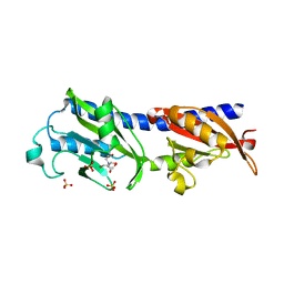
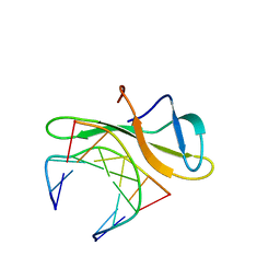 | | Crystal structure of Cren7-dsDNA complex | | 分子名称: | Chromatin protein Cren7, DNA (5'-D(*GP*TP*AP*AP*TP*TP*AP*C)-3') | | 著者 | Zhang, Z.F, Gong, Y, Guo, L, Jiang, T, Huang, L. | | 登録日 | 2010-02-23 | | 公開日 | 2010-05-26 | | 最終更新日 | 2023-11-01 | | 実験手法 | X-RAY DIFFRACTION (1.9 Å) | | 主引用文献 | Structural insights into the interaction of the crenarchaeal chromatin protein Cren7 with DNA
Mol.Microbiol., 76, 2010
|
|
3JAB
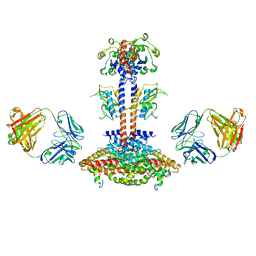 | | Domain organization and conformational plasticity of the G protein effector, PDE6 | | 分子名称: | 3-ISOBUTYL-1-METHYLXANTHINE, GafA domain of cone phosphodiesterase 6C, GafB domain of phosphodiesterase 2A, ... | | 著者 | Zhang, Z, He, F, Constantine, R, Baker, M.L, Baehr, W, Schmid, M.F, Wensel, T.G, Agosto, M.A. | | 登録日 | 2015-05-26 | | 公開日 | 2015-06-10 | | 最終更新日 | 2018-07-18 | | 実験手法 | ELECTRON MICROSCOPY (11 Å) | | 主引用文献 | Domain Organization and Conformational Plasticity of the G Protein Effector, PDE6.
J.Biol.Chem., 290, 2015
|
|
2JX0
 | | The paxillin-binding domain (PBD) of G Protein Coupled Receptor (GPCR)-kinase (GRK) interacting protein 1 (GIT1) | | 分子名称: | ARF GTPase-activating protein GIT1 | | 著者 | Zhang, Z, Guibao, C.D, Simmerman, J.A, Zheng, J. | | 登録日 | 2007-11-01 | | 公開日 | 2008-04-29 | | 最終更新日 | 2024-05-29 | | 実験手法 | SOLUTION NMR | | 主引用文献 | GIT1 paxillin-binding domain is a four-helix bundle, and it binds to both paxillin LD2 and LD4 motifs.
J.Biol.Chem., 283, 2008
|
|
3JBQ
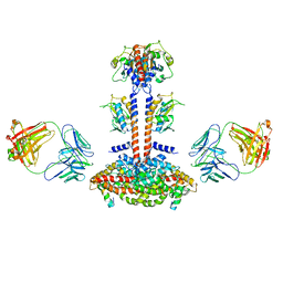 | | Domain Organization and Conformational Plasticity of the G Protein Effector, PDE6 | | 分子名称: | GafA domain of cone phosphodiesterase 6C, GafB domain of phosphodiesterase 2A, IgG1-kappa 2E8 heavy chain, ... | | 著者 | Zhang, Z, He, F, Constantine, R, Baker, M.L, Baehr, W, Schmid, M.F, Wensel, T.G, Agosto, M.A. | | 登録日 | 2015-09-17 | | 公開日 | 2015-09-30 | | 最終更新日 | 2019-12-18 | | 実験手法 | ELECTRON MICROSCOPY (11 Å) | | 主引用文献 | Domain Organization and Conformational Plasticity of the G Protein Effector, PDE6.
J.Biol.Chem., 290, 2015
|
|


