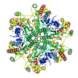
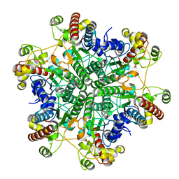
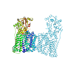
3AP9
 
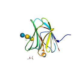 | | Crystal structure of the galectin-8 N-terminal carbohydrate recognition domain in complex with Lacto-N-fucopentaose III | | 分子名称: | (4S)-2-METHYL-2,4-PENTANEDIOL, CHLORIDE ION, Galectin-8, ... | | 著者 | Matsuzaka, T, Ideo, H, Yamashita, K, Nonaka, T. | | 登録日 | 2010-10-12 | | 公開日 | 2011-02-02 | | 最終更新日 | 2023-11-01 | | 実験手法 | X-RAY DIFFRACTION (1.33 Å) | | 主引用文献 | Galectin-8-N-domain recognition mechanism for sialylated and sulfated glycans
J.Biol.Chem., 286, 2011
|
|
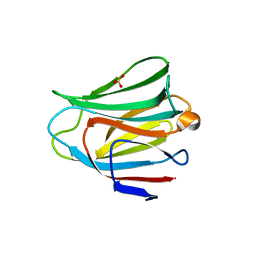
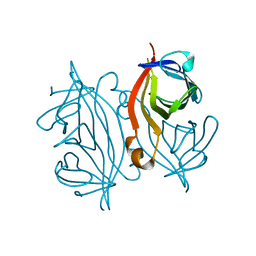
3AP5
 | |
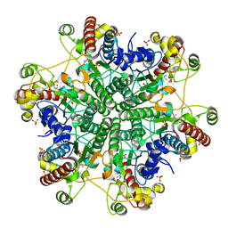
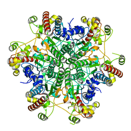
3AP6
 | | Crystal structure of the galectin-8 N-terminal carbohydrate recognition domain in complex with lactose 3'-sulfate | | 分子名称: | Galectin-8, SULFATE ION, beta-D-galactopyranose-(1-4)-beta-D-glucopyranose | | 著者 | Matsuzaka, T, Ideo, H, Yamashita, K, Nonaka, T. | | 登録日 | 2010-10-12 | | 公開日 | 2011-01-26 | | 最終更新日 | 2023-11-01 | | 実験手法 | X-RAY DIFFRACTION (1.58 Å) | | 主引用文献 | Galectin-8-N-domain recognition mechanism for sialylated and sulfated glycans
J.Biol.Chem., 286, 2011
|
|
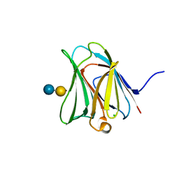
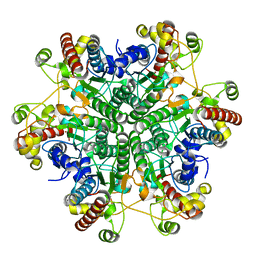
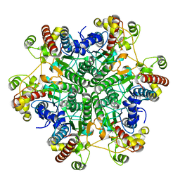
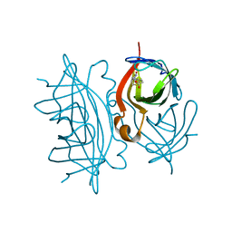
3AP4
 | | Crystal structure of the galectin-8 N-terminal carbohydrate recognition domain in complex with lactose | | 分子名称: | Galectin-8, beta-D-galactopyranose-(1-4)-alpha-D-glucopyranose | | 著者 | Matsuzaka, T, Ideo, H, Yamashita, K, Nonaka, T. | | 登録日 | 2010-10-11 | | 公開日 | 2011-01-26 | | 最終更新日 | 2024-03-13 | | 実験手法 | X-RAY DIFFRACTION (2.33 Å) | | 主引用文献 | Galectin-8-N-domain recognition mechanism for sialylated and sulfated glycans
J.Biol.Chem., 286, 2011
|
|
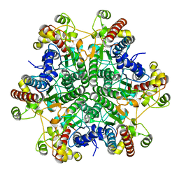
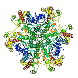
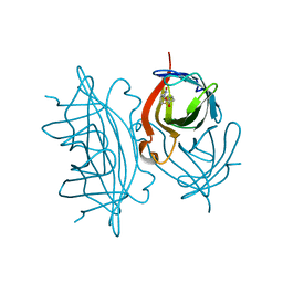
3AP7
 | | Crystal structure of the galectin-8 N-terminal carbohydrate recognition domain in complex with lactose sialic acid | | 分子名称: | CHLORIDE ION, Galectin-8, N-acetyl-alpha-neuraminic acid-(2-3)-beta-D-galactopyranose-(1-4)-beta-D-glucopyranose | | 著者 | Matsuzaka, T, Ideo, H, Yamashita, K, Nonaka, T. | | 登録日 | 2010-10-12 | | 公開日 | 2011-01-26 | | 最終更新日 | 2023-11-01 | | 実験手法 | X-RAY DIFFRACTION (1.53 Å) | | 主引用文献 | Galectin-8-N-domain recognition mechanism for sialylated and sulfated glycans
J.Biol.Chem., 286, 2011
|
|
3A6J
 | | E122Q mutant creatininase complexed with creatine | | 分子名称: | Creatinine amidohydrolase, N-[(E)-AMINO(IMINO)METHYL]-N-METHYLGLYCINE, SULFATE ION, ... | | 著者 | Nakajima, Y, Yamashita, K, Ito, K, Yoshimoto, T. | | 登録日 | 2009-09-02 | | 公開日 | 2010-02-09 | | 最終更新日 | 2023-11-01 | | 実験手法 | X-RAY DIFFRACTION (2 Å) | | 主引用文献 | Substitution of Glu122 by glutamine revealed the function of the second water molecule as a proton donor in the binuclear metal enzyme creatininase
J.Mol.Biol., 396, 2010
|
|
3A6D
 | | Creatininase complexed with 1-methylguanidine | | 分子名称: | 1-METHYLGUANIDINE, Creatinine amidohydrolase, MANGANESE (II) ION, ... | | 著者 | Nakajima, Y, Yamashita, K, Ito, K, Yoshimoto, T. | | 登録日 | 2009-08-31 | | 公開日 | 2010-02-09 | | 最終更新日 | 2023-11-01 | | 実験手法 | X-RAY DIFFRACTION (1.9 Å) | | 主引用文献 | Substitution of Glu122 by glutamine revealed the function of the second water molecule as a proton donor in the binuclear metal enzyme creatininase
J.Mol.Biol., 396, 2010
|
|
3A6L
 | | E122Q mutant creatininase, Zn-Zn type | | 分子名称: | CHLORIDE ION, Creatinine amidohydrolase, ZINC ION | | 著者 | Nakajima, Y, Yamashita, K, Ito, K, Yoshimoto, T. | | 登録日 | 2009-09-02 | | 公開日 | 2010-02-09 | | 最終更新日 | 2023-11-01 | | 実験手法 | X-RAY DIFFRACTION (2 Å) | | 主引用文献 | Substitution of Glu122 by glutamine revealed the function of the second water molecule as a proton donor in the binuclear metal enzyme creatininase
J.Mol.Biol., 396, 2010
|
|
3A6E
 | | W174F mutant creatininase, type I | | 分子名称: | CACODYLATE ION, Creatinine amidohydrolase, MANGANESE (II) ION, ... | | 著者 | Nakajima, Y, Yamashita, K, Ito, K, Yoshimoto, T. | | 登録日 | 2009-08-31 | | 公開日 | 2010-02-09 | | 最終更新日 | 2023-11-01 | | 実験手法 | X-RAY DIFFRACTION (2 Å) | | 主引用文献 | Substitution of Glu122 by glutamine revealed the function of the second water molecule as a proton donor in the binuclear metal enzyme creatininase
J.Mol.Biol., 396, 2010
|
|
3A6H
 | | W154A mutant creatininase | | 分子名称: | CHLORIDE ION, Creatinine amidohydrolase, MANGANESE (II) ION, ... | | 著者 | Nakajima, Y, Yamashita, K, Ito, K, Yoshimoto, T. | | 登録日 | 2009-08-31 | | 公開日 | 2010-02-09 | | 最終更新日 | 2023-11-01 | | 実験手法 | X-RAY DIFFRACTION (2 Å) | | 主引用文献 | Substitution of Glu122 by glutamine revealed the function of the second water molecule as a proton donor in the binuclear metal enzyme creatininase
J.Mol.Biol., 396, 2010
|
|
3A6G
 | | W154F mutant creatininase | | 分子名称: | Creatinine amidohydrolase, MANGANESE (II) ION, ZINC ION | | 著者 | Nakajima, Y, Yamashita, K, Ito, K, Yoshimoto, T. | | 登録日 | 2009-08-31 | | 公開日 | 2010-02-09 | | 最終更新日 | 2023-11-01 | | 実験手法 | X-RAY DIFFRACTION (2 Å) | | 主引用文献 | Substitution of Glu122 by glutamine revealed the function of the second water molecule as a proton donor in the binuclear metal enzyme creatininase
J.Mol.Biol., 396, 2010
|
|
3A6F
 | | W174F mutant creatininase, Type II | | 分子名称: | CACODYLATE ION, Creatinine amidohydrolase, MANGANESE (II) ION, ... | | 著者 | Nakajima, Y, Yamashita, K, Ito, K, Yoshimoto, T. | | 登録日 | 2009-08-31 | | 公開日 | 2010-02-09 | | 最終更新日 | 2023-11-01 | | 実験手法 | X-RAY DIFFRACTION (1.78 Å) | | 主引用文献 | Substitution of Glu122 by glutamine revealed the function of the second water molecule as a proton donor in the binuclear metal enzyme creatininase
J.Mol.Biol., 396, 2010
|
|
3A6K
 | | The E122Q mutant creatininase, Mn-Zn type | | 分子名称: | CHLORIDE ION, Creatinine amidohydrolase, MANGANESE (II) ION, ... | | 著者 | Nakajima, Y, Yamashita, K, Ito, K, Yoshimoto, T. | | 登録日 | 2009-09-02 | | 公開日 | 2010-02-09 | | 最終更新日 | 2023-11-01 | | 実験手法 | X-RAY DIFFRACTION (2.2 Å) | | 主引用文献 | Substitution of Glu122 by glutamine revealed the function of the second water molecule as a proton donor in the binuclear metal enzyme creatininase
J.Mol.Biol., 396, 2010
|
|
7A4M
 | | Cryo-EM structure of mouse heavy-chain apoferritin at 1.22 A | | 分子名称: | FE (III) ION, Ferritin heavy chain, ZINC ION | | 著者 | Nakane, T, Kotecha, A, Sente, A, Yamashita, K, McMullan, G, Masiulis, S, Brown, P.M.G.E, Grigoras, I.T, Malinauskaite, L, Malinauskas, T, Miehling, J, Yu, L, Karia, D, Pechnikova, E.V, de Jong, E, Keizer, J, Bischoff, M, McCormack, J, Tiemeijer, P, Hardwick, S.W, Chirgadze, D.Y, Murshudov, G, Aricescu, A.R, Scheres, S.H.W. | | 登録日 | 2020-08-20 | | 公開日 | 2020-10-28 | | 最終更新日 | 2024-07-10 | | 実験手法 | ELECTRON MICROSCOPY (1.22 Å) | | 主引用文献 | Single-particle cryo-EM at atomic resolution.
Nature, 587, 2020
|
|
7A5V
 | | CryoEM structure of a human gamma-aminobutyric acid receptor, the GABA(A)R-beta3 homopentamer, in complex with histamine and megabody Mb25 in lipid nanodisc | | 分子名称: | 2-acetamido-2-deoxy-beta-D-glucopyranose, 2-acetamido-2-deoxy-beta-D-glucopyranose-(1-4)-2-acetamido-2-deoxy-beta-D-glucopyranose, CHLORIDE ION, ... | | 著者 | Nakane, T, Kotecha, A, Sente, A, Yamashita, K, McMullan, G, Masiulis, S, Brown, P.M.G.E, Grigoras, I.T, Malinauskaite, L, Malinauskas, T, Miehling, J, Yu, L, Karia, D, Pechnikova, E.V, de Jong, E, Keizer, J, Bischoff, M, McCormack, J, Tiemeijer, P, Hardwick, S.W, Chirgadze, D.Y, Murshudov, G, Aricescu, A.R, Scheres, S.H.W. | | 登録日 | 2020-08-22 | | 公開日 | 2020-11-18 | | 最終更新日 | 2020-11-25 | | 実験手法 | ELECTRON MICROSCOPY (1.7 Å) | | 主引用文献 | Single-particle cryo-EM at atomic resolution.
Nature, 587, 2020
|
|
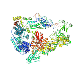
7F47
 | | Cryo-EM structure of Rhizobium etli MprF | | 分子名称: | (1R)-2-{[(S)-{[(2S)-2,3-dihydroxypropyl]oxy}(hydroxy)phosphoryl]oxy}-1-[(hexadecanoyloxy)methyl]ethyl (9Z)-octadec-9-enoate, Hypothetical conserved protein, [(2R)-1-[[(2R)-3-[(2S)-2,6-bis(azanyl)hexanoyl]oxy-2-oxidanyl-propoxy]-oxidanyl-phosphoryl]oxy-3-hexadecanoyloxy-propan-2-yl] (E)-octadec-9-enoate | | 著者 | Nishimura, M, Hirano, H, Kobayashi, K, Gill, C.P, Phan, C.N.K, Kise, Y, Kusakizako, T, Yamashita, K, Ito, Y, Roy, H, Nishizawa, T, Nureki, O. | | 登録日 | 2021-06-17 | | 公開日 | 2022-06-22 | | 最終更新日 | 2024-06-12 | | 実験手法 | ELECTRON MICROSCOPY (2.99 Å) | | 主引用文献 | Cryo-EM structure of Rhizobium etli MprF
To Be Published
|
|
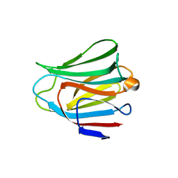
3APB
 | |
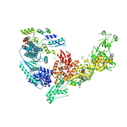
3B0V
 | | tRNA-dihydrouridine synthase from Thermus thermophilus in complex with tRNA | | 分子名称: | FLAVIN MONONUCLEOTIDE, tRNA, tRNA-dihydrouridine synthase | | 著者 | Yu, F, Tanaka, Y, Yamashita, K, Nakamura, A, Yao, M, Tanaka, I. | | 登録日 | 2011-06-14 | | 公開日 | 2011-12-14 | | 最終更新日 | 2017-10-11 | | 実験手法 | X-RAY DIFFRACTION (3.51 Å) | | 主引用文献 | Molecular basis of dihydrouridine formation on tRNA
Proc.Natl.Acad.Sci.USA, 108, 2011
|
|
7DY0
 | |
7EFD
 | | 1.77 A cryo-EM structure of Streptavidin using first 40 frames (corresponding to about 40 e/A^2 total dose) | | 分子名称: | BIOTIN, Streptavidin | | 著者 | Hiraizumi, M, Yamashita, K, Nishizawa, T, Kotecha, A, Nureki, O. | | 登録日 | 2021-03-21 | | 公開日 | 2021-04-28 | | 最終更新日 | 2024-06-05 | | 実験手法 | ELECTRON MICROSCOPY (1.77 Å) | | 主引用文献 | 1.77 A cryo-EM structure of Streptavidin using first 40 frames (corresponding to about 40e/A^2 total dose)
To Be Published
|
|
7EFC
 | | 1.70 A cryo-EM structure of streptavidin | | 分子名称: | BIOTIN, Streptavidin | | 著者 | Hiraizumi, M, Yamashita, K, Nishizawa, T, Kotecha, A, Nureki, O. | | 登録日 | 2021-03-21 | | 公開日 | 2021-04-28 | | 最終更新日 | 2024-06-05 | | 実験手法 | ELECTRON MICROSCOPY (1.7 Å) | | 主引用文献 | 1.70 A cryo-EM structure of streptavidin using all frames (corresponding to 70 e/A^2 total dose)
To Be Published
|
|
7V6B
 | | Structure of the Dicer-2-R2D2 heterodimer | | 分子名称: | Dicer-2, isoform A, R2D2 | | 著者 | Yamaguchi, S, Nishizawa, T, Kusakizako, T, Yamashita, K, Tomita, A, Hirano, H, Nishimasu, H, Nureki, O. | | 登録日 | 2021-08-20 | | 公開日 | 2022-03-23 | | 最終更新日 | 2024-06-12 | | 実験手法 | ELECTRON MICROSCOPY (3.3 Å) | | 主引用文献 | Structure of the Dicer-2-R2D2 heterodimer bound to a small RNA duplex.
Nature, 607, 2022
|
|
7V6C
 | | Structure of the Dicer-2-R2D2 heterodimer bound to small RNA duplex | | 分子名称: | Dicer-2, isoform A, R2D2, ... | | 著者 | Yamaguchi, S, Nishizawa, T, Kusakizako, T, Yamashita, K, Tomita, A, Hirano, H, Nishimasu, H, Nureki, O. | | 登録日 | 2021-08-20 | | 公開日 | 2022-03-23 | | 最終更新日 | 2024-06-12 | | 実験手法 | ELECTRON MICROSCOPY (3.3 Å) | | 主引用文献 | Structure of the Dicer-2-R2D2 heterodimer bound to a small RNA duplex.
Nature, 607, 2022
|
|


