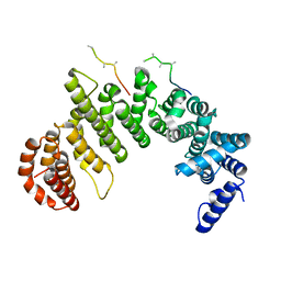
3ZFM
 
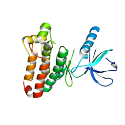 | | Crystal structure of EphB2 | | 分子名称: | EPHRIN TYPE-B RECEPTOR 2 | | 著者 | Debreczeni, J.E, Overman, R, Truman, C, McAlister, M, Attwood, T.K. | | 登録日 | 2012-12-12 | | 公開日 | 2014-01-08 | | 最終更新日 | 2024-05-08 | | 実験手法 | X-RAY DIFFRACTION (2.27 Å) | | 主引用文献 | Completing the Structural Family Portrait of the Human Ephb Tyrosine Kinase Domains
Protein Sci., 23, 2014
|
|
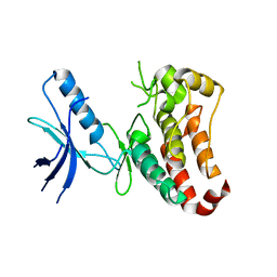
3ZEW
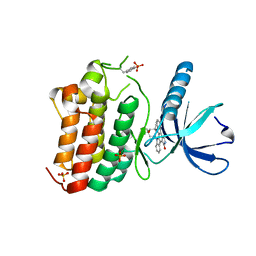 | | Crystal structure of EphB4 in complex with staurosporine | | 分子名称: | EPHRIN TYPE-B RECEPTOR 4, STAUROSPORINE, SULFATE ION | | 著者 | Debreczeni, J.E, Overman, R, Truman, C, McAlister, M, Attwood, T.K. | | 登録日 | 2012-12-07 | | 公開日 | 2013-12-25 | | 最終更新日 | 2017-06-28 | | 実験手法 | X-RAY DIFFRACTION (2.5 Å) | | 主引用文献 | Completing the Structural Family Portrait of the Human Ephb Tyrosine Kinase Domains
Protein Sci., 23, 2014
|
|
1NYS
 | |
1NYU
 | |
1QQ3
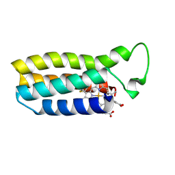 | | THE SOLUTION STRUCTURE OF THE HEME BINDING VARIANT ARG98CYS OF OXIDIZED ESCHERICHIA COLI CYTOCHROME B562 | | 分子名称: | CYTOCHROME B562, HEME B/C | | 著者 | Arnesano, F, Banci, L, Bertini, I, Ciofi-Baffoni, S, Barker, P.D, Woodyear, T. | | 登録日 | 1999-06-10 | | 公開日 | 2000-05-24 | | 最終更新日 | 2021-11-03 | | 実験手法 | SOLUTION NMR | | 主引用文献 | Structural consequences of b- to c-type heme conversion in oxidized Escherichia coli cytochrome b562.
Biochemistry, 39, 2000
|
|
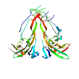
2F9N
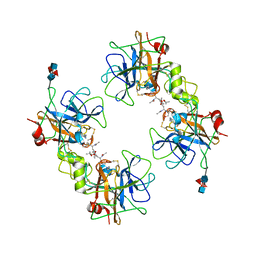 | | Crystal Structure of the Recombinant Human Alpha I Tryptase Mutant K192Q/D216G in Complex with Leupeptin | | 分子名称: | (R,R)-2,3-BUTANEDIOL, 2-acetamido-2-deoxy-beta-D-glucopyranose, Leupeptin, ... | | 著者 | Rohr, K.B, Selwood, T, Marquardt, U, Huber, R, Schechter, N.M, Bode, W, Than, M.E. | | 登録日 | 2005-12-06 | | 公開日 | 2006-01-31 | | 最終更新日 | 2023-08-30 | | 実験手法 | X-RAY DIFFRACTION (1.6 Å) | | 主引用文献 | X-ray Structures of Free and Leupeptin-complexed Human alpha I-Tryptase Mutants: Indication for an alpha to beta-Tryptase Transition
J.Mol.Biol., 357, 2005
|
|
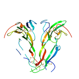
2F9P
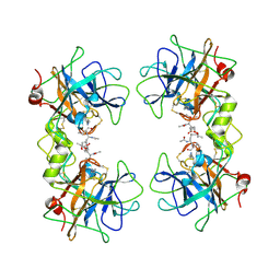 | | Crystal Structure of the Recombinant Human Alpha I Tryptase Mutant D216G in Complex with Leupeptin | | 分子名称: | (R,R)-2,3-BUTANEDIOL, 2-acetamido-2-deoxy-beta-D-glucopyranose, Leupeptin, ... | | 著者 | Rohr, K.B, Selwood, T, Marquardt, U, Huber, R, Schechter, N.M, Bode, W, Than, M.E. | | 登録日 | 2005-12-06 | | 公開日 | 2006-01-31 | | 最終更新日 | 2023-08-30 | | 実験手法 | X-RAY DIFFRACTION (2.3 Å) | | 主引用文献 | X-ray Structures of Free and Leupeptin-complexed Human alpha I-Tryptase Mutants: Indication for an alpha to beta-Tryptase Transition
J.Mol.Biol., 357, 2005
|
|
2F9O
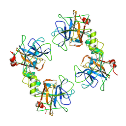 | | Crystal Structure of the Recombinant Human Alpha I Tryptase Mutant D216G | | 分子名称: | Tryptase alpha-1, alpha-L-fucopyranose-(1-3)-[2-acetamido-2-deoxy-beta-D-glucopyranose-(1-4)]2-acetamido-2-deoxy-beta-D-glucopyranose | | 著者 | Rohr, K.B, Selwood, T, Marquardt, U, Huber, R, Schechter, N.M, Bode, W, Than, M.E. | | 登録日 | 2005-12-06 | | 公開日 | 2006-01-31 | | 最終更新日 | 2023-08-30 | | 実験手法 | X-RAY DIFFRACTION (2.1 Å) | | 主引用文献 | X-ray Structures of Free and Leupeptin-complexed Human alpha I-Tryptase Mutants: Indication for an alpha to beta-Tryptase Transition
J.Mol.Biol., 357, 2005
|
|
1F59
 | |
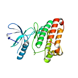
3ZFY
 | | Crystal structure of EphB3 | | 分子名称: | EPHRIN TYPE-B RECEPTOR 3 | | 著者 | Debreczeni, J.E, Overman, R, Truman, C, McAlister, M, Attwood, T.K. | | 登録日 | 2012-12-12 | | 公開日 | 2014-01-08 | | 最終更新日 | 2024-05-08 | | 実験手法 | X-RAY DIFFRACTION (2.2 Å) | | 主引用文献 | Completing the Structural Family Portrait of the Human Ephb Tyrosine Kinase Domains
Protein Sci., 23, 2014
|
|
3ZFX
 | | Crystal structure of EphB1 | | 分子名称: | EPHRIN TYPE-B RECEPTOR 1, SULFATE ION | | 著者 | Debreczeni, J.E, Overman, R, Truman, C, McAlister, M, Attwood, T.K. | | 登録日 | 2012-12-12 | | 公開日 | 2014-01-08 | | 最終更新日 | 2024-05-08 | | 実験手法 | X-RAY DIFFRACTION (2.5 Å) | | 主引用文献 | Completing the Structural Family Portrait of the Human Ephb Tyrosine Kinase Domains
Protein Sci., 23, 2014
|
|


