3F6D
 
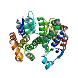
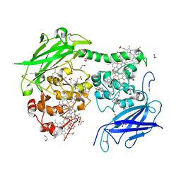
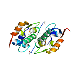
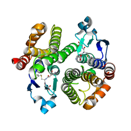 | | Crystal Structure of a Genetically Modified Delta Class GST (adGSTD4-4) from Anopheles dirus, F123A, in Complex with S-Hexyl Glutathione | | 分子名称: | Glutathione transferase GST1-4, S-HEXYLGLUTATHIONE | | 著者 | Wongsantichon, J, Robinson, R.C, Ketterman, A.J. | | 登録日 | 2008-11-05 | | 公開日 | 2009-10-27 | | 最終更新日 | 2023-11-01 | | 実験手法 | X-RAY DIFFRACTION (1.7 Å) | | 主引用文献 | Structural contributions of delta class glutathione transferase active-site residues to catalysis
Biochem.J., 428, 2010
|
|
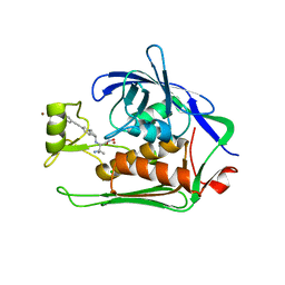
3F6F
 | |
3GH6
 | |
3MAK
 | |
3G7I
 | |
3OMY
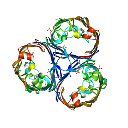
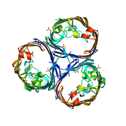
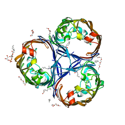
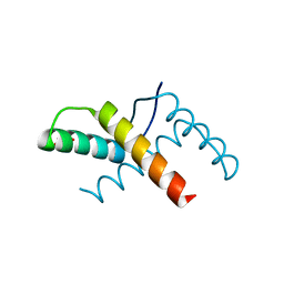 | | Crystal structure of the pED208 TraM N-terminal domain | | 分子名称: | GLYCEROL, MAGNESIUM ION, Protein traM | | 著者 | Wong, J.J.W, Lu, J, Edwards, R.A, Frost, L.S, Mark Glover, J.N. | | 登録日 | 2010-08-27 | | 公開日 | 2011-05-25 | | 最終更新日 | 2023-09-06 | | 実験手法 | X-RAY DIFFRACTION (1.3 Å) | | 主引用文献 | Structural basis of cooperative DNA recognition by the plasmid conjugation factor, TraM.
Nucleic Acids Res., 39, 2011
|
|
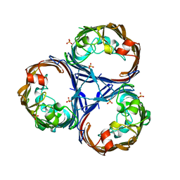
3ON0
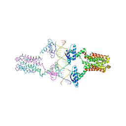 | | Crystal structure of the pED208 TraM-sbmA complex | | 分子名称: | Protein traM, sbmA | | 著者 | Wong, J.J.W, Lu, J, Edwards, R.A, Frost, L.S, Mark Glover, J.N. | | 登録日 | 2010-08-27 | | 公開日 | 2011-05-25 | | 最終更新日 | 2023-09-06 | | 実験手法 | X-RAY DIFFRACTION (2.874 Å) | | 主引用文献 | Structural basis of cooperative DNA recognition by the plasmid conjugation factor, TraM.
Nucleic Acids Res., 39, 2011
|
|
5UKE
 | |
7O7G
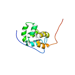
 | | Crystal structure of the Shewanella oneidensis MR1 MtrC mutant H561M | | 分子名称: | 1,2-ETHANEDIOL, ACETATE ION, CALCIUM ION, ... | | 著者 | Edwards, M.J, van Wonderen, J.H, Newton-Payne, S.E, Butt, J.N, Clarke, T.A. | | 登録日 | 2021-04-13 | | 公開日 | 2021-10-06 | | 最終更新日 | 2024-01-31 | | 実験手法 | X-RAY DIFFRACTION (1.6 Å) | | 主引用文献 | Nanosecond heme-to-heme electron transfer rates in a multiheme cytochrome nanowire reported by a spectrally unique His/Met-ligated heme.
Proc.Natl.Acad.Sci.USA, 118, 2021
|
|
4QPQ
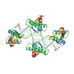 | | Mechanistic basis of plasmid-specific DNA binding of the F plasmid regulatory protein, TraM | | 分子名称: | Relaxosome protein TraM, sbmA DNA1, sbmA DNA2 | | 著者 | Peng, Y, Lu, J, Wong, J, Edwards, R.A, Frost, L.S, Glover, J.N.M. | | 登録日 | 2014-06-24 | | 公開日 | 2014-09-24 | | 最終更新日 | 2024-02-28 | | 実験手法 | X-RAY DIFFRACTION (3.106 Å) | | 主引用文献 | Mechanistic Basis of Plasmid-Specific DNA Binding of the F Plasmid Regulatory Protein, TraM.
J.Mol.Biol., 426, 2014
|
|
4QPO
 | | Mechanistic basis of plasmid-specific DNA binding of the F plasmid regulatory protein, TraM | | 分子名称: | PHOSPHATE ION, Relaxosome protein TraM | | 著者 | Peng, Y, Lu, J, Wong, J, Edwards, R.A, Frost, L.S, Glover, J.N.M. | | 登録日 | 2014-06-24 | | 公開日 | 2014-09-03 | | 最終更新日 | 2024-02-28 | | 実験手法 | X-RAY DIFFRACTION (1.999 Å) | | 主引用文献 | Mechanistic Basis of Plasmid-Specific DNA Binding of the F Plasmid Regulatory Protein, TraM.
J.Mol.Biol., 426, 2014
|
|
7T7A
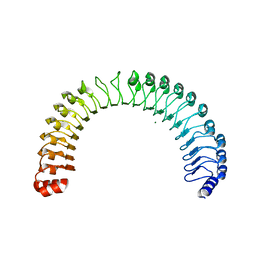 | | Crystal Structure of Human SHOC2: A Leucine-Rich Repeat Protein | | 分子名称: | Leucine-rich repeat protein SHOC-2, MAGNESIUM ION, NITRATE ION | | 著者 | Hajian, B, Lemke, C, Kwon, J, Bian, Y, Fuller, C, Aguirre, J. | | 登録日 | 2021-12-14 | | 公開日 | 2022-05-04 | | 最終更新日 | 2024-05-22 | | 実験手法 | X-RAY DIFFRACTION (1.79 Å) | | 主引用文献 | Structure-function analysis of the SHOC2-MRAS-PP1C holophosphatase complex.
Nature, 609, 2022
|
|
4UMN
 | | Structure of a stapled peptide antagonist bound to Nutlin-resistant Mdm2. | | 分子名称: | E3 ubiquitin-protein ligase Mdm2, M06 | | 著者 | Chee, S, Wongsantichon, J, Quah, S, Robinson, R.C, Verma, C, Lane, D.P, Brown, C.J, Ghadessy, F.J. | | 登録日 | 2014-05-20 | | 公開日 | 2014-05-28 | | 最終更新日 | 2024-02-07 | | 実験手法 | X-RAY DIFFRACTION (1.99 Å) | | 主引用文献 | Structure of a stapled peptide antagonist bound to nutlin-resistant Mdm2.
PLoS ONE, 9, 2014
|
|
5N8C
 | | Crystal structure of Pseudomonas aeruginosa LpxC complexed with inhibitor | | 分子名称: | (2~{S})-3-azanyl-2-[[(1~{R})-5-[2-[4-[[2-(hydroxymethyl)imidazol-1-yl]methyl]phenyl]ethynyl]-2,3-dihydro-1~{H}-inden-1-yl]amino]-3-methyl-~{N}-oxidanyl-butanamide, CHLORIDE ION, UDP-3-O-acyl-N-acetylglucosamine deacetylase, ... | | 著者 | Cross, J.B, Ryan, M.D, Zhang, J, Cheng, R.K, Wood, M, Andersen, O.A, Brooks, M, Kwong, J, Barker, J. | | 登録日 | 2017-02-23 | | 公開日 | 2017-03-29 | | 最終更新日 | 2024-01-17 | | 実験手法 | X-RAY DIFFRACTION (1.9 Å) | | 主引用文献 | Structure-based discovery of LpxC inhibitors.
Bioorg. Med. Chem. Lett., 27, 2017
|
|
7UPI
 | | Cryo-EM structure of SHOC2-PP1c-MRAS holophosphatase complex | | 分子名称: | CHLORIDE ION, GUANOSINE-5'-TRIPHOSPHATE, Leucine-rich repeat protein SHOC-2, ... | | 著者 | Fuller, J.R, Hajian, B, Lemke, C, Kwon, J, Bian, Y, Aguirre, A. | | 登録日 | 2022-04-15 | | 公開日 | 2022-05-04 | | 最終更新日 | 2024-06-12 | | 実験手法 | ELECTRON MICROSCOPY (2.89 Å) | | 主引用文献 | Structure-function analysis of the SHOC2-MRAS-PP1C holophosphatase complex.
Nature, 609, 2022
|
|
8QVV
 | | Crystal structure of Ompk36 GD at 3500 eV based on analytical absorption corrections | | 分子名称: | OmpK36, SULFATE ION | | 著者 | Duman, R, Wagner, A, Beis, K, Wong, J. | | 登録日 | 2023-10-18 | | 公開日 | 2024-06-19 | | 実験手法 | X-RAY DIFFRACTION (2.34 Å) | | 主引用文献 | Ray-tracing analytical absorption correction for X-ray crystallography based on tomographic reconstructions.
J.Appl.Crystallogr., 57, 2024
|
|
8QUQ
 | | Crystal structure of Ompk36 GD at 3500 eV based on spherical harmonics absorption corrections | | 分子名称: | OmpK36, SULFATE ION | | 著者 | Duman, R, Wagner, A, Beis, K, Wong, J. | | 登録日 | 2023-10-16 | | 公開日 | 2024-06-19 | | 実験手法 | X-RAY DIFFRACTION (2.34 Å) | | 主引用文献 | Ray-tracing analytical absorption correction for X-ray crystallography based on tomographic reconstructions.
J.Appl.Crystallogr., 57, 2024
|
|
8QVS
 | | Crystal structure of Ompk36 GD at 3500 eV based on a combination of spherical harmonics and analytical absorption corrections | | 分子名称: | OmpK36, SULFATE ION | | 著者 | Duman, R, Wagner, A, Beis, K, Wong, J. | | 登録日 | 2023-10-18 | | 公開日 | 2024-06-19 | | 実験手法 | X-RAY DIFFRACTION (2.34 Å) | | 主引用文献 | Ray-tracing analytical absorption correction for X-ray crystallography based on tomographic reconstructions.
J.Appl.Crystallogr., 57, 2024
|
|
8QUR
 | | Crystal structure of Ompk36 GD at 3500 eV with no absorption corrections | | 分子名称: | OmpK36, SULFATE ION | | 著者 | Duman, R, Wagner, A, Beis, K, Wong, J. | | 登録日 | 2023-10-16 | | 公開日 | 2024-06-19 | | 実験手法 | X-RAY DIFFRACTION (2.34 Å) | | 主引用文献 | Ray-tracing analytical absorption correction for X-ray crystallography based on tomographic reconstructions.
J.Appl.Crystallogr., 57, 2024
|
|
6RCP
 | |
6RD3
 | |
3D8A
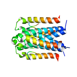 | | Co-crystal structure of TraM-TraD complex. | | 分子名称: | Protein traD, Relaxosome protein TraM | | 著者 | Glover, J.N.M, Lu, J, Wong, J.J, Edwards, R.A. | | 登録日 | 2008-05-22 | | 公開日 | 2008-09-09 | | 最終更新日 | 2023-08-30 | | 実験手法 | X-RAY DIFFRACTION (2.55 Å) | | 主引用文献 | Structural basis of specific TraD-TraM recognition during F plasmid-mediated bacterial conjugation.
Mol.Microbiol., 70, 2008
|
|
6RCK
 | | Crystal structure of the OmpK36 GD insertion chimera from Klebsiella pneumonia | | 分子名称: | (2S)-3-{[{[(2S)-2,3-DIHYDROXYPROPYL]OXY}(HYDROXY)PHOSPHORYL]OXY}-2-[(6E)-HEXADEC-6-ENOYLOXY]PROPYL (8E)-OCTADEC-8-ENOATE, (HYDROXYETHYLOXY)TRI(ETHYLOXY)OCTANE, LAURYL DIMETHYLAMINE-N-OXIDE, ... | | 著者 | Beis, K, Romano, M, Kwong, J. | | 登録日 | 2019-04-11 | | 公開日 | 2019-09-11 | | 最終更新日 | 2024-01-24 | | 実験手法 | X-RAY DIFFRACTION (2.029 Å) | | 主引用文献 | OmpK36-mediated Carbapenem resistance attenuates ST258 Klebsiella pneumoniae in vivo.
Nat Commun, 10, 2019
|
|
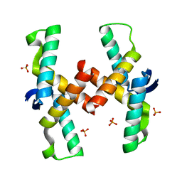
2G7O
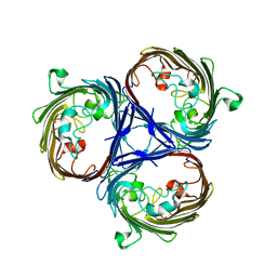
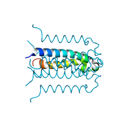 | | Protonation-mediated structural flexibility in the F conjugation regulatory protein, TraM | | 分子名称: | Protein traM | | 著者 | Lu, J, Edwards, R.A, Wong, J.J, Manchak, J, Scott, P.G, Frost, L.S, Glover, J.N. | | 登録日 | 2006-02-28 | | 公開日 | 2006-06-13 | | 最終更新日 | 2024-02-14 | | 実験手法 | X-RAY DIFFRACTION (1.4 Å) | | 主引用文献 | Protonation-mediated structural flexibility in the F conjugation regulatory protein, TraM.
Embo J., 25, 2006
|
|
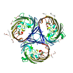
2G9E
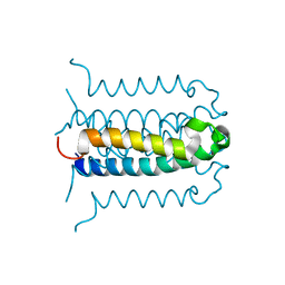 | | Protonation-mediated structural flexibility in the F conjugation regulatory protein, TRAM | | 分子名称: | Protein traM | | 著者 | Lu, J, Edwards, R.A, Wong, J.J, Manchak, J, Scott, P.G, Frost, L.S, Glover, J.N. | | 登録日 | 2006-03-06 | | 公開日 | 2006-06-13 | | 最終更新日 | 2023-08-30 | | 実験手法 | X-RAY DIFFRACTION (1.8 Å) | | 主引用文献 | Protonation-mediated structural flexibility in the F conjugation regulatory protein, TraM.
Embo J., 25, 2006
|
|


