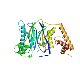
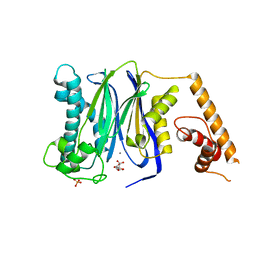
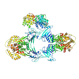
5G0I
 
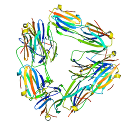
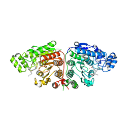 | | Crystal structure of Danio rerio HDAC6 CD1 and CD2 (linker cleaved) in complex with Nexturastat A | | 分子名称: | 1,2-ETHANEDIOL, CHLORIDE ION, HDAC6, ... | | 著者 | Miyake, Y, Keusch, J.J, Wang, L, Saito, M, Hess, D, Wang, X, Melancon, B.J, Helquist, P, Gut, H, Matthias, P. | | 登録日 | 2016-03-18 | | 公開日 | 2016-07-27 | | 最終更新日 | 2024-01-10 | | 実験手法 | X-RAY DIFFRACTION (1.99 Å) | | 主引用文献 | Structural Insights Into Hdac6 Tubulin Deacetylation and its Selective Inhibition
Nat.Chem.Biol., 12, 2016
|
|
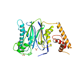
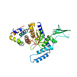
5G0F
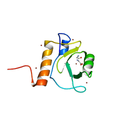 | | Crystal structure of Danio rerio HDAC6 ZnF-UBP domain | | 分子名称: | GLYCINE, HDAC6, NICKEL (II) ION, ... | | 著者 | Miyake, Y, Keusch, J.J, Wang, L, Saito, M, Hess, D, Wang, X, Melancon, B.J, Helquist, P, Gut, H, Matthias, P. | | 登録日 | 2016-03-18 | | 公開日 | 2016-07-27 | | 最終更新日 | 2024-01-10 | | 実験手法 | X-RAY DIFFRACTION (1.9 Å) | | 主引用文献 | Structural Insights Into Hdac6 Tubulin Deacetylation and its Selective Inhibition
Nat.Chem.Biol., 12, 2016
|
|
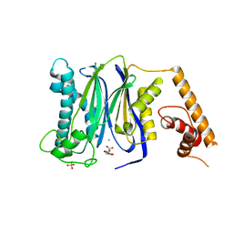
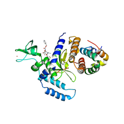
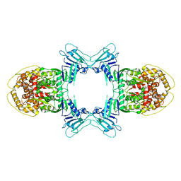
5G0J
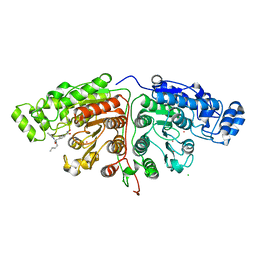 | | Crystal structure of Danio rerio HDAC6 CD1 and CD2 (linker intact) in complex with Nexturastat A | | 分子名称: | CHLORIDE ION, HDAC6, NEXTURASTAT A, ... | | 著者 | Miyake, Y, Keusch, J.J, Wang, L, Saito, M, Hess, D, Wang, X, Melancon, B.J, Helquist, P, Gut, H, Matthias, P. | | 登録日 | 2016-03-18 | | 公開日 | 2016-07-27 | | 最終更新日 | 2024-01-10 | | 実験手法 | X-RAY DIFFRACTION (2.88 Å) | | 主引用文献 | Structural Insights Into Hdac6 Tubulin Deacetylation and its Selective Inhibition
Nat.Chem.Biol., 12, 2016
|
|
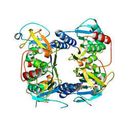
5G0G
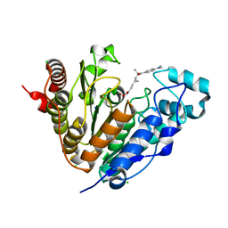 | | Crystal structure of Danio rerio HDAC6 CD1 in complex with trichostatin A | | 分子名称: | CHLORIDE ION, HDAC6, SODIUM ION, ... | | 著者 | Miyake, Y, Keusch, J.J, Wang, L, Saito, M, Hess, D, Wang, X, Melancon, B.J, Helquist, P, Gut, H, Matthias, P. | | 登録日 | 2016-03-18 | | 公開日 | 2016-07-27 | | 最終更新日 | 2024-05-01 | | 実験手法 | X-RAY DIFFRACTION (1.499 Å) | | 主引用文献 | Structural Insights Into Hdac6 Tubulin Deacetylation and its Selective Inhibition
Nat.Chem.Biol., 12, 2016
|
|
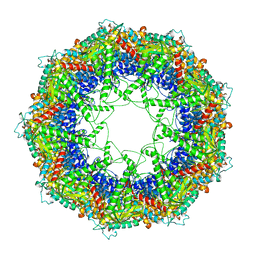
2BYU
 | | Negative stain EM reconstruction of M.tuberculosis Acr1(Hsp 16.3) fitted with wheat sHSP dimer | | 分子名称: | HEAT SHOCK PROTEIN 16.9B | | 著者 | Kennaway, C.K, Benesch, J.L.P, Gohlke, U, Wang, L, Robinson, C.V, Orlova, E.V, Saibil, H.R, Keep, N.H. | | 登録日 | 2005-08-05 | | 公開日 | 2005-08-22 | | 最終更新日 | 2024-05-08 | | 実験手法 | ELECTRON MICROSCOPY (16.5 Å) | | 主引用文献 | Dodecameric Structure of the Small Heat Shock Protein Acr1 from Mycobacterium Tuberculosis.
J.Biol.Chem., 280, 2005
|
|
6PAF
 | |
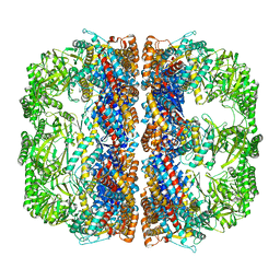
6OMM
 | | Cryo-EM structure of formyl peptide receptor 2/lipoxin A4 receptor in complex with Gi | | 分子名称: | CHOLESTEROL, Guanine nucleotide-binding protein G(I)/G(S)/G(O) subunit gamma-2, Guanine nucleotide-binding protein G(I)/G(S)/G(T) subunit beta-1, ... | | 著者 | Zhuang, Y, Liu, H, de Waal, P.W, Zhou, X.E, Wang, L, Meng, X, Zhao, G, Kang, Y, Melcher, K, Xu, H.E, Zhang, C. | | 登録日 | 2019-04-19 | | 公開日 | 2020-02-26 | | 実験手法 | ELECTRON MICROSCOPY (3.17 Å) | | 主引用文献 | Structure of formylpeptide receptor 2-Gicomplex reveals insights into ligand recognition and signaling.
Nat Commun, 11, 2020
|
|
3J1E
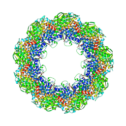 | | Cryo-EM structure of 9-fold symmetric rATcpn-beta in apo state | | 分子名称: | Chaperonin beta subunit | | 著者 | Zhang, K, Wang, L, Liu, Y.X, Wang, X, Gao, B, Hu, Z.J, Ji, G, Chan, K.Y, Schulten, K, Dong, Z.Y, Sun, F. | | 登録日 | 2012-02-06 | | 公開日 | 2013-08-07 | | 最終更新日 | 2024-02-21 | | 実験手法 | ELECTRON MICROSCOPY (8.3 Å) | | 主引用文献 | Flexible interwoven termini determine the thermal stability of thermosomes.
Protein Cell, 4, 2013
|
|
3J1B
 | | Cryo-EM structure of 8-fold symmetric rATcpn-alpha in apo state | | 分子名称: | Chaperonin alpha subunit | | 著者 | Zhang, K, Wang, L, Liu, Y.X, Wang, X, Gao, B, Hu, Z.J, Ji, G, Chan, K.Y, Schulten, K, Dong, Z.Y, Sun, F. | | 登録日 | 2012-02-06 | | 公開日 | 2013-08-07 | | 最終更新日 | 2024-02-21 | | 実験手法 | ELECTRON MICROSCOPY (4.9 Å) | | 主引用文献 | Flexible interwoven termini determine the thermal stability of thermosomes.
Protein Cell, 4, 2013
|
|
3J1C
 | | Cryo-EM structure of 9-fold symmetric rATcpn-alpha in apo state | | 分子名称: | Chaperonin alpha subunit | | 著者 | Zhang, K, Wang, L, Liu, Y.X, Wang, X, Gao, B, Hu, Z.J, Ji, G, Chan, K.Y, Schulten, K, Dong, Z.Y, Sun, F. | | 登録日 | 2012-02-06 | | 公開日 | 2013-08-07 | | 最終更新日 | 2024-02-21 | | 実験手法 | ELECTRON MICROSCOPY (9.1 Å) | | 主引用文献 | Flexible interwoven termini determine the thermal stability of thermosomes.
Protein Cell, 4, 2013
|
|
5G0H
 | | Crystal structure of Danio rerio HDAC6 CD2 in complex with (S)- trichostatin A | | 分子名称: | 1,2-ETHANEDIOL, HDAC6, POTASSIUM ION, ... | | 著者 | Miyake, Y, Keusch, J.J, Wang, L, Saito, M, Hess, D, Wang, X, Melancon, B.J, Helquist, P, Gut, H, Matthias, P. | | 登録日 | 2016-03-18 | | 公開日 | 2016-07-27 | | 最終更新日 | 2024-05-01 | | 実験手法 | X-RAY DIFFRACTION (1.6 Å) | | 主引用文献 | Structural Insights Into Hdac6 Tubulin Deacetylation and its Selective Inhibition
Nat.Chem.Biol., 12, 2016
|
|
3J1F
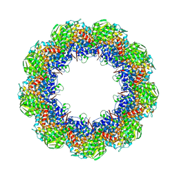 | | Cryo-EM structure of 9-fold symmetric rATcpn-beta in ATP-binding state | | 分子名称: | ADENOSINE-5'-TRIPHOSPHATE, Chaperonin beta subunit, MAGNESIUM ION | | 著者 | Zhang, K, Wang, L, Liu, Y.X, Wang, X, Gao, B, Hu, Z.J, Ji, G, Chan, K.Y, Schulten, K, Dong, Z.Y, Sun, F. | | 登録日 | 2012-02-06 | | 公開日 | 2013-08-07 | | 最終更新日 | 2024-02-21 | | 実験手法 | ELECTRON MICROSCOPY (6.2 Å) | | 主引用文献 | Flexible interwoven termini determine the thermal stability of thermosomes.
Protein Cell, 4, 2013
|
|
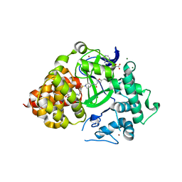
1QBQ
 | | STRUCTURE OF RAT FARNESYL PROTEIN TRANSFERASE COMPLEXED WITH A CVIM PEPTIDE AND ALPHA-HYDROXYFARNESYLPHOSPHONIC ACID. | | 分子名称: | ACETATE ION, ACETYL-CYS-VAL-ILE-SELENOMET-COOH PEPTIDE, ALPHA-HYDROXYFARNESYLPHOSPHONIC ACID, ... | | 著者 | Strickland, C.L, Windsor, W.T, Syto, R, Wang, L, Bond, R, Wu, Z, Schwartz, J, Le, H.V, Beese, L.S, Weber, P.C. | | 登録日 | 1999-04-27 | | 公開日 | 1999-06-18 | | 最終更新日 | 2024-03-13 | | 実験手法 | X-RAY DIFFRACTION (2.4 Å) | | 主引用文献 | Crystal structure of farnesyl protein transferase complexed with a CaaX peptide and farnesyl diphosphate analogue
Biochemistry, 37, 1998
|
|
3FXK
 | | Crystal Structure of Human Protein phosphatase 1A (PPM1A) Bound with Phosphate at 10 mM of Mn2+ | | 分子名称: | MANGANESE (II) ION, PHOSPHATE ION, Protein phosphatase 1A | | 著者 | Hu, T, Wang, L, Wang, K, Jiang, H, Shen, X. | | 登録日 | 2009-01-21 | | 公開日 | 2010-01-26 | | 最終更新日 | 2024-03-20 | | 実験手法 | X-RAY DIFFRACTION (2.1 Å) | | 主引用文献 | Structural basis for the Mn2+-dependent activation of human PPM1A
To be published
|
|
3FXJ
 | | Crystal Structure of Human Protein phosphatase 1A (PPM1A) Bound with Phosphate at 3 mM of Mn2+ | | 分子名称: | MANGANESE (II) ION, PHOSPHATE ION, Protein phosphatase 1A | | 著者 | Hu, T, Wang, L, Wang, K, Jiang, H, Shen, X. | | 登録日 | 2009-01-21 | | 公開日 | 2010-01-26 | | 最終更新日 | 2024-03-20 | | 実験手法 | X-RAY DIFFRACTION (2.5 Å) | | 主引用文献 | Structural basis for the Mn2+-dependent activation of human PPM1A
To be published
|
|
3FXL
 | | Crystal Structure of Human Protein phosphatase 1A (PPM1A) Bound with Citrate at 1 mM of Mn2+ | | 分子名称: | CITRATE ANION, MANGANESE (II) ION, PHOSPHATE ION, ... | | 著者 | Hu, T, Wang, L, Wang, K, Jiang, H, Shen, X. | | 登録日 | 2009-01-21 | | 公開日 | 2010-01-26 | | 最終更新日 | 2024-03-20 | | 実験手法 | X-RAY DIFFRACTION (2.3 Å) | | 主引用文献 | Structural basis for the Mn2+-dependent activation of human PPM1A
To be published
|
|
2B8T
 | | Crystal structure of Thymidine Kinase from U.urealyticum in complex with thymidine | | 分子名称: | 2-AMINO-2-HYDROXYMETHYL-PROPANE-1,3-DIOL, THYMIDINE, Thymidine kinase, ... | | 著者 | Kosinska, U, Carnrot, C, Eriksson, S, Wang, L, Eklund, H. | | 登録日 | 2005-10-10 | | 公開日 | 2005-12-20 | | 最終更新日 | 2023-08-23 | | 実験手法 | X-RAY DIFFRACTION (2 Å) | | 主引用文献 | Structure of the substrate complex of thymidine kinase from Ureaplasma urealyticum and investigations of possible drug targets for the enzyme
FEBS Lett., 272, 2005
|
|
3FXO
 | | Crystal Structure of Human Protein phosphatase 1A (PPM1A) Bound with Phosphate at 1 mM of Mn2+ | | 分子名称: | MANGANESE (II) ION, PHOSPHATE ION, Protein phosphatase 1A | | 著者 | Hu, T, Wang, L, Wang, K, Jiang, H, Shen, X. | | 登録日 | 2009-01-21 | | 公開日 | 2010-01-26 | | 最終更新日 | 2024-03-20 | | 実験手法 | X-RAY DIFFRACTION (2.5 Å) | | 主引用文献 | Structural basis for the Mn2+-dependent activation of human PPM1A
To be published
|
|
3FXM
 | | Crystal Structure of Human Protein phosphatase 1A (PPM1A) Bound with Citrate at 10 mM of Mn2+ | | 分子名称: | CITRATE ANION, MANGANESE (II) ION, PHOSPHATE ION, ... | | 著者 | Hu, T, Wang, L, Wang, K, Jiang, H, Shen, X. | | 登録日 | 2009-01-21 | | 公開日 | 2010-01-26 | | 最終更新日 | 2024-03-20 | | 実験手法 | X-RAY DIFFRACTION (2.5 Å) | | 主引用文献 | Structural basis for the Mn2+-dependent activation of human PPM1A
To be published
|
|
2F4M
 | | The Mouse PNGase-HR23 Complex Reveals a Complete Remodulation of the Protein-Protein Interface Compared to its Yeast Orthologs | | 分子名称: | CHLORIDE ION, UV excision repair protein RAD23 homolog B, ZINC ION, ... | | 著者 | Zhao, G, Zhou, X, Wang, L, Kisker, C, Lennarz, W.J, Schindelin, H. | | 登録日 | 2005-11-23 | | 公開日 | 2006-03-07 | | 最終更新日 | 2011-07-13 | | 実験手法 | X-RAY DIFFRACTION (1.85 Å) | | 主引用文献 | Structure of the mouse peptide N-glycanase-HR23 complex suggests co-evolution of the endoplasmic reticulum-associated degradation and DNA repair pathways.
J.Biol.Chem., 281, 2006
|
|
2F4O
 | | The Mouse PNGase-HR23 Complex Reveals a Complete Remodulation of the Protein-Protein Interface Compared to its Yeast Orthologs | | 分子名称: | CHLORIDE ION, PHQ-VAL-ALA-ASP-CF0, XP-C repair complementing complex 58 kDa protein, ... | | 著者 | Zhao, G, Zhou, X, Wang, L, Kisker, C, Lennarz, W.J, Schindelin, H. | | 登録日 | 2005-11-23 | | 公開日 | 2006-03-07 | | 最終更新日 | 2023-08-23 | | 実験手法 | X-RAY DIFFRACTION (2.26 Å) | | 主引用文献 | Structure of the mouse peptide N-glycanase-HR23 complex suggests co-evolution of the endoplasmic reticulum-associated degradation and DNA repair pathways.
J.Biol.Chem., 281, 2006
|
|
8JQB
 | |
8JQC
 | | Novel Anti-phage System | | 分子名称: | Endonuclease GajA, Gabija protein GajB | | 著者 | Li, J, Wang, Z, Wang, L. | | 登録日 | 2023-06-13 | | 公開日 | 2024-02-28 | | 最終更新日 | 2024-05-22 | | 実験手法 | ELECTRON MICROSCOPY (3.39 Å) | | 主引用文献 | Structures and activation mechanism of the Gabija anti-phage system.
Nature, 629, 2024
|
|
8JQ9
 | | Novel Anti-phage System | | 分子名称: | Endonuclease GajA | | 著者 | Li, J, Wang, Z, Wang, L. | | 登録日 | 2023-06-13 | | 公開日 | 2024-02-28 | | 最終更新日 | 2024-05-22 | | 実験手法 | ELECTRON MICROSCOPY (2.66 Å) | | 主引用文献 | Structures and activation mechanism of the Gabija anti-phage system.
Nature, 629, 2024
|
|
1J4I
 | | crystal structure analysis of the FKBP12 complexed with 000308 small molecule | | 分子名称: | 4-METHYL-2-{[4-(TOLUENE-4-SULFONYL)-THIOMORPHOLINE-3-CARBONYL]-AMINO}-PENTANOIC ACID, FKBP12 | | 著者 | Li, P, Ding, Y, Wang, L, Wu, B, Shu, C, Li, S, Shen, B, Rao, Z. | | 登録日 | 2001-09-30 | | 公開日 | 2003-06-03 | | 最終更新日 | 2023-12-27 | | 実験手法 | X-RAY DIFFRACTION (1.8 Å) | | 主引用文献 | Design and structure-based study of new potential FKBP12 inhibitors.
Biophys.J., 85, 2003
|
|


