3OSY
 
 | |
7R77
 | |
7R78
 | |
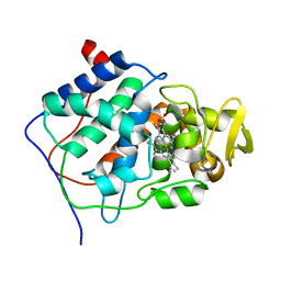
6HK6
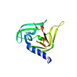
 | | Human RIOK2 bound to inhibitor | | 分子名称: | 1,2-ETHANEDIOL, 2-naphthalen-2-yl-~{N}-pyridin-2-yl-ethanamide, 4-(2-HYDROXYETHYL)-1-PIPERAZINE ETHANESULFONIC ACID, ... | | 著者 | Wang, J, Krojer, T, Bountra, C, Edwards, A.M, Arrowsmith, C, Knapp, S, Elkins, J.M. | | 登録日 | 2018-09-05 | | 公開日 | 2018-10-24 | | 最終更新日 | 2024-01-17 | | 実験手法 | X-RAY DIFFRACTION (2.35 Å) | | 主引用文献 | Crystal structure of human RIOK2 bound to a specific inhibitor.
Open Biology, 9, 2019
|
|
7XZ9
 | |
7XZD
 | |
7XZF
 | | Wild type of the N-terminal domain of fucoidan lyase FdlA | | 分子名称: | Fucoidan lyase, GLYCEROL, IMIDAZOLE, ... | | 著者 | Wang, J, Li, M, Pan, X. | | 登録日 | 2022-06-02 | | 公開日 | 2022-09-28 | | 最終更新日 | 2024-05-29 | | 実験手法 | X-RAY DIFFRACTION (1.3 Å) | | 主引用文献 | Structural and Biochemical Analysis Reveals Catalytic Mechanism of Fucoidan Lyase from Flavobacterium sp. SA-0082.
Mar Drugs, 20, 2022
|
|
7XZ8
 | |
7XZB
 | |
7XZA
 | |
7XZE
 | |
7XZ7
 | |
7XZC
 | |
3CCP
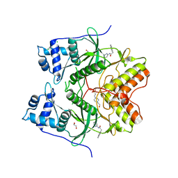
 | | X-RAY STRUCTURES OF RECOMBINANT YEAST CYTOCHROME C PEROXIDASE AND THREE HEME-CLEFT MUTANTS PREPARED BY SITE-DIRECTED MUTAGENESIS | | 分子名称: | PROTOPORPHYRIN IX CONTAINING FE, YEAST CYTOCHROME C PEROXIDASE | | 著者 | Wang, J, Mauro, J.M, Edwards, S.L, Oatley, S.J, Fishel, L.A, Ashford, V.A, Xuong, N.-H, Kraut, J. | | 登録日 | 1990-02-28 | | 公開日 | 1991-07-15 | | 最終更新日 | 2024-02-21 | | 実験手法 | X-RAY DIFFRACTION (2.2 Å) | | 主引用文献 | X-ray structures of recombinant yeast cytochrome c peroxidase and three heme-cleft mutants prepared by site-directed mutagenesis.
Biochemistry, 29, 1990
|
|
8HQG
 | |
8HVM
 | | Crystal structure of SARS-Cov-2 main protease K90R mutant in complex with PF07321332 | | 分子名称: | (1R,2S,5S)-N-{(1E,2S)-1-imino-3-[(3S)-2-oxopyrrolidin-3-yl]propan-2-yl}-6,6-dimethyl-3-[3-methyl-N-(trifluoroacetyl)-L-valyl]-3-azabicyclo[3.1.0]hexane-2-carboxamide, 3C-like proteinase nsp5 | | 著者 | Wang, J, Zhang, J, Li, J. | | 登録日 | 2022-12-27 | | 公開日 | 2023-12-27 | | 実験手法 | X-RAY DIFFRACTION (1.48 Å) | | 主引用文献 | Crystal structure of SARS-Cov-2 main protease
K90R mutant in complex with PF07321332
To Be Published
|
|
8HVY
 | |
7CPY
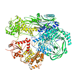
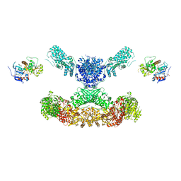 | | Lovastatin nonaketide synthase with LovC | | 分子名称: | Lovastatin nonaketide synthase, enoyl reductase component lovC, polyketide synthase component, ... | | 著者 | Wang, J, Wang, Z. | | 登録日 | 2020-08-08 | | 公開日 | 2021-01-13 | | 最終更新日 | 2024-05-29 | | 実験手法 | ELECTRON MICROSCOPY (3.6 Å) | | 主引用文献 | Structural basis for the biosynthesis of lovastatin.
Nat Commun, 12, 2021
|
|
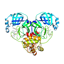
3R0F
 | | Human enterovirus 71 3C protease mutant H133G in complex with rupintrivir | | 分子名称: | 1,2-ETHANEDIOL, 3C protein, 4-{2-(4-FLUORO-BENZYL)-6-METHYL-5-[(5-METHYL-ISOXAZOLE-3-CARBONYL)-AMINO]-4-OXO-HEPTANOYLAMINO}-5-(2-OXO-PYRROLIDIN-3-YL)-PENTANOIC ACID ETHYL ESTER | | 著者 | Wang, J, Fan, T, Yao, X, Wu, Z, Guo, L, Lei, X, Wang, J, Wang, M, Jin, Q, Cui, S. | | 登録日 | 2011-03-08 | | 公開日 | 2011-08-10 | | 最終更新日 | 2023-09-13 | | 実験手法 | X-RAY DIFFRACTION (1.3083 Å) | | 主引用文献 | Crystal Structures of Enterovirus 71 3C Protease Complexed with Rupintrivir Reveal the Roles of Catalytically Important Residues.
J.Virol., 85, 2011
|
|
6OPF
 | |
4HJX
 | | Crystal structure of F2YRS complexed with F2Y | | 分子名称: | 3,5-difluoro-L-tyrosine, Tyrosine-tRNA ligase | | 著者 | Wang, J, Tian, C, Gong, W, Li, F, Shi, P, Li, J, Ding, W. | | 登録日 | 2012-10-14 | | 公開日 | 2013-03-13 | | 最終更新日 | 2023-12-06 | | 実験手法 | X-RAY DIFFRACTION (2.91 Å) | | 主引用文献 | A genetically encoded 19F NMR probe for tyrosine phosphorylation.
Angew.Chem.Int.Ed.Engl., 52, 2013
|
|
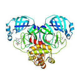
3QZR
 | | Human enterovirus 71 3C protease mutant E71A in complex with rupintrivir | | 分子名称: | 1,2-ETHANEDIOL, 3C protein, 4-{2-(4-FLUORO-BENZYL)-6-METHYL-5-[(5-METHYL-ISOXAZOLE-3-CARBONYL)-AMINO]-4-OXO-HEPTANOYLAMINO}-5-(2-OXO-PYRROLIDIN-3-YL)-PENTANOIC ACID ETHYL ESTER | | 著者 | Wang, J, Fan, T, Yao, X, Wu, Z, Guo, L, Lei, X, Wang, J, Wang, M, Jin, Q, Cui, S. | | 登録日 | 2011-03-07 | | 公開日 | 2011-08-10 | | 最終更新日 | 2024-02-21 | | 実験手法 | X-RAY DIFFRACTION (1.039 Å) | | 主引用文献 | Crystal Structures of Enterovirus 71 3C Protease Complexed with Rupintrivir Reveal the Roles of Catalytically Important Residues.
J.Virol., 85, 2011
|
|
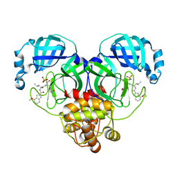
3QZQ
 | | Human enterovirus 71 3C protease mutant E71D in complex with rupintrivir | | 分子名称: | 3C protein, 4-{2-(4-FLUORO-BENZYL)-6-METHYL-5-[(5-METHYL-ISOXAZOLE-3-CARBONYL)-AMINO]-4-OXO-HEPTANOYLAMINO}-5-(2-OXO-PYRROLIDIN-3-YL)-PENTANOIC ACID ETHYL ESTER | | 著者 | Wang, J, Fan, T, Yao, X, Wu, Z, Guo, L, Lei, X, Wang, J, Wang, M, Jin, Q, Cui, S. | | 登録日 | 2011-03-07 | | 公開日 | 2011-08-10 | | 最終更新日 | 2024-02-21 | | 実験手法 | X-RAY DIFFRACTION (1.7001 Å) | | 主引用文献 | Crystal Structures of Enterovirus 71 3C Protease Complexed with Rupintrivir Reveal the Roles of Catalytically Important Residues.
J.Virol., 85, 2011
|
|
1EQ1
 | |
7CPX
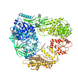
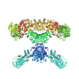 | | Lovastatin nonaketide synthase | | 分子名称: | Lovastatin nonaketide synthase, polyketide synthase component, NADP NICOTINAMIDE-ADENINE-DINUCLEOTIDE PHOSPHATE | | 著者 | Wang, J, Wang, Z. | | 登録日 | 2020-08-08 | | 公開日 | 2021-01-13 | | 最終更新日 | 2024-05-29 | | 実験手法 | ELECTRON MICROSCOPY (2.91 Å) | | 主引用文献 | Structural basis for the biosynthesis of lovastatin.
Nat Commun, 12, 2021
|
|


