7WK4
 
 | |
7WK6
 | |
6IFU
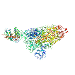
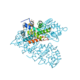
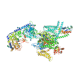
 | | Cryo-EM structure of type III-A Csm-CTR2-dsDNA complex | | 分子名称: | CTR2, Type III-A CRISPR-associated RAMP protein Csm3, Type III-A CRISPR-associated RAMP protein Csm4, ... | | 著者 | You, L, Ma, J, Wang, J, Zhang, X, Wang, Y. | | 登録日 | 2018-09-21 | | 公開日 | 2018-12-12 | | 最終更新日 | 2024-03-27 | | 実験手法 | ELECTRON MICROSCOPY (3.05 Å) | | 主引用文献 | Structure Studies of the CRISPR-Csm Complex Reveal Mechanism of Co-transcriptional Interference
Cell, 176, 2019
|
|
6IFR
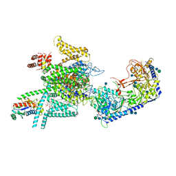
 | | Type III-A Csm complex, Cryo-EM structure of Csm-NTR, ATP bound | | 分子名称: | ADENOSINE-5'-TRIPHOSPHATE, MAGNESIUM ION, Type III-A CRISPR-associated RAMP protein Csm3, ... | | 著者 | You, L, Ma, J, Wang, J, Zhang, X, Wang, Y. | | 登録日 | 2018-09-21 | | 公開日 | 2018-12-12 | | 最終更新日 | 2019-01-23 | | 実験手法 | ELECTRON MICROSCOPY (3.4 Å) | | 主引用文献 | Structure Studies of the CRISPR-Csm Complex Reveal Mechanism of Co-transcriptional Interference
Cell, 176, 2019
|
|
2WOQ
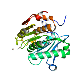
 | | Porphobilinogen Synthase (HemB) in Complex with 5-acetamido-4- oxohexanoic acid (Alaremycin 2) | | 分子名称: | 3,6,9,12,15,18,21,24-OCTAOXAHEXACOSAN-1-OL, 4-(2-HYDROXYETHYL)-1-PIPERAZINE ETHANESULFONIC ACID, ALAREMYCIN 2, ... | | 著者 | Heinemann, I.U, Schulz, C, Schubert, W.-D, Heinz, D.W, Wang, Y.-G, Kobayashi, Y, Awa, Y, Wachi, M, Jahn, D, Jahn, M. | | 登録日 | 2009-07-27 | | 公開日 | 2009-10-27 | | 最終更新日 | 2023-12-20 | | 実験手法 | X-RAY DIFFRACTION (1.75 Å) | | 主引用文献 | Structure of the heme biosynthetic Pseudomonas aeruginosa porphobilinogen synthase in complex with the antibiotic alaremycin.
Antimicrob. Agents Chemother., 54, 2010
|
|
2ZEM
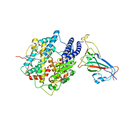
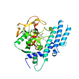
 | | Crystal structure of the human glutaminyl cyclase mutant D248Q at 2.18 angstrom resolution | | 分子名称: | Glutaminyl-peptide cyclotransferase, SULFATE ION, ZINC ION | | 著者 | Huang, K.F, Wang, Y.R, Chang, E.C, Chou, T.L, Wang, A.H. | | 登録日 | 2007-12-13 | | 公開日 | 2008-04-22 | | 最終更新日 | 2023-11-01 | | 実験手法 | X-RAY DIFFRACTION (2.18 Å) | | 主引用文献 | A conserved hydrogen-bond network in the catalytic centre of animal glutaminyl cyclases is critical for catalysis.
Biochem.J., 411, 2008
|
|
2ZEP
 | | Crystal structure of the human glutaminyl cyclase mutant H319L at 2.1 angstrom resolution | | 分子名称: | Glutaminyl-peptide cyclotransferase, SULFATE ION, ZINC ION | | 著者 | Huang, K.F, Wang, Y.R, Chang, E.C, Chou, T.L, Wang, A.H. | | 登録日 | 2007-12-13 | | 公開日 | 2008-04-22 | | 最終更新日 | 2023-11-01 | | 実験手法 | X-RAY DIFFRACTION (2.1 Å) | | 主引用文献 | A conserved hydrogen-bond network in the catalytic centre of animal glutaminyl cyclases is critical for catalysis.
Biochem.J., 411, 2008
|
|
2ZEE
 | | Crystal structure of the human glutaminyl cyclase mutant S160G at 1.99 angstrom resolution | | 分子名称: | Glutaminyl-peptide cyclotransferase, SULFATE ION, ZINC ION | | 著者 | Huang, K.F, Wang, Y.R, Chang, E.C, Chou, T.L, Wang, A.H. | | 登録日 | 2007-12-12 | | 公開日 | 2008-04-22 | | 最終更新日 | 2023-11-01 | | 実験手法 | X-RAY DIFFRACTION (1.99 Å) | | 主引用文献 | A conserved hydrogen-bond network in the catalytic centre of animal glutaminyl cyclases is critical for catalysis.
Biochem.J., 411, 2008
|
|
2ZEO
 | | Crystal structure of the human glutaminyl cyclase mutant D305E at 1.66 angstrom resolution | | 分子名称: | Glutaminyl-peptide cyclotransferase, SULFATE ION, ZINC ION | | 著者 | Huang, K.F, Wang, Y.R, Chang, E.C, Chou, T.L, Wang, A.H. | | 登録日 | 2007-12-13 | | 公開日 | 2008-04-22 | | 最終更新日 | 2023-11-01 | | 実験手法 | X-RAY DIFFRACTION (1.66 Å) | | 主引用文献 | A conserved hydrogen-bond network in the catalytic centre of animal glutaminyl cyclases is critical for catalysis.
Biochem.J., 411, 2008
|
|
2ZEF
 | | Crystal structure of the human glutaminyl cyclase mutant E201D at 1.67 angstrom resolution | | 分子名称: | Glutaminyl-peptide cyclotransferase, SULFATE ION, ZINC ION | | 著者 | Huang, K.F, Wang, Y.R, Chang, E.C, Chou, T.L, Wang, A.H. | | 登録日 | 2007-12-12 | | 公開日 | 2008-04-22 | | 最終更新日 | 2023-11-01 | | 実験手法 | X-RAY DIFFRACTION (1.67 Å) | | 主引用文献 | A conserved hydrogen-bond network in the catalytic centre of animal glutaminyl cyclases is critical for catalysis.
Biochem.J., 411, 2008
|
|
2ZEL
 | | Crystal structure of the human glutaminyl cyclase mutant D248A at 1.97 angstrom resolution | | 分子名称: | Glutaminyl-peptide cyclotransferase, SULFATE ION, ZINC ION | | 著者 | Huang, K.F, Wang, Y.R, Chang, E.C, Chou, T.L, Wang, A.H. | | 登録日 | 2007-12-13 | | 公開日 | 2008-04-22 | | 最終更新日 | 2023-11-01 | | 実験手法 | X-RAY DIFFRACTION (1.97 Å) | | 主引用文献 | A conserved hydrogen-bond network in the catalytic centre of animal glutaminyl cyclases is critical for catalysis.
Biochem.J., 411, 2008
|
|
2ZEG
 | | Crystal structure of the human glutaminyl cyclase mutant E201L at 2.08 angstrom resolution | | 分子名称: | Glutaminyl-peptide cyclotransferase, IMIDAZOLE, SULFATE ION, ... | | 著者 | Huang, K.F, Wang, Y.R, Chang, E.C, Chou, T.L, Wang, A.H. | | 登録日 | 2007-12-12 | | 公開日 | 2008-04-22 | | 最終更新日 | 2023-11-01 | | 実験手法 | X-RAY DIFFRACTION (2.08 Å) | | 主引用文献 | A conserved hydrogen-bond network in the catalytic centre of animal glutaminyl cyclases is critical for catalysis.
Biochem.J., 411, 2008
|
|
8IAG
 | |
7EOA
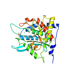
 | | HR-PETase from Bacterium HR29 | | 分子名称: | ETHANOL, Poly(Ethylene terephthalate) hydrolase | | 著者 | Wang, J, Wang, Y.H. | | 登録日 | 2021-04-21 | | 公開日 | 2022-04-27 | | 最終更新日 | 2023-11-29 | | 実験手法 | X-RAY DIFFRACTION (1.24 Å) | | 主引用文献 | Crystal structure of Bomgl, a monoacylglycerol lipase from marine Bacillus
To Be Published
|
|
5KPO
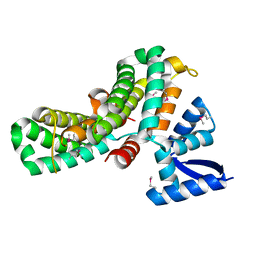
 | | Structure of human PARP1 catalytic domain bound to a quinazoline-2,4(1H,3H)-dione inhibitor | | 分子名称: | 1-[[3-(4-ethyl-3-oxidanylidene-piperazin-1-yl)carbonyl-4-fluoranyl-phenyl]methyl]quinazoline-2,4-dione, Poly [ADP-ribose] polymerase 1 | | 著者 | Cao, R, Wang, Y.L, Zhou, J, Yao, H.P, Huang, N, Xu, B.L. | | 登録日 | 2016-07-05 | | 公開日 | 2016-12-21 | | 最終更新日 | 2024-03-20 | | 実験手法 | X-RAY DIFFRACTION (2.65 Å) | | 主引用文献 | Structure of human PARP1 catalytic domain bound to a quinazoline-2,4(1H,3H)-dione inhibitor
To Be Published
|
|
5KPQ
 | | Structure of human PARP1 catalytic domain bound to a quinazoline-2,4(1H,3H)-dione inhibitor | | 分子名称: | 1-[[4-fluoranyl-3-[(3R)-3-methyl-4-propyl-piperazin-1-yl]carbonyl-phenyl]methyl]quinazoline-2,4-dione, Poly [ADP-ribose] polymerase 1 | | 著者 | Cao, R, Wang, Y.L, Zhou, J, Huang, N, Xu, B.L. | | 登録日 | 2016-07-05 | | 公開日 | 2016-12-14 | | 最終更新日 | 2023-11-08 | | 実験手法 | X-RAY DIFFRACTION (2.55 Å) | | 主引用文献 | Structure of human PARP1 catalytic domain bound to a quinazoline-2,4(1H,3H)-dione inhibitor
To Be Published
|
|
1LR6
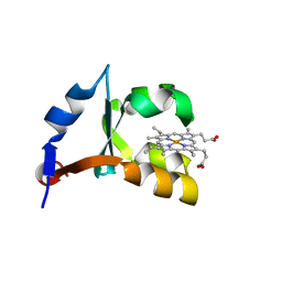
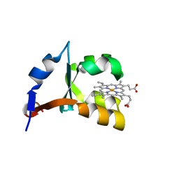 | | Crystal structure of V45Y mutant of cytochrome b5 | | 分子名称: | PROTOPORPHYRIN IX CONTAINING FE, cytochrome b5 | | 著者 | Gan, J.-H, Wu, J, Wang, Z.-Q, Wang, Y.-H, Huang, Z.-X, Xia, Z.-X. | | 登録日 | 2002-05-15 | | 公開日 | 2002-09-04 | | 最終更新日 | 2023-10-25 | | 実験手法 | X-RAY DIFFRACTION (1.9 Å) | | 主引用文献 | Structures of V45E and V45Y mutants and structure comparison of a variety of cytochrome b5 mutants.
Acta Crystallogr.,Sect.D, 58, 2002
|
|
5KPN
 | | Structure of human PARP1 catalytic domain bound to a quinazoline-2,4(1H,3H)-dione inhibitor | | 分子名称: | 1-[[4-fluoranyl-3-(3-oxidanylidene-4-propyl-piperazin-1-yl)carbonyl-phenyl]methyl]quinazoline-2,4-dione, Poly [ADP-ribose] polymerase 1 | | 著者 | Cao, R, Wang, Y.L, Zhou, J, Yao, H.P, Huang, N, Xu, B.L. | | 登録日 | 2016-07-05 | | 公開日 | 2016-12-21 | | 最終更新日 | 2024-03-20 | | 実験手法 | X-RAY DIFFRACTION (2.3 Å) | | 主引用文献 | Structure of human PARP1 catalytic domain bound to a quinazoline-2,4(1H,3H)-dione inhibitor
To Be Published
|
|
6X2Q
 | |
6DDA
 | | Nurr1 Covalently Modified by a Dopamine Metabolite | | 分子名称: | 5-hydroxy-1,2-dihydro-6H-indol-6-one, BROMIDE ION, Nuclear receptor subfamily 4 group A member 2, ... | | 著者 | Bruning, J.M, Wang, Y, Otrabella, F, Boxue, T, Liu, H, Bhattacharya, P, Guo, S, Holton, J.M, Fletterick, R.J, Jacobson, M.P, England, P.M. | | 登録日 | 2018-05-09 | | 公開日 | 2019-03-20 | | 最終更新日 | 2019-11-06 | | 実験手法 | X-RAY DIFFRACTION (3.2 Å) | | 主引用文献 | Covalent Modification and Regulation of the Nuclear Receptor Nurr1 by a Dopamine Metabolite.
Cell Chem Biol, 26, 2019
|
|
8JDI
 | |
8JDH
 | |
1M20
 | | Crystal Structure of F35Y Mutant of Trypsin-solubilized Fragment of Cytochrome b5 | | 分子名称: | Cytochrome b5, PROTOPORPHYRIN IX CONTAINING FE | | 著者 | Yao, P, Wu, J, Wang, Y.-H, Sun, B.-Y, Xia, Z.-X, Huang, Z.-X. | | 登録日 | 2002-06-20 | | 公開日 | 2002-09-11 | | 最終更新日 | 2023-10-25 | | 実験手法 | X-RAY DIFFRACTION (1.8 Å) | | 主引用文献 | X-ray crystallography, CD and kinetic studies revealed the essence of the abnormal behaviors of the cytochrome b5 Phe35-->Tyr mutant.
Eur.J.Biochem., 269, 2002
|
|
7VFV
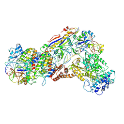
 | | Human N-type voltage gated calcium channel CaV2.2-alpha2/delta1-beta1 complex, bound to PD173212 | | 分子名称: | (2~{S})-~{N}-[(2~{S})-1-(~{tert}-butylamino)-1-oxidanylidene-3-(4-phenylmethoxyphenyl)propan-2-yl]-2-[(4-~{tert}-butylphenyl)methyl-methyl-amino]-4-methyl-pentanamide, 2-acetamido-2-deoxy-beta-D-glucopyranose, CALCIUM ION, ... | | 著者 | Dong, Y, Gao, Y, Wang, Y, Zhao, Y. | | 登録日 | 2021-09-13 | | 公開日 | 2021-11-03 | | 最終更新日 | 2022-02-16 | | 実験手法 | ELECTRON MICROSCOPY (3 Å) | | 主引用文献 | Closed-state inactivation and pore-blocker modulation mechanisms of human Ca V 2.2.
Cell Rep, 37, 2021
|
|
7VFU
 | | Human N-type voltage gated calcium channel CaV2.2-alpha2/delta1-beta1 complex, bound to ziconotide | | 分子名称: | 2-acetamido-2-deoxy-beta-D-glucopyranose, CALCIUM ION, CHOLESTEROL HEMISUCCINATE, ... | | 著者 | Dong, Y, Gao, Y, Wang, Y, Zhao, Y. | | 登録日 | 2021-09-13 | | 公開日 | 2021-11-03 | | 最終更新日 | 2022-02-16 | | 実験手法 | ELECTRON MICROSCOPY (3 Å) | | 主引用文献 | Closed-state inactivation and pore-blocker modulation mechanisms of human Ca V 2.2.
Cell Rep, 37, 2021
|
|


