3A0O
 
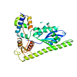
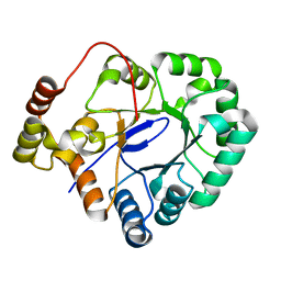
 | | Crystal structure of alginate lyase from Agrobacterium tumefaciens C58 | | 分子名称: | CHLORIDE ION, Oligo alginate lyase | | 著者 | Ochiai, A, Yamasaki, M, Mikami, B, Hashimoto, W, Murata, K. | | 登録日 | 2009-03-23 | | 公開日 | 2010-03-31 | | 最終更新日 | 2024-03-13 | | 実験手法 | X-RAY DIFFRACTION (2.11 Å) | | 主引用文献 | Crystal structure of exotype alginate lyase Atu3025 from Agrobacterium tumefaciens
J.Biol.Chem., 285, 2010
|
|
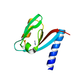
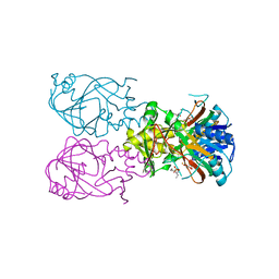
4XLG
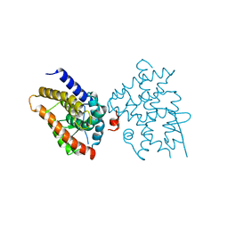
 | | C. glabrata Slx1 in complex with Slx4CCD. | | 分子名称: | CHLORIDE ION, Structure-specific endonuclease subunit SLX1, Structure-specific endonuclease subunit SLX4, ... | | 著者 | Gaur, V, Wyatt, H.D.M, Komorowska, W, Szczepanowski, R.H, de Sanctis, D, Gorecka, K.M, West, S.C, Nowotny, M. | | 登録日 | 2015-01-13 | | 公開日 | 2015-03-25 | | 最終更新日 | 2024-01-10 | | 実験手法 | X-RAY DIFFRACTION (1.78 Å) | | 主引用文献 | Structural and Mechanistic Analysis of the Slx1-Slx4 Endonuclease.
Cell Rep, 10, 2015
|
|
6ET1
 | |
6EU9
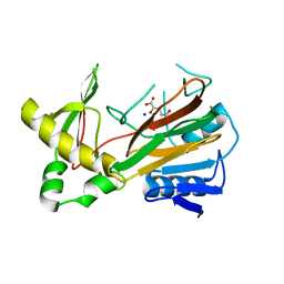
 | | Crystal structure of Platynereis dumerilii RAR ligand-binding domain in complex with all-trans retinoic acid | | 分子名称: | RETINOIC ACID, Retinoic acid receptor | | 著者 | Handberg-Thorsager, M, Gutierrez-Mazariegos, J, Arold, S.T, Nadendla, E.K, Bertucci, P.Y, Germain, P, Tomancak, P, Pierzchalski, K, Jones, J.W, Albalat, R, Kane, M.A, Bourguet, W, Laudet, V, Arendt, D, Schubert, M. | | 登録日 | 2017-10-29 | | 公開日 | 2018-03-14 | | 最終更新日 | 2024-05-08 | | 実験手法 | X-RAY DIFFRACTION (2.69 Å) | | 主引用文献 | The ancestral retinoic acid receptor was a low-affinity sensor triggering neuronal differentiation.
Sci Adv, 4, 2018
|
|
6OZJ
 | |
6OZQ
 | |
1NDA
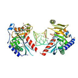
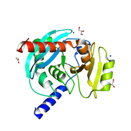
 | | THE STRUCTURE OF TRYPANOSOMA CRUZI TRYPANOTHIONE REDUCTASE IN THE OXIDIZED AND NADPH REDUCED STATE | | 分子名称: | FLAVIN-ADENINE DINUCLEOTIDE, TRYPANOTHIONE OXIDOREDUCTASE | | 著者 | Lantwin, C.B, Kabsch, W, Pai, E.F, Schlichting, I, Krauth-Siegel, R.L. | | 登録日 | 1993-07-02 | | 公開日 | 1994-09-30 | | 最終更新日 | 2017-11-29 | | 実験手法 | X-RAY DIFFRACTION (3.3 Å) | | 主引用文献 | The structure of Trypanosoma cruzi trypanothione reductase in the oxidized and NADPH reduced state.
Proteins, 18, 1994
|
|
6EYS
 | |
4Y05
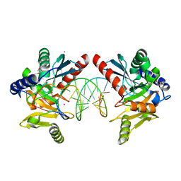
 | | KIF2C short Loop2 construct | | 分子名称: | ADENOSINE-5'-DIPHOSPHATE, Kinesin-like protein KIF2C, MAGNESIUM ION, ... | | 著者 | Wang, W, Knossow, M, Gigant, B. | | 登録日 | 2015-02-05 | | 公開日 | 2015-06-17 | | 最終更新日 | 2024-01-10 | | 実験手法 | X-RAY DIFFRACTION (2.59 Å) | | 主引用文献 | New Insights into the Coupling between Microtubule Depolymerization and ATP Hydrolysis by Kinesin-13 Protein Kif2C.
J.Biol.Chem., 290, 2015
|
|
6ET2
 | |
6OZP
 | |
4XPN
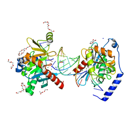
 | | Crystal Structure of Protein Phosphate 1 complexed with PP1 binding domain of GADD34 | | 分子名称: | GLYCEROL, MANGANESE (II) ION, PHOSPHATE ION, ... | | 著者 | Choy, M.S, Peti, W, Page, R. | | 登録日 | 2015-01-17 | | 公開日 | 2015-07-01 | | 最終更新日 | 2023-09-27 | | 実験手法 | X-RAY DIFFRACTION (2.285 Å) | | 主引用文献 | Structural and Functional Analysis of the GADD34:PP1 eIF2 alpha Phosphatase.
Cell Rep, 11, 2015
|
|
6P0Z
 | | Crystal structure of N-acetylated KRAS (2-169) bound to GDP and Mg | | 分子名称: | ACETYL GROUP, DI(HYDROXYETHYL)ETHER, GTPase KRas, ... | | 著者 | Dharmaiah, S, Tran, T.H, Yan, W, Simanshu, D.K. | | 登録日 | 2019-05-17 | | 公開日 | 2019-07-31 | | 最終更新日 | 2023-10-11 | | 実験手法 | X-RAY DIFFRACTION (1.011 Å) | | 主引用文献 | Structures of N-terminally processed KRAS provide insight into the role of N-acetylation.
Sci Rep, 9, 2019
|
|
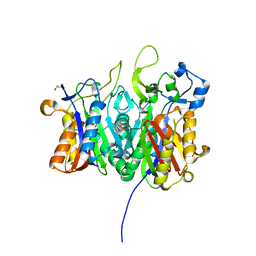
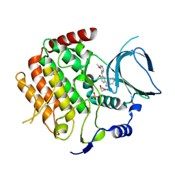
4XAP
 | | Crystal structure of aldo-keto reductase from Sinorhizobium meliloti 1021 | | 分子名称: | Aldo-keto reductase | | 著者 | Gasiorowska, O.A, Handing, K.B, Shabalin, I.G, Bonanno, J, Almo, S.C, Minor, W, New York Structural Genomics Research Consortium (NYSGRC) | | 登録日 | 2014-12-15 | | 公開日 | 2014-12-31 | | 最終更新日 | 2023-09-27 | | 実験手法 | X-RAY DIFFRACTION (2.21 Å) | | 主引用文献 | Crystal structure of aldo-keto reductase from Sinorhizobium meliloti 1021
to be published
|
|
6P6J
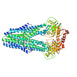 | | Structure of YbtPQ importer with substrate Ybt-Fe bound | | 分子名称: | ABC transporter protein, FE (III) ION, inner membrane ABC-transporter, ... | | 著者 | Wang, Z, Hu, W, Zheng, H. | | 登録日 | 2019-06-04 | | 公開日 | 2020-03-04 | | 最終更新日 | 2024-03-13 | | 実験手法 | ELECTRON MICROSCOPY (3.4 Å) | | 主引用文献 | Pathogenic siderophore ABC importer YbtPQ adopts a surprising fold of exporter.
Sci Adv, 6, 2020
|
|
4DU0
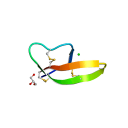 | | Crystal structure of human alpha-defensin 1, HNP1 (G17A mutant) | | 分子名称: | CHLORIDE ION, GLYCEROL, Neutrophil defensin 1 | | 著者 | Wu, X, Lu, W, Pazgier, M. | | 登録日 | 2012-02-21 | | 公開日 | 2012-04-11 | | 最終更新日 | 2023-09-13 | | 実験手法 | X-RAY DIFFRACTION (1.9 Å) | | 主引用文献 | Invariant gly residue is important for alpha-defensin folding, dimerization, and function: a case study of the human neutrophil alpha-defensin HNP1
J.Biol.Chem., 287, 2012
|
|
6PAX
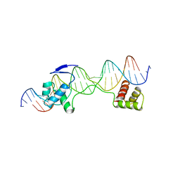 | | CRYSTAL STRUCTURE OF THE HUMAN PAX-6 PAIRED DOMAIN-DNA COMPLEX REVEALS A GENERAL MODEL FOR PAX PROTEIN-DNA INTERACTIONS | | 分子名称: | 26 NUCLEOTIDE DNA, HOMEOBOX PROTEIN PAX-6 | | 著者 | Xu, H.E, Rould, M.A, Xu, W, Epstein, J.A, Maas, R.L, Pabo, C.O. | | 登録日 | 1999-04-22 | | 公開日 | 1999-07-13 | | 最終更新日 | 2024-04-03 | | 実験手法 | X-RAY DIFFRACTION (2.5 Å) | | 主引用文献 | Crystal structure of the human Pax6 paired domain-DNA complex reveals specific roles for the linker region and carboxy-terminal subdomain in DNA binding.
Genes Dev., 13, 1999
|
|
4XH7
 | | Crystal structure of MUPP1 PDZ4 | | 分子名称: | IMIDAZOLE, Multiple PDZ domain protein | | 著者 | Liu, Z, Zhu, H, Liu, W. | | 登録日 | 2015-01-05 | | 公開日 | 2015-03-04 | | 最終更新日 | 2023-11-08 | | 実験手法 | X-RAY DIFFRACTION (1.65 Å) | | 主引用文献 | Biochemical and structural characterization of MUPP1-PDZ4 domain from Mus musculus.
Acta Biochim.Biophys.Sin., 47, 2015
|
|
1OIJ
 | | Crystal structure of the alkylsulfatase AtsK, a non-heme Fe(II) alphaketoglutarate dependent Dioxygenase in complex with alphaketoglutarate | | 分子名称: | 2-OXOGLUTARIC ACID, PUTATIVE ALKYLSULFATASE ATSK, SODIUM ION | | 著者 | Mueller, I, Kahnert, A, Pape, T, Dierks, T, Meyer-Klauke, W, Kertesz, M.A, Uson, I. | | 登録日 | 2003-06-18 | | 公開日 | 2004-03-30 | | 最終更新日 | 2023-12-13 | | 実験手法 | X-RAY DIFFRACTION (2.1 Å) | | 主引用文献 | Crystal Structure of the Alkylsulfatase Atsk: Insights Into the Catalytic Mechanism of the Fe(II) Alpha-Ketoglutarate-Dependent Dioxygenase Superfamily
Biochemistry, 42, 2004
|
|
6OZE
 | |
6OZI
 | |
1OIK
 | | Crystal structure of the alkylsulfatase AtsK, a non-heme Fe(II) alphaketoglutarate dependent Dioxygenase in complex with fe, alphaketoglutarate and 2-ethyl-1-hexanesulfuric acid | | 分子名称: | (2R)-2-ETHYL-1-HEXANESULFONIC ACID, 2-OXOGLUTARIC ACID, FE (II) ION, ... | | 著者 | Mueller, I, Kahnert, A, Pape, T, Dierks, T, Meyer-Klauke, W, Kertesz, M.A, Uson, I. | | 登録日 | 2003-06-18 | | 公開日 | 2004-03-30 | | 最終更新日 | 2023-12-13 | | 実験手法 | X-RAY DIFFRACTION (2.06 Å) | | 主引用文献 | Crystal Structure of the Alkylsulfatase Atsk: Insights Into the Catalytic Mechanism of the Fe(II) Alpha-Ketoglutarate-Dependent Dioxygenase Superfamily
Biochemistry, 42, 2004
|
|
6OZS
 | |
6F7B
 | | Crystal structure of the human Bub1 kinase domain in complex with BAY 1816032 | | 分子名称: | 2-[3,5-bis(fluoranyl)-4-[[3-[5-methoxy-4-[(3-methoxypyridin-4-yl)amino]pyrimidin-2-yl]indazol-1-yl]methyl]phenoxy]ethanol, MAGNESIUM ION, Mitotic checkpoint serine/threonine-protein kinase BUB1 | | 著者 | Holton, S.J, Siemeister, G, Mengel, A, Bone, W, Schroeder, J, Zitzmann-Kolbe, S, Briem, H, Fernandez-Montalvan, A, Prechtl, S, Moenning, U, von Ahsen, O, Johanssen, J, Cleve, A, Puetter, V, Hitchcock, M, von Nussbaum, F, Brands, M, Mumberg, D, Ziegelbauer, K. | | 登録日 | 2017-12-08 | | 公開日 | 2018-12-19 | | 最終更新日 | 2021-05-05 | | 実験手法 | X-RAY DIFFRACTION (2 Å) | | 主引用文献 | Inhibition of BUB1 Kinase by BAY 1816032 Sensitizes Tumor Cells toward Taxanes, ATR, and PARP InhibitorsIn VitroandIn Vivo.
Clin.Cancer Res., 25, 2019
|
|
3AFA
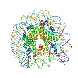 | | The human nucleosome structure | | 分子名称: | 146-MER DNA, CHLORIDE ION, Histone H2A type 1-B/E, ... | | 著者 | Tachiwana, H, Kagawa, W, Osakabe, A, Koichiro, K, Shiga, T, Kimura, H, Kurumizaka, H. | | 登録日 | 2010-02-24 | | 公開日 | 2010-05-26 | | 最終更新日 | 2023-11-01 | | 実験手法 | X-RAY DIFFRACTION (2.5 Å) | | 主引用文献 | Structural basis of instability of the nucleosome containing a testis-specific histone variant, human H3T
Proc.Natl.Acad.Sci.USA, 107, 2010
|
|


