Loading
PDBj
MenuPDBj@FacebookPDBj@X(formerly Twitter)PDBj@BlueSkyPDBj@YouTubewwPDB FoundationwwPDB
RCSB PDBPDBeBMRBAdv. SearchSearch help
PDB: 37 results

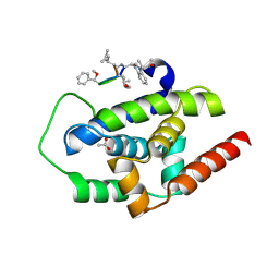
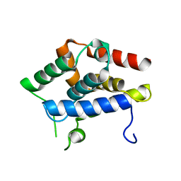
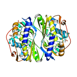
3RFY
DownloadVisualize
BU of 3rfy by Molmil
Crystal structure of arabidopsis thaliana cyclophilin 38 (ATCYP38)
Descriptor: Peptidyl-prolyl cis-trans isomerase CYP38, chloroplastic
Authors:Vasudevan, D, Swaminathan, K.
Deposit date:2011-04-07
Release date:2012-06-13
Last modified:2024-03-20
Method:X-RAY DIFFRACTION (2.39 Å)
Cite:Crystal structure of Arabidopsis cyclophilin38 reveals a previously uncharacterized immunophilin fold and a possible autoinhibitory mechanism.
Plant Cell, 24, 2012
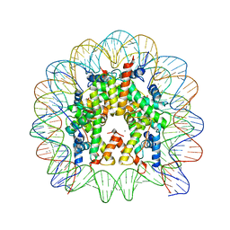
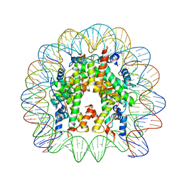
3LZ0
DownloadVisualize
BU of 3lz0 by Molmil
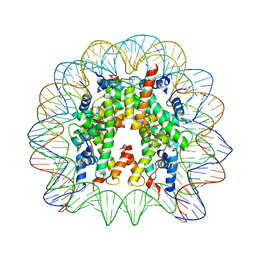
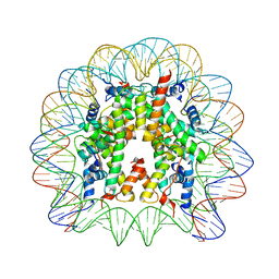
Crystal Structure of Nucleosome Core Particle Composed of the Widom 601 DNA Sequence (orientation 1)
Descriptor: CHLORIDE ION, DNA (145-MER), Histone H2A, ...
Authors:Vasudevan, D, Chua, E.Y.D, Davey, C.A.
Deposit date:2010-03-01
Release date:2010-09-15
Last modified:2023-11-01
Method:X-RAY DIFFRACTION (2.5 Å)
Cite:Crystal structures of nucleosome core particles containing the '601' strong positioning sequence
J.Mol.Biol., 403, 2010
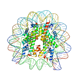
3LZ1
DownloadVisualize
BU of 3lz1 by Molmil
Crystal Structure of Nucleosome Core Particle Composed of the Widom 601 DNA Sequence (orientation 2)
Descriptor: CHLORIDE ION, DNA (145-MER), Histone H2A, ...
Authors:Vasudevan, D, Chua, E.Y.D, Davey, C.A.
Deposit date:2010-03-01
Release date:2010-09-15
Last modified:2023-11-01
Method:X-RAY DIFFRACTION (2.5 Å)
Cite:Crystal structures of nucleosome core particles containing the '601' strong positioning sequence
J.Mol.Biol., 403, 2010
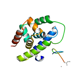
3WDB
DownloadVisualize
BU of 3wdb by Molmil
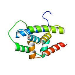
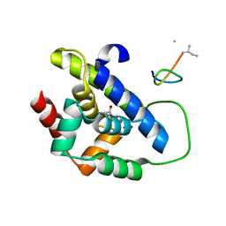
N-terminal domain of Mycobacterium tuberculosis ClpC1
Descriptor: FORMIC ACID, Probable ATP-dependent Clp protease ATP-binding subunit
Authors:Vasudevan, D, Noble, C.G.
Deposit date:2013-06-14
Release date:2013-09-18
Last modified:2023-11-08
Method:X-RAY DIFFRACTION (1.37 Å)
Cite:Structural basis of mycobacterial inhibition by cyclomarin A
J.Biol.Chem., 288, 2013
3WDD
DownloadVisualize
BU of 3wdd by Molmil
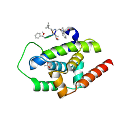
Mutant N-terminal domain of Mycobacterium tuberculosis ClpC1, F2Y, bound to Cyclomarin A
Descriptor: ACETATE ION, Cyclomarin A, Probable ATP-dependent Clp protease ATP-binding subunit
Authors:Vasudevan, D, Noble, C.G.
Deposit date:2013-06-14
Release date:2013-09-18
Last modified:2024-10-30
Method:X-RAY DIFFRACTION (1.18 Å)
Cite:Structural basis of mycobacterial inhibition by cyclomarin A
J.Biol.Chem., 288, 2013
3WDC
DownloadVisualize
BU of 3wdc by Molmil
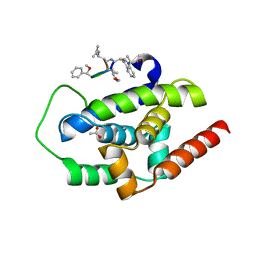
N-terminal domain of Mycobacterium tuberculosis ClpC1 bound to Cyclomarin A
Descriptor: ACETATE ION, Cyclomarin A, Probable ATP-dependent Clp protease ATP-binding subunit
Authors:Vasudevan, D, Noble, C.G.
Deposit date:2013-06-14
Release date:2013-09-18
Last modified:2024-11-06
Method:X-RAY DIFFRACTION (1.18 Å)
Cite:Structural basis of mycobacterial inhibition by cyclomarin A
J.Biol.Chem., 288, 2013
3WDE
DownloadVisualize
BU of 3wde by Molmil
Mutant N-terminal domain of Mycobacterium tuberculosis ClpC1, F80Y, bound to Cyclomarin A
Descriptor: ACETATE ION, Cyclomarin A, Probable ATP-dependent Clp protease ATP-binding subunit
Authors:Vasudevan, D, Noble, C.G.
Deposit date:2013-06-14
Release date:2013-09-18
Last modified:2024-11-20
Method:X-RAY DIFFRACTION (1.44 Å)
Cite:Structural basis of mycobacterial inhibition by cyclomarin A
J.Biol.Chem., 288, 2013
5XHO
DownloadVisualize
BU of 5xho by Molmil
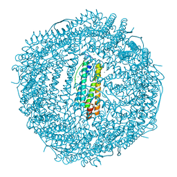
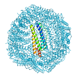
Crystal structure of Frog M-ferritin E135K mutant
Descriptor: CHLORIDE ION, Ferritin, middle subunit, ...
Authors:Jagdev, M.K, Vasudevan, D.
Deposit date:2017-04-21
Release date:2017-08-09
Last modified:2023-11-22
Method:X-RAY DIFFRACTION (1.73 Å)
Cite:Surface charge dependent separation of modified and hybrid ferritin in native PAGE: Impact of lysine 104
Biochim. Biophys. Acta, 1865, 2017
8IBP
DownloadVisualize
BU of 8ibp by Molmil
Crystal structure of Mycobacterium tuberculosis R21K ClpC1 N-terminal domain in complex with Lassomycin
Descriptor: ACETATE ION, Lassomycin, Negative regulator of genetic competence ClpC/mecB
Authors:Jagdev, M.K, Vasudevan, D.
Deposit date:2023-02-10
Release date:2023-09-20
Last modified:2024-05-08
Method:X-RAY DIFFRACTION (1.45 Å)
Cite:Crystal structure of the N-terminal domain of MtClpC1 in complex with the anti-mycobacterial natural peptide Lassomycin.
Int.J.Biol.Macromol., 253, 2023
8IBO
DownloadVisualize
BU of 8ibo by Molmil
Crystal structure of Wild-Type Mycobacterium tuberculosis ClpC1 N-terminal domain in complex with Lassomycin
Descriptor: ACETATE ION, Lassomycin, Negative regulator of genetic competence ClpC/mecB
Authors:Jagdev, M.K, Vasudevan, D.
Deposit date:2023-02-10
Release date:2023-09-20
Last modified:2024-05-08
Method:X-RAY DIFFRACTION (1.83 Å)
Cite:Crystal structure of the N-terminal domain of MtClpC1 in complex with the anti-mycobacterial natural peptide Lassomycin.
Int.J.Biol.Macromol., 253, 2023
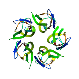
7EP8
DownloadVisualize
BU of 7ep8 by Molmil
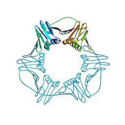
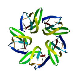
Crystal structure of PCNA from Neurospora crassa
Descriptor: Proliferating cell nuclear antigen
Authors:Sundaram, R, Vasudevan, D.
Deposit date:2021-04-26
Release date:2021-06-30
Last modified:2023-11-29
Method:X-RAY DIFFRACTION (1.95 Å)
Cite:Interdomain connecting loop and J loop structures determine cross-species compatibility of PCNA.
J.Biol.Chem., 297, 2021
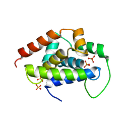
8WZL
DownloadVisualize
BU of 8wzl by Molmil
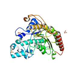
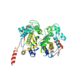
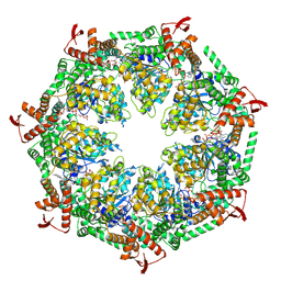
Arabidopsis thaliana Histone deacetylase 7 (HDA7)
Descriptor: Histone deacetylase 7, SULFATE ION
Authors:Saharan, K, Vasudevan, D.
Deposit date:2023-11-02
Release date:2024-05-29
Method:X-RAY DIFFRACTION (1.5 Å)
Cite:Structure-function analyses reveal Arabidopsis thaliana HDA7 to be an inactive histone deacetylase.
Curr Res Struct Biol, 7, 2024
7BUP
DownloadVisualize
BU of 7bup by Molmil
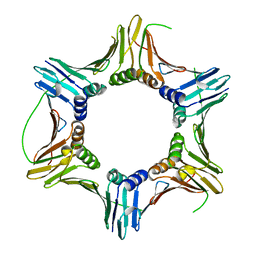
Crystal structure of PCNA from pathogenic yeast Candida albicans
Descriptor: Proliferating cell nuclear antigen
Authors:sundaram, R, Vasudevan, D.
Deposit date:2020-04-07
Release date:2021-02-17
Last modified:2023-11-29
Method:X-RAY DIFFRACTION (2.7 Å)
Cite:Structural analyses of PCNA from the fungal pathogen Candida albicans identify three regions with species-specific conformations.
Febs Lett., 595, 2021
7F2J
DownloadVisualize
BU of 7f2j by Molmil
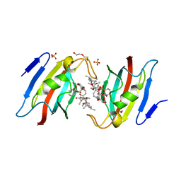
Crystal structure of AtFKBP53 FKBD in complex with rapamycin
Descriptor: DI(HYDROXYETHYL)ETHER, Peptidyl-prolyl cis-trans isomerase FKBP53, RAPAMYCIN IMMUNOSUPPRESSANT DRUG, ...
Authors:Singh, A.K, Saharan, K, Vasudevan, D.
Deposit date:2021-06-11
Release date:2022-03-09
Last modified:2023-11-29
Method:X-RAY DIFFRACTION (1.6 Å)
Cite:Crystal packing reveals rapamycin-mediated homodimerization of an FK506-binding domain.
Int.J.Biol.Macromol., 206, 2022
7CC8
DownloadVisualize
BU of 7cc8 by Molmil
Crystal structure of White Spot Syndrome Virus Thymidylate Synthase - Apo form
Descriptor: SULFATE ION, TRIETHYLENE GLYCOL, Thymidylate Synthase
Authors:Kumar, S, Panchal, N.V, Shaikh, N, Vasudevan, D.
Deposit date:2020-06-16
Release date:2020-11-25
Last modified:2023-11-29
Method:X-RAY DIFFRACTION (2.3 Å)
Cite:Structure analysis of thymidylate synthase from white spot syndrome virus reveals WSSV-specific structural elements.
Int.J.Biol.Macromol., 167, 2021
5GUI
DownloadVisualize
BU of 5gui by Molmil
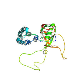
Crystal structure of the N-terminal Domain of Caseinolytic protease associated chaperone ClpC1 from Arabidopsis thaliana
Descriptor: Chaperone protein ClpC1, chloroplastic, PHOSPHATE ION
Authors:Jagdev, M.K, Vasudevan, D.
Deposit date:2016-08-29
Release date:2017-03-22
Last modified:2023-11-08
Method:X-RAY DIFFRACTION (1.2 Å)
Cite:Crystal structures reveal N-terminal Domain of Arabidopsis thaliana ClpD to be highly divergent from that of ClpC1.
Sci Rep, 7, 2017
5GKM
DownloadVisualize
BU of 5gkm by Molmil
Crystal structure of the N-terminal Domain of Caseinolytic protease associated chaperone ClpD from Arabidopsis thaliana
Descriptor: AT5g51070/K3K7_27
Authors:Mohapatra, C, Vasudevan, D.
Deposit date:2016-07-04
Release date:2017-03-22
Last modified:2023-11-08
Method:X-RAY DIFFRACTION (1.6 Å)
Cite:Crystal structures reveal N-terminal Domain of Arabidopsis thaliana ClpD to be highly divergent from that of ClpC1.
Sci Rep, 7, 2017
7CCA
DownloadVisualize
BU of 7cca by Molmil
Crystal structure of White Spot Syndrome Virus Thymidylate Synthase - ternary complex with Methotrexate and dUMP
Descriptor: 2'-DEOXYURIDINE 5'-MONOPHOSPHATE, METHOTREXATE, Thymidylate Synthase
Authors:Panchal, N.V, Kumar, S, Shaikh, N, Vasudevan, D.
Deposit date:2020-06-16
Release date:2020-11-25
Last modified:2023-11-29
Method:X-RAY DIFFRACTION (2.75 Å)
Cite:Structure analysis of thymidylate synthase from white spot syndrome virus reveals WSSV-specific structural elements.
Int.J.Biol.Macromol., 167, 2021
7VRR
DownloadVisualize
BU of 7vrr by Molmil
Crystal structure of Arabidopsis thaliana HDT1
Descriptor: Histone deacetylase HDT1
Authors:Kumar, A, Bobde, R.C, Vasudevan, D.
Deposit date:2021-10-23
Release date:2022-09-21
Last modified:2023-11-29
Method:X-RAY DIFFRACTION (2.95 Å)
Cite:Plant-specific HDT family histone deacetylases are nucleoplasmins.
Plant Cell, 34, 2022
7WIM
DownloadVisualize
BU of 7wim by Molmil
Crystal structure of Arabidopsis thaliana FKBP43 N-terminal domain
Descriptor: Peptidyl-prolyl cis-trans isomerase FKBP43
Authors:Singh, A.K, Saharan, K, Baral, S, Vasudevan, D.
Deposit date:2022-01-03
Release date:2022-09-21
Last modified:2023-11-29
Method:X-RAY DIFFRACTION (2.29 Å)
Cite:The plant nucleoplasmin AtFKBP43 needs its extended arms for histone interaction.
Biochim Biophys Acta Gene Regul Mech, 1865, 2022
5FOE
DownloadVisualize
BU of 5foe by Molmil
Crystal structure of the C. elegans Protein O-fucosyltransferase 2 (CePOFUT2) double mutant (R298K-R299K) in complex with GDP and the human TSR1 from thrombospondin 1
Descriptor: 1,2-ETHANEDIOL, 2-acetamido-2-deoxy-beta-D-glucopyranose, GDP-fucose protein O-fucosyltransferase 2,Thrombospondin-1, ...
Authors:Valero-Gonzalez, J, Leonhard-Melief, C, Lira-Navarrete, E, Jimenez-Oses, G, Hernandez-Ruiz, C, Pallares, M.C, Yruela, I, Vasudevan, D, Lostao, A, Corzana, F, Takeuchi, H, Haltiwanger, R.S, Hurtado-Guerrero, R.
Deposit date:2015-11-19
Release date:2016-01-27
Last modified:2024-11-20
Method:X-RAY DIFFRACTION (1.98 Å)
Cite:A Proactive Role of Water Molecules in Acceptor Recognition of Thrombospondin Type 1 Repeats by Protein-O-Fucosyltransferase 2
Nat.Chem.Biol., 12, 2016
3LEL
DownloadVisualize
BU of 3lel by Molmil
Structural Insight into the Sequence-Dependence of Nucleosome Positioning
Descriptor: 147-MER DNA, Histone H2A, Histone H2B 1.1, ...
Authors:Wu, B, Vasudevan, D, Davey, C.A.
Deposit date:2010-01-15
Release date:2010-05-19
Last modified:2023-11-01
Method:X-RAY DIFFRACTION (2.95 Å)
Cite:Structural insight into the sequence dependence of nucleosome positioning
Structure, 18, 2010
7VMH
DownloadVisualize
BU of 7vmh by Molmil
Crystal structure of Arabidopsis thaliana HDT4
Descriptor: Histone deacetylase HDT4
Authors:Bobde, R.C, Kumar, A, Vasudevan, D.
Deposit date:2021-10-08
Release date:2022-09-21
Last modified:2023-11-29
Method:X-RAY DIFFRACTION (1.85 Å)
Cite:Plant-specific HDT family histone deacetylases are nucleoplasmins.
Plant Cell, 34, 2022
7VMI
DownloadVisualize
BU of 7vmi by Molmil
Crystal structure of Arabidopsis thaliana HDT3
Descriptor: Histone deacetylase HDT3
Authors:Bobde, R.C, Kumar, A, Vasudevan, D.
Deposit date:2021-10-08
Release date:2022-09-21
Last modified:2023-11-29
Method:X-RAY DIFFRACTION (1.8 Å)
Cite:Plant-specific HDT family histone deacetylases are nucleoplasmins.
Plant Cell, 34, 2022
7VMF
DownloadVisualize
BU of 7vmf by Molmil
Crystal structure of Arabidopsis thaliana HDT2
Descriptor: Histone deacetylase HDT2, SODIUM ION
Authors:Kumar, A, Bobde, R.C, Vasudevan, D.
Deposit date:2021-10-08
Release date:2022-09-21
Last modified:2023-11-29
Method:X-RAY DIFFRACTION (1.32 Å)
Cite:Plant-specific HDT family histone deacetylases are nucleoplasmins.
Plant Cell, 34, 2022


12>

227561

PDB entries from 2024-11-20

PDB statisticsPDBj update infoContact PDBjnumon