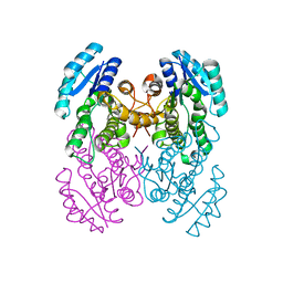
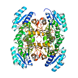
5XAM
 
 | |
5XAP
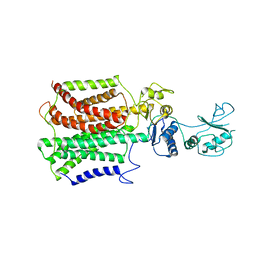
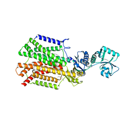
 | | Crystal structure of SecDF in I form (C2 space group) | | 分子名称: | (2R)-2,3-dihydroxypropyl (9Z)-octadec-9-enoate, DI(HYDROXYETHYL)ETHER, Protein translocase subunit SecD | | 著者 | Tsukazaki, T, Tanaka, Y, Furukwa, A. | | 登録日 | 2017-03-14 | | 公開日 | 2017-05-17 | | 最終更新日 | 2023-11-22 | | 実験手法 | X-RAY DIFFRACTION (2.605 Å) | | 主引用文献 | Tunnel Formation Inferred from the I-Form Structures of the Proton-Driven Protein Secretion Motor SecDF
Cell Rep, 19, 2017
|
|
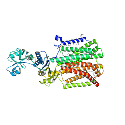
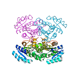
5XAN
 | | Crystal structure of SecDF in I form (P212121 space group) | | 分子名称: | (2R)-2,3-dihydroxypropyl (9Z)-octadec-9-enoate, POLYETHYLENE GLYCOL (N=34), Protein translocase subunit SecD | | 著者 | Tsukazaki, T, Tanaka, Y, Furukwa, A. | | 登録日 | 2017-03-14 | | 公開日 | 2017-05-17 | | 最終更新日 | 2023-11-22 | | 実験手法 | X-RAY DIFFRACTION (2.75 Å) | | 主引用文献 | Tunnel Formation Inferred from the I-Form Structures of the Proton-Driven Protein Secretion Motor SecDF
Cell Rep, 19, 2017
|
|
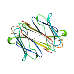
6KX0
 | | Crystal structure of SN-101 mAb non-liganded form | | 分子名称: | 2-AMINO-2-HYDROXYMETHYL-PROPANE-1,3-DIOL, Fab Fragment-SN-101-Heavy chain, Fab Fragment-SN-101-Light chain | | 著者 | Wakui, H, Tanaka, Y, Kato, K, Ose, T, Matsumoto, I, Min, Y, Tachibana, T, Nishimura, S.-I. | | 登録日 | 2019-09-09 | | 公開日 | 2020-07-29 | | 最終更新日 | 2023-11-22 | | 実験手法 | X-RAY DIFFRACTION (2.404 Å) | | 主引用文献 | A straightforward approach to antibodies recognising cancer specific glycopeptidic neoepitopes
Chem Sci, 11, 2020
|
|
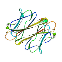
6KX1
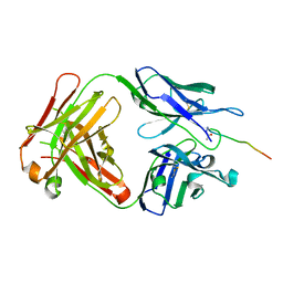 | | Crystal structure of SN-101 mAb in complex with MUC1 glycopeptide | | 分子名称: | 2-acetamido-2-deoxy-alpha-D-galactopyranose, Fab Fragment-SN-101-Heavy chain, Fab Fragment-SN-101-Light chain, ... | | 著者 | Wakui, H, Tanaka, Y, Kato, K, Ose, T, Matsumoto, I, Min, Y, Tachibana, T, Nishimura, S.-I. | | 登録日 | 2019-09-09 | | 公開日 | 2020-07-29 | | 最終更新日 | 2023-11-22 | | 実験手法 | X-RAY DIFFRACTION (1.773 Å) | | 主引用文献 | A straightforward approach to antibodies recognising cancer specific glycopeptidic neoepitopes
Chem Sci, 11, 2020
|
|
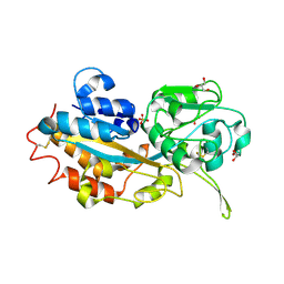
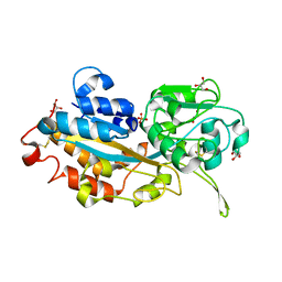
3W9Z
 | | Crystal structure of DusC | | 分子名称: | FLAVIN MONONUCLEOTIDE, tRNA-dihydrouridine synthase C | | 著者 | Chen, M, Yu, J, Tanaka, Y, Tanaka, I, Yao, M. | | 登録日 | 2013-04-19 | | 公開日 | 2013-07-31 | | 最終更新日 | 2024-03-20 | | 実験手法 | X-RAY DIFFRACTION (2.1 Å) | | 主引用文献 | Structure of dihydrouridine synthase C (DusC) from Escherichia coli
Acta Crystallogr.,Sect.F, 69, 2013
|
|
2ZKL
 | | Crystal Structure of capsular polysaccharide assembling protein CapF from staphylococcus aureus | | 分子名称: | Capsular polysaccharide synthesis enzyme Cap5F, GLYCEROL, ZINC ION | | 著者 | Miyafusa, T, Tanaka, Y, Yao, M, Tanaka, I, Tsumoto, K. | | 登録日 | 2008-03-25 | | 公開日 | 2009-03-31 | | 最終更新日 | 2024-03-13 | | 実験手法 | X-RAY DIFFRACTION (2.61 Å) | | 主引用文献 | Crystal structure of capsular polysaccharide assembling protein from Staphylococcus aureus
to be published
|
|
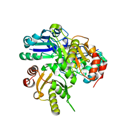
3AXG
 | | Structure of 6-aminohexanoate-oligomer hydrolase | | 分子名称: | Endotype 6-aminohexanoat-oligomer hydrolase, SODIUM ION | | 著者 | Negoro, S, Shibata, N, Tanaka, Y, Yasuhira, K, Shibata, H, Hashimoto, H, Lee, Y.H, Ohshima, S, Santa, R, Mochiji, K, Goto, Y, Ikegami, T, Nagai, K, Kato, D, Takeo, M, Higuchi, Y. | | 登録日 | 2011-04-04 | | 公開日 | 2011-12-21 | | 最終更新日 | 2024-03-13 | | 実験手法 | X-RAY DIFFRACTION (2 Å) | | 主引用文献 | Three-dimensional structure of nylon hydrolase and mechanism of nylon-6 hydrolysis
J.Biol.Chem., 287, 2012
|
|
3B0V
 | | tRNA-dihydrouridine synthase from Thermus thermophilus in complex with tRNA | | 分子名称: | FLAVIN MONONUCLEOTIDE, tRNA, tRNA-dihydrouridine synthase | | 著者 | Yu, F, Tanaka, Y, Yamashita, K, Nakamura, A, Yao, M, Tanaka, I. | | 登録日 | 2011-06-14 | | 公開日 | 2011-12-14 | | 最終更新日 | 2017-10-11 | | 実験手法 | X-RAY DIFFRACTION (3.51 Å) | | 主引用文献 | Molecular basis of dihydrouridine formation on tRNA
Proc.Natl.Acad.Sci.USA, 108, 2011
|
|
3B0U
 | | tRNA-dihydrouridine synthase from Thermus thermophilus in complex with tRNA fragment | | 分子名称: | FLAVIN MONONUCLEOTIDE, RNA (5'-R(*GP*GP*(H2U)P*A)-3'), tRNA-dihydrouridine synthase | | 著者 | Yu, F, Tanaka, Y, Yamashita, K, Nakamura, A, Yao, M, Tanaka, I. | | 登録日 | 2011-06-14 | | 公開日 | 2011-12-14 | | 最終更新日 | 2023-11-01 | | 実験手法 | X-RAY DIFFRACTION (1.948 Å) | | 主引用文献 | Molecular basis of dihydrouridine formation on tRNA
Proc.Natl.Acad.Sci.USA, 108, 2011
|
|
3B0P
 | | tRNA-dihydrouridine synthase from Thermus thermophilus | | 分子名称: | FLAVIN MONONUCLEOTIDE, tRNA-dihydrouridine synthase | | 著者 | Yu, F, Tanaka, Y, Yamashita, K, Nakamura, A, Yao, M, Tanaka, I. | | 登録日 | 2011-06-12 | | 公開日 | 2011-12-14 | | 最終更新日 | 2024-03-13 | | 実験手法 | X-RAY DIFFRACTION (1.7 Å) | | 主引用文献 | Molecular basis of dihydrouridine formation on tRNA
Proc.Natl.Acad.Sci.USA, 108, 2011
|
|
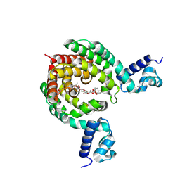
2E8H
 | | Crystal structure of PH0725 from Pyrococcus horikoshii OT3 | | 分子名称: | Probable diphthine synthase, S-ADENOSYL-L-HOMOCYSTEINE, SODIUM ION | | 著者 | Sugahara, M, Taketa, M, Tanaka, Y, Matsuura, Y, Kunishima, N, RIKEN Structural Genomics/Proteomics Initiative (RSGI) | | 登録日 | 2007-01-19 | | 公開日 | 2007-07-24 | | 最終更新日 | 2023-10-25 | | 実験手法 | X-RAY DIFFRACTION (2.1 Å) | | 主引用文献 | Crystal structure of PH0725 from Pyrococcus horikoshii OT3
To be Published
|
|
3ST7
 | | Crystal Structure of capsular polysaccharide assembling protein CapF from staphylococcus aureus | | 分子名称: | Capsular polysaccharide synthesis enzyme Cap5F, GLYCEROL, ZINC ION | | 著者 | Miyafusa, T, Tanaka, Y, Kuroda, M, Yao, M, Watanabe, M, Ohta, T, Tanaka, I, Caaveiro, J.M.M, Tsumoto, K. | | 登録日 | 2011-07-09 | | 公開日 | 2012-02-15 | | 最終更新日 | 2023-12-06 | | 実験手法 | X-RAY DIFFRACTION (2.45 Å) | | 主引用文献 | Crystal structure of the enzyme CapF of Staphylococcus aureus reveals a unique architecture composed of two functional domains.
Biochem.J., 443, 2012
|
|
7EQF
 | | Crystal Structure of a Transcription Factor in complex with Ligand | | 分子名称: | (6~{R})-3-methyl-8-[(2~{S},4~{R},5~{S},6~{R})-6-methyl-5-[(2~{S},4~{R},5~{R},6~{R})-6-methyl-4-[(2~{S},5~{S},6~{S})-6-methyl-5-[(2~{S},4~{R},5~{S},6~{R})-6-methyl-5-[(2~{S},4~{S},5~{S},6~{R})-6-methyl-4-[(2~{S},5~{S},6~{S})-6-methyl-5-oxidanyl-oxan-2-yl]oxy-5-oxidanyl-oxan-2-yl]oxy-4-oxidanyl-oxan-2-yl]oxy-oxan-2-yl]oxy-5-oxidanyl-oxan-2-yl]oxy-4-oxidanyl-oxan-2-yl]oxy-1,6,11-tris(oxidanyl)-5,6-dihydrobenzo[a]anthracene-7,12-dione, TetR/AcrR family transcriptional regulator | | 著者 | Uehara, S, Tsugita, A, Matsui, T, Yokoyama, T, Ostash, I, Ostash, B, Tanaka, Y. | | 登録日 | 2021-05-01 | | 公開日 | 2022-04-27 | | 最終更新日 | 2023-11-29 | | 実験手法 | X-RAY DIFFRACTION (2.91 Å) | | 主引用文献 | The carbohydrate tail of landomycin A is responsible for its interaction with the repressor protein LanK.
Febs J., 289, 2022
|
|
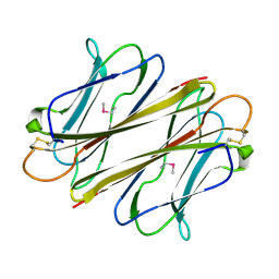
2E27
 | | Crystal structure of Fv fragment of anti-ciguatoxin antibody complexed with ABC-ring of ciguatoxin | | 分子名称: | 3,7:6,10:9,14-TRIANHYDRO-2,5,11,12,13-PENTADEOXY-4-O-(METHOXYMETHYL)-L-ARABINO-L-ALLO-TETRADEC-12-ENONIC ACID, DIMETHYL SULFOXIDE, GLYCEROL, ... | | 著者 | Yokota, A, Tanaka, Y, Tsumoto, K. | | 登録日 | 2006-11-08 | | 公開日 | 2007-11-13 | | 最終更新日 | 2023-10-25 | | 実験手法 | X-RAY DIFFRACTION (1.7 Å) | | 主引用文献 | Critical contribution of aromatic rings to specific recognition of polyether rings. The case of ciguatoxin CTX3C-ABC and its specific antibody 1C49.
J.Biol.Chem., 283, 2008
|
|
3VZR
 | |
3VZQ
 | |
3VZP
 | | Crystal structure of PhaB from Ralstonia eutropha | | 分子名称: | 1,4-DIETHYLENE DIOXIDE, Acetoacetyl-CoA reductase, GLYCEROL, ... | | 著者 | Ikeda, K, Tanaka, Y, Tanaka, I, Yao, M. | | 登録日 | 2012-10-15 | | 公開日 | 2013-08-28 | | 最終更新日 | 2023-11-08 | | 実験手法 | X-RAY DIFFRACTION (1.792 Å) | | 主引用文献 | Directed evolution and structural analysis of NADPH-dependent Acetoacetyl Coenzyme A (Acetoacetyl-CoA) reductase from Ralstonia eutropha reveals two mutations responsible for enhanced kinetics
Appl.Environ.Microbiol., 79, 2013
|
|
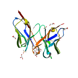
3VSE
 | | Crystal structure of methyltransferase | | 分子名称: | Putative uncharacterized protein, S-ADENOSYL-L-HOMOCYSTEINE | | 著者 | Kita, S, Tanaka, Y, Yao, M, Tanaka, I. | | 登録日 | 2012-04-25 | | 公開日 | 2013-04-10 | | 最終更新日 | 2024-03-20 | | 実験手法 | X-RAY DIFFRACTION (2.099 Å) | | 主引用文献 | Crystal structure of a putative methyltransferase SAV1081 from Staphylococcus aureus
Protein Pept.Lett., 20, 2012
|
|
3VZS
 | | Crystal structure of PhaB from Ralstonia eutropha in complex with Acetoacetyl-CoA and NADP | | 分子名称: | ACETOACETYL-COENZYME A, Acetoacetyl-CoA reductase, NADP NICOTINAMIDE-ADENINE-DINUCLEOTIDE PHOSPHATE, ... | | 著者 | Ikeda, K, Tanaka, Y, Tanaka, I, Yao, M. | | 登録日 | 2012-10-15 | | 公開日 | 2013-08-28 | | 最終更新日 | 2023-11-08 | | 実験手法 | X-RAY DIFFRACTION (2.14 Å) | | 主引用文献 | Directed evolution and structural analysis of NADPH-dependent Acetoacetyl Coenzyme A (Acetoacetyl-CoA) reductase from Ralstonia eutropha reveals two mutations responsible for enhanced kinetics
Appl.Environ.Microbiol., 79, 2013
|
|
3W9V
 | | Crystal structure of refolded DING protein | | 分子名称: | GLYCEROL, PHOSPHATE ION, Phosphate-binding protein | | 著者 | Gai, Z.Q, Nakamura, A, Tanaka, Y, Hirano, N, Tanaka, I, Yao, M. | | 登録日 | 2013-04-17 | | 公開日 | 2013-10-30 | | 最終更新日 | 2023-11-08 | | 実験手法 | X-RAY DIFFRACTION (1.031 Å) | | 主引用文献 | Crystal structure analysis, overexpression and refolding behaviour of a DING protein with single mutation.
J.SYNCHROTRON RADIAT., 20, 2013
|
|
3W9W
 | | Crystal structure of DING protein | | 分子名称: | DING protein, GLYCEROL, PHOSPHATE ION | | 著者 | Gai, Z.Q, Nakamura, A, Tanaka, Y, Hirano, N, Tanaka, I, Yao, M. | | 登録日 | 2013-04-17 | | 公開日 | 2013-10-30 | | 最終更新日 | 2023-11-08 | | 実験手法 | X-RAY DIFFRACTION (1.35 Å) | | 主引用文献 | Crystal structure analysis, overexpression and refolding behaviour of a DING protein with single mutation.
J.SYNCHROTRON RADIAT., 20, 2013
|
|
7F91
 | |
7F9F
 | | Thrombocorticin | | 分子名称: | MAGNESIUM ION, Thrombocorticin | | 著者 | Kageyama, H, Onodera, K, Sakai, R, Tanaka, Y, Freymann, D.M. | | 登録日 | 2021-07-04 | | 公開日 | 2022-07-06 | | 最終更新日 | 2023-11-29 | | 実験手法 | X-RAY DIFFRACTION (1.411 Å) | | 主引用文献 | A marine sponge-derived lectin reveals hidden pathway for thrombopoietin receptor activation.
Nat Commun, 13, 2022
|
|
7F9J
 | | Thrombocorticin Q25K in complex with Ca2+ | | 分子名称: | CALCIUM ION, Thrombocorticin Q25K mutant | | 著者 | Kageyama, H, Onodera, K, Sakai, R, Tanaka, Y. | | 登録日 | 2021-07-04 | | 公開日 | 2022-07-06 | | 最終更新日 | 2023-11-29 | | 実験手法 | X-RAY DIFFRACTION (1.1 Å) | | 主引用文献 | A marine sponge-derived lectin reveals hidden pathway for thrombopoietin receptor activation.
Nat Commun, 13, 2022
|
|


