6VU6
 
 | |
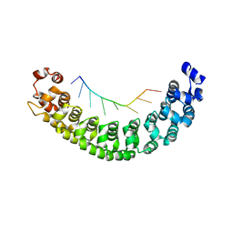
6VL9
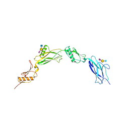
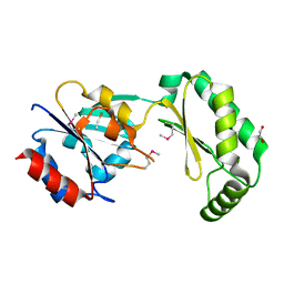
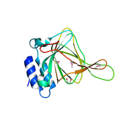
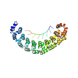
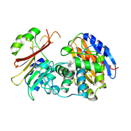
 | | Anti-PEG antibody 6-3 Fab fragment in complex with PEG | | 分子名称: | 3,6,9,12,15,18,21,24,27,30,33,36,39,42,45,48,51,54,57-nonadecaoxanonapentacontane-1,59-diol, 6-3 Fab heavy chain, 6-3 Fab light chain | | 著者 | Nicely, N.I, Huckaby, J.T, Lai, S.K, Jacobs, T.M. | | 登録日 | 2020-01-23 | | 公開日 | 2020-09-09 | | 最終更新日 | 2023-10-11 | | 実験手法 | X-RAY DIFFRACTION (2.63 Å) | | 主引用文献 | Structure of an anti-PEG antibody reveals an open ring that captures highly flexible PEG polymers
Commun Chem, 3, 2020
|
|
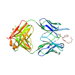
2B76
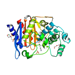
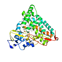
 | | E. coli Quinol fumarate reductase FrdA E49Q mutation | | 分子名称: | CITRATE ANION, FE2/S2 (INORGANIC) CLUSTER, FE3-S4 CLUSTER, ... | | 著者 | Maklashina, E, Iverson, T.M, Sher, Y, Kotlyar, V, Mirza, O, Andrell, J, Hudson, J.M, Armstrong, F.A, Cecchini, G. | | 登録日 | 2005-10-03 | | 公開日 | 2006-02-21 | | 最終更新日 | 2023-08-23 | | 実験手法 | X-RAY DIFFRACTION (3.3 Å) | | 主引用文献 | Fumarate Reductase and Succinate Oxidase Activity of Escherichia coli Complex II Homologs Are Perturbed Differently by Mutation of the Flavin Binding Domain
J.Biol.Chem., 281, 2006
|
|
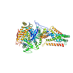
2AL1
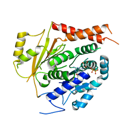
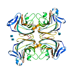
 | | Crystal Structure Analysis of Enolase Mg Subunit Complex at pH 8.0 | | 分子名称: | 2-PHOSPHOGLYCERIC ACID, CHLORIDE ION, MAGNESIUM ION, ... | | 著者 | Sims, P.A, Menefee, A.L, Larsen, T.M, Mansoorabadi, S.O, Reed, G.H. | | 登録日 | 2005-08-04 | | 公開日 | 2006-01-24 | | 最終更新日 | 2024-02-14 | | 実験手法 | X-RAY DIFFRACTION (1.5 Å) | | 主引用文献 | Structure and catalytic properties of an engineered heterodimer of enolase composed of one active and one inactive subunit
J.Mol.Biol., 355, 2006
|
|
6W8N
 | |
6W8P
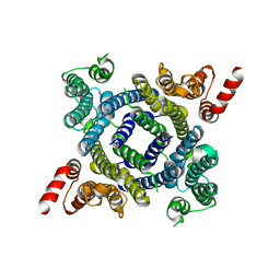
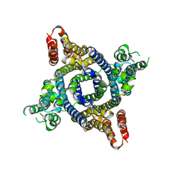
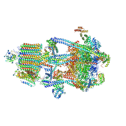
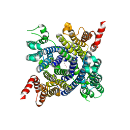 | | Structure of membrane protein with ions | | 分子名称: | Endosomal/lysosomal potassium channel TMEM175, POTASSIUM ION | | 著者 | Shen, C, Fu, T.M, Wang, L.F, Rawson, S, Wu, H. | | 登録日 | 2020-03-21 | | 公開日 | 2021-08-04 | | 実験手法 | ELECTRON MICROSCOPY (3.6 Å) | | 主引用文献 | Structure of membrane protein with ions
To Be Published
|
|
6W8O
 | |
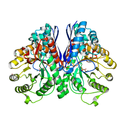
2AL2
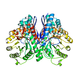 | | Crystal Structure Analysis of Enolase Mg Subunit Complex at pH 8.0 | | 分子名称: | 2-PHOSPHOGLYCERIC ACID, CHLORIDE ION, MAGNESIUM ION, ... | | 著者 | Sims, P.A, Menefee, A.L, Larsen, T.M, Mansoorabadi, S.O, Reed, G.H. | | 登録日 | 2005-08-04 | | 公開日 | 2006-01-24 | | 最終更新日 | 2021-10-20 | | 実験手法 | X-RAY DIFFRACTION (1.85 Å) | | 主引用文献 | Structure and catalytic properties of an engineered heterodimer of enolase composed of one active and one inactive subunit
J.Mol.Biol., 355, 2006
|
|
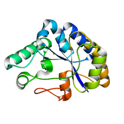
2AMY
 | | X-Ray Structure of Human Phosphomannomutase 2 (PMM2) | | 分子名称: | 1,2-ETHANEDIOL, GLYCINE, Phosphomannomutase 2 | | 著者 | Wesenberg, G.E, Phillips Jr, G.N, McCoy, J.G, Bitto, E, Bingman, C.A, Allard, S.T.M, Center for Eukaryotic Structural Genomics (CESG) | | 登録日 | 2005-08-10 | | 公開日 | 2005-08-23 | | 最終更新日 | 2018-01-24 | | 実験手法 | X-RAY DIFFRACTION (2.09 Å) | | 主引用文献 | X-Ray Structure of Human Phosphomannomutase 2 (PMM2)
To be Published
|
|
6TPM
 | | Crystal structure of AmpC from E.coli with Relebactam (MK-7655) | | 分子名称: | (2S,5R)-1-formyl-N-(piperidin-4-yl)-5-[(sulfooxy)amino]piperidine-2-carboxamide, 2-(N-MORPHOLINO)-ETHANESULFONIC ACID, Beta-lactamase, ... | | 著者 | Lang, P.A, Leissing, T.M, Schofield, C.J, Brem, J. | | 登録日 | 2019-12-13 | | 公開日 | 2020-11-25 | | 最終更新日 | 2024-01-24 | | 実験手法 | X-RAY DIFFRACTION (1.72 Å) | | 主引用文献 | Structural Investigations of the Inhibition of Escherichia coli AmpC beta-Lactamase by Diazabicyclooctanes.
Antimicrob.Agents Chemother., 65, 2021
|
|
6V5V
 | | Structure of gamma-tubulin in the native human gamma-tubulin ring complex | | 分子名称: | GUANOSINE-5'-DIPHOSPHATE, Tubulin gamma-1 chain | | 著者 | Wieczorek, M, Urnavicius, L, Ti, S, Molloy, K.R, Chait, B.T, Kapoor, T.M. | | 登録日 | 2019-12-04 | | 公開日 | 2020-01-01 | | 最終更新日 | 2024-03-06 | | 実験手法 | ELECTRON MICROSCOPY (3.8 Å) | | 主引用文献 | Asymmetric Molecular Architecture of the Human gamma-Tubulin Ring Complex.
Cell, 180, 2020
|
|
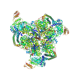
6WM4
 | | Human V-ATPase in state 3 with SidK and ADP | | 分子名称: | 2-acetamido-2-deoxy-beta-D-glucopyranose, ADENOSINE-5'-DIPHOSPHATE, Renin receptor, ... | | 著者 | Wang, L, Wu, H, Fu, T.M. | | 登録日 | 2020-04-20 | | 公開日 | 2020-11-11 | | 最終更新日 | 2020-11-18 | | 実験手法 | ELECTRON MICROSCOPY (3.6 Å) | | 主引用文献 | Structures of a Complete Human V-ATPase Reveal Mechanisms of Its Assembly.
Mol.Cell, 80, 2020
|
|
6WLZ
 | | The V1 region of human V-ATPase in state 1 (focused refinement) | | 分子名称: | ADENOSINE-5'-DIPHOSPHATE, SidK, V-type proton ATPase catalytic subunit A, ... | | 著者 | Wang, L, Wu, H, Fu, T.M. | | 登録日 | 2020-04-20 | | 公開日 | 2020-11-11 | | 最終更新日 | 2024-03-06 | | 実験手法 | ELECTRON MICROSCOPY (2.9 Å) | | 主引用文献 | Structures of a Complete Human V-ATPase Reveal Mechanisms of Its Assembly.
Mol.Cell, 80, 2020
|
|
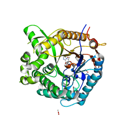
2ATF
 | | X-RAY STRUCTURE OF cysteine dioxygenase type I FROM MUS MUSCULUS MM.241056 | | 分子名称: | 1,2-ETHANEDIOL, Cysteine dioxygenase type I, NICKEL (II) ION | | 著者 | Wesenberg, G.E, Phillips Jr, G.N, Mccoy, J.G, Bitto, E, Bingman, C.A, Allard, S.T.M, Center for Eukaryotic Structural Genomics (CESG) | | 登録日 | 2005-08-24 | | 公開日 | 2005-10-18 | | 最終更新日 | 2017-10-11 | | 実験手法 | X-RAY DIFFRACTION (1.75 Å) | | 主引用文献 | Structure and mechanism of mouse cysteine dioxygenase.
Proc.Natl.Acad.Sci.Usa, 103, 2006
|
|
2C71
 | | The structure of a family 4 acetyl xylan esterase from Clostridium thermocellum in complex with a magnesium ion. | | 分子名称: | GLYCOSIDE HYDROLASE, FAMILY 11:CLOSTRIDIUM CELLULOSOME ENZYME, DOCKERIN TYPE I:POLYSACCHARIDE, ... | | 著者 | Taylor, E.J, Turkenburg, P.J, Vincent, F, Brzozowski, A.M, Gloster, T.M, Dupont, C, Shareck, F, Centeno, M.S.J, Prates, J.A.M, Ferreira, L.M.A, Fontes, C.M.G.A, Biely, P, Davies, G.J. | | 登録日 | 2005-11-17 | | 公開日 | 2006-01-23 | | 最終更新日 | 2024-05-08 | | 実験手法 | X-RAY DIFFRACTION (1.05 Å) | | 主引用文献 | Structure and Activity of Two Metal-Ion Dependent Acetyl Xylan Esterases Involved in Plant Cell-Wall Degradation Reveals a Close Similarity to Peptidoglycan Deacetylases.
J.Biol.Chem., 281, 2006
|
|
2CES
 | | Beta-glucosidase from Thermotoga maritima in complex with glucoimidazole | | 分子名称: | ACETATE ION, BETA-GLUCOSIDASE A, CALCIUM ION, ... | | 著者 | Gloster, T.M, Roberts, S, Vasella, A, Davies, G.J. | | 登録日 | 2006-02-10 | | 公開日 | 2006-09-27 | | 最終更新日 | 2023-12-13 | | 実験手法 | X-RAY DIFFRACTION (2.15 Å) | | 主引用文献 | Structural, Kinetic, and Thermodynamic Analysis of Glucoimidazole-Derived Glycosidase Inhibitors.
Biochemistry, 45, 2006
|
|
2FER
 | | P450CAM from Pseudomonas putida reconstituted with manganic protoporphyrin IX | | 分子名称: | Cytochrome P450-cam, POTASSIUM ION, PROTOPORPHYRIN IX CONTAINING MN | | 著者 | von Koenig, K, Makris, T.M, Sligar, S.G, Schlichting, I. | | 登録日 | 2005-12-16 | | 公開日 | 2006-03-14 | | 最終更新日 | 2023-08-30 | | 実験手法 | X-RAY DIFFRACTION (1.7 Å) | | 主引用文献 | The status of high-valent metal oxo complexes in the P450 cytochromes.
J.Inorg.Biochem., 100, 2006
|
|
3OYP
 | | HCV NS3/4A in complex with ligand 3 | | 分子名称: | Non-structural protein 4A, Peptidomimetic inhibitor, Serine protease NS3, ... | | 著者 | Hagel, M, Niu, D, St.Martin, T, Sheets, M.P, Qiao, L, Bernard, H, Karp, R.M, Zhu, Z, Labenski, M.T, Chaturvedi, P.C, Nacht, M, Westlin, W.F, Petter, R.C, Singh, J. | | 登録日 | 2010-09-23 | | 公開日 | 2010-12-01 | | 最終更新日 | 2023-11-15 | | 実験手法 | X-RAY DIFFRACTION (2.76 Å) | | 主引用文献 | Selective irreversible inhibition of a protease by targeting a noncatalytic cysteine.
Nat.Chem.Biol., 7, 2011
|
|
2FEY
 | | The structure of stem loop IV of Tetrahymena telomerase RNA | | 分子名称: | stem-loop IV of Tetrahymena telomerase RNA | | 著者 | Chen, Y, Fender, J, Legassie, J.D, Jarstfer, M.B, Bryan, T.M, Varani, G. | | 登録日 | 2005-12-16 | | 公開日 | 2006-06-27 | | 最終更新日 | 2024-05-29 | | 実験手法 | SOLUTION NMR | | 主引用文献 | Structure of stem-loop IV of Tetrahymena telomerase RNA.
Embo J., 25, 2006
|
|
2FEU
 | | P450CAM from Pseudomonas putida reconstituted with manganic protoporphyrin IX | | 分子名称: | 2-AMINO-2-HYDROXYMETHYL-PROPANE-1,3-DIOL, CAMPHOR, Cytochrome P450-cam, ... | | 著者 | von Koenig, K, Makris, T.M, Sligar, S.G, Schlichting, I. | | 登録日 | 2005-12-16 | | 公開日 | 2006-03-14 | | 最終更新日 | 2023-08-30 | | 実験手法 | X-RAY DIFFRACTION (1.7 Å) | | 主引用文献 | The status of high-valent metal oxo complexes in the P450 cytochromes.
J.Inorg.Biochem., 100, 2006
|
|
3Q0L
 | |
3Q0M
 | |
2FE6
 | | P450CAM from Pseudomonas putida reconstituted with manganic protoporphyrin IX | | 分子名称: | Cytochrome P450-cam, POTASSIUM ION, PROTOPORPHYRIN IX CONTAINING MN | | 著者 | von Koenig, K, Makris, T.M, Sligar, S.G, Schlichting, I. | | 登録日 | 2005-12-15 | | 公開日 | 2006-03-14 | | 最終更新日 | 2023-08-30 | | 実験手法 | X-RAY DIFFRACTION (1.5 Å) | | 主引用文献 | The status of high-valent metal oxo complexes in the P450 cytochromes.
J.Inorg.Biochem., 100, 2006
|
|
2EIG
 | | Lotus tetragonolobus seed lectin (Isoform) | | 分子名称: | 2-acetamido-2-deoxy-beta-D-glucopyranose, CALCIUM ION, MANGANESE (II) ION, ... | | 著者 | Moreno, F.B.M.B, Vicoti, M.M, Abrego, J.R.B, de Oliveira, T.M, Bezerra, G.A, Cavada, B.S, Filgueira de Azevedo Jr, W. | | 登録日 | 2007-03-13 | | 公開日 | 2008-03-04 | | 最終更新日 | 2020-07-29 | | 実験手法 | X-RAY DIFFRACTION (2 Å) | | 主引用文献 | Identification of a new quaternary association for legume lectins
J.Struct.Biol., 161, 2008
|
|
2G2W
 | | Crystal Structure of the SHV D104K Beta-lactamase/Beta-lactamase inhibitor protein (BLIP) complex | | 分子名称: | Beta-lactamase SHV-1, Beta-lactamase inhibitory protein | | 著者 | Reynolds, K.A, Thomson, J.M, Corbett, K.D, Bethel, C.R, Berger, J.M, Kirsch, J.F, Bonomo, R.A, Handel, T.M. | | 登録日 | 2006-02-16 | | 公開日 | 2006-07-04 | | 最終更新日 | 2023-08-30 | | 実験手法 | X-RAY DIFFRACTION (1.8 Å) | | 主引用文献 | Structural and Computational Characterization of the SHV-1 beta-Lactamase-beta-Lactamase Inhibitor Protein Interface.
J.Biol.Chem., 281, 2006
|
|


