3VWV
 
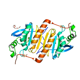 | |
3W8J
 | | Crystal structure of P5 a0 in a complex with Prx4 c-term | | 分子名称: | C-terminal peptide from Peroxiredoxin-4, GLYCEROL, POTASSIUM ION, ... | | 著者 | Inaba, K, Suzuki, M, Kojima, R. | | 登録日 | 2013-03-13 | | 公開日 | 2013-09-04 | | 実験手法 | X-RAY DIFFRACTION (2.1 Å) | | 主引用文献 | Synergistic cooperation of PDI family members in peroxiredoxin 4-driven oxidative protein folding
Sci Rep, 3, 2013
|
|
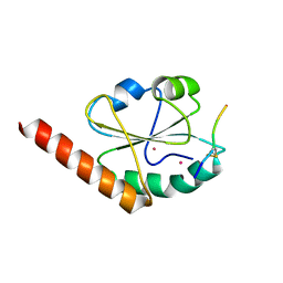
3WUM
 | |
3WXU
 | |
3WGE
 | | Crystal structure of ERp46 Trx2 | | 分子名称: | GLYCEROL, Thioredoxin domain-containing protein 5 | | 著者 | Inaba, K, Suzuki, M, Kojima, R. | | 登録日 | 2013-08-04 | | 公開日 | 2014-06-25 | | 実験手法 | X-RAY DIFFRACTION (0.95 Å) | | 主引用文献 | Radically different thioredoxin domain arrangement of ERp46, an efficient disulfide bond introducer of the mammalian PDI family
Structure, 22, 2014
|
|
3WGD
 | | Crystal structure of ERp46 Trx1 | | 分子名称: | GLYCEROL, PHOSPHATE ION, POTASSIUM ION, ... | | 著者 | Inaba, K, Suzuki, M, Kojima, R. | | 登録日 | 2013-08-04 | | 公開日 | 2014-06-25 | | 実験手法 | X-RAY DIFFRACTION (2.5 Å) | | 主引用文献 | Radically different thioredoxin domain arrangement of ERp46, an efficient disulfide bond introducer of the mammalian PDI family
Structure, 22, 2014
|
|
3WGX
 | | Crystal structure of ERp46 Trx2 in a complex with Prx4 C-term | | 分子名称: | GLYCEROL, Peroxiredoxin-4, Thioredoxin domain-containing protein 5 | | 著者 | Inaba, K, Suzuki, M, Kojima, R. | | 登録日 | 2013-08-13 | | 公開日 | 2014-06-25 | | 実験手法 | X-RAY DIFFRACTION (0.92 Å) | | 主引用文献 | Radically different thioredoxin domain arrangement of ERp46, an efficient disulfide bond introducer of the mammalian PDI family
Structure, 22, 2014
|
|
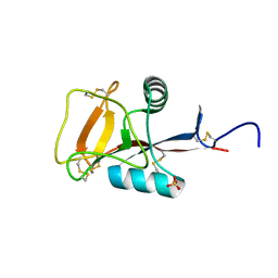
3WUL
 | |
3WXS
 | | Thaumatin structure determined by SPring-8 Angstrom Compact free electron Laser (SACLA) | | 分子名称: | L(+)-TARTARIC ACID, thaumatin I | | 著者 | Masuda, T, Nango, E, Sugahara, M, Mizohata, E, Tanaka, T, Tanaka, R, Suzuki, M, Mikami, B, Iwata, S. | | 登録日 | 2014-08-07 | | 公開日 | 2014-11-05 | | 最終更新日 | 2023-11-08 | | 実験手法 | X-RAY DIFFRACTION (2 Å) | | 主引用文献 | Grease matrix as a versatile carrier of proteins for serial crystallography
Nat. Methods, 12, 2015
|
|
3WXT
 | |
3VK9
 | | Crystal structure of delta-class glutathione transferase from silkmoth | | 分子名称: | GLYCEROL, Glutathione S-transferase delta | | 著者 | Kakuta, Y, Usuda, K, Higashiura, A, Suzuki, M, Nakagawa, A, Kimura, M, Yamamoto, K. | | 登録日 | 2011-11-10 | | 公開日 | 2012-10-03 | | 最終更新日 | 2024-03-20 | | 実験手法 | X-RAY DIFFRACTION (2.001 Å) | | 主引用文献 | Structural basis for catalytic activity of a silkworm Delta-class glutathione transferase
Biochim.Biophys.Acta, 1820, 2012
|
|
2ZUQ
 | | Crystal structure of DsbB-Fab complex | | 分子名称: | Disulfide bond formation protein B, Fab fragment heavy chain, Fab fragment light chain, ... | | 著者 | Inaba, K, Suzuki, M, Murakami, S. | | 登録日 | 2008-10-28 | | 公開日 | 2009-04-14 | | 最終更新日 | 2023-11-01 | | 実験手法 | X-RAY DIFFRACTION (3.3 Å) | | 主引用文献 | Dynamic nature of disulphide bond formation catalysts revealed by crystal structures of DsbB
Embo J., 28, 2009
|
|
3APQ
 | |
3APO
 | |
3APS
 | |
2ZUP
 | | Updated crystal structure of DsbB-DsbA complex from E. coli | | 分子名称: | Disulfide bond formation protein B, Thiol:disulfide interchange protein dsbA, UBIQUINONE-1, ... | | 著者 | Inaba, K, Suzuki, M, Murakami, S, Nakagawa, A. | | 登録日 | 2008-10-28 | | 公開日 | 2009-04-14 | | 最終更新日 | 2023-11-01 | | 実験手法 | X-RAY DIFFRACTION (3.7 Å) | | 主引用文献 | Dynamic nature of disulphide bond formation catalysts revealed by crystal structures of DsbB
Embo J., 28, 2009
|
|
2ZIB
 | | Crystal structure analysis of calcium-independent type II antifreeze protein | | 分子名称: | SULFATE ION, Type II antifreeze protein | | 著者 | Nishimiya, Y, Sato, R, Kondo, H, Noro, N, Sugimoto, H, Suzuki, M, Tsuda, S. | | 登録日 | 2008-02-14 | | 公開日 | 2008-08-19 | | 最終更新日 | 2011-07-13 | | 実験手法 | X-RAY DIFFRACTION (1.34 Å) | | 主引用文献 | Crystal structure and mutational analysis of Ca2+-independent type II antifreeze protein from longsnout poacher, Brachyopsis rostratus
J.Mol.Biol., 382, 2008
|
|
3AXA
 | | Crystal structure of afadin PDZ domain in complex with the C-terminal peptide from nectin-3 | | 分子名称: | Afadin, Nectin-3 | | 著者 | Fujiwara, Y, Goda, N, Narita, H, Satomura, K, Nakagawa, A, Sakisaka, T, Suzuki, M, Hiroaki, H. | | 登録日 | 2011-03-31 | | 公開日 | 2012-04-25 | | 最終更新日 | 2023-11-01 | | 実験手法 | X-RAY DIFFRACTION (2.78 Å) | | 主引用文献 | Crystal structure of afadin PDZ domain-nectin-3 complex shows the structural plasticity of the ligand-binding site.
Protein Sci., 24, 2015
|
|
3VQF
 | | Crystal Structure Analysis of the PDZ Domain Derived from the Tight Junction Regulating Protein | | 分子名称: | E3 ubiquitin-protein ligase LNX | | 著者 | Akiyoshi, Y, Hamada, D, Goda, N, Tenno, T, Narita, H, Nakagawa, A, Furuse, M, Suzuki, M, Hiroaki, H. | | 登録日 | 2012-03-22 | | 公開日 | 2013-03-27 | | 最終更新日 | 2023-11-08 | | 実験手法 | X-RAY DIFFRACTION (1.199 Å) | | 主引用文献 | Structural basis for down regulation of tight junction by PDZ-domain containing E3-Ubiquitin ligase
To be Published
|
|
3VQG
 | | Crystal Structure Analysis of the PDZ Domain Derived from the Tight Junction Regulating Protein | | 分子名称: | C-terminal peptide from Immunoglobulin superfamily member 5, E3 ubiquitin-protein ligase LNX, SULFATE ION | | 著者 | Akiyoshi, Y, Hamada, D, Goda, N, Tenno, T, Narita, H, Nakagawa, A, Furuse, M, Suzuki, M, Hiroaki, H. | | 登録日 | 2012-03-23 | | 公開日 | 2013-03-27 | | 最終更新日 | 2023-11-08 | | 実験手法 | X-RAY DIFFRACTION (1.35 Å) | | 主引用文献 | Structural basis for down regulation of tight junction by PDZ-domain containing E3-Ubiquitin ligase
To be Published
|
|
3WUN
 | |
3VRP
 | | Crystal structure of the tyrosine kinase binding domain of Cbl-c in complex with phospho-EGFR peptide | | 分子名称: | CALCIUM ION, Epidermal growth factor receptor, Signal transduction protein CBL-C | | 著者 | Takeshita, K, Tezuka, T, Isozaki, Y, Yamashita, E, Suzuki, M, Yamanashi, Y, Yamamoto, T, Nakagawa, A. | | 登録日 | 2012-04-13 | | 公開日 | 2013-03-06 | | 実験手法 | X-RAY DIFFRACTION (1.52 Å) | | 主引用文献 | Structural flexibility regulates phosphopeptide-binding activity of the tyrosine kinase binding domain of Cbl-c.
J.Biochem., 152, 2012
|
|
3VRN
 | | Crystal structure of the tyrosine kinase binding domain of Cbl-c | | 分子名称: | CALCIUM ION, Signal transduction protein CBL-C | | 著者 | Takeshita, K, Tezuka, T, Isozaki, Y, Yamashita, E, Suzuki, M, Yamanashi, Y, Yamamoto, T, Nakagawa, A. | | 登録日 | 2012-04-13 | | 公開日 | 2013-03-06 | | 最終更新日 | 2023-11-08 | | 実験手法 | X-RAY DIFFRACTION (1.64 Å) | | 主引用文献 | Structural flexibility regulates phosphopeptide-binding activity of the tyrosine kinase binding domain of Cbl-c
J.Biochem., 152, 2012
|
|
3VRR
 | | Crystal structure of the tyrosine kinase binding domain of Cbl-c (PL mutant) in complex with phospho-EGFR peptide | | 分子名称: | CALCIUM ION, Epidermal growth factor receptor, Signal transduction protein CBL-C | | 著者 | Takeshita, K, Tezuka, T, Isozaki, Y, Yamashita, E, Suzuki, M, Yamanashi, Y, Yamamoto, T, Nakagawa, A. | | 登録日 | 2012-04-13 | | 公開日 | 2013-03-06 | | 実験手法 | X-RAY DIFFRACTION (2 Å) | | 主引用文献 | Structural flexibility regulates phosphopeptide-binding activity of the tyrosine kinase binding domain of Cbl-c.
J.Biochem., 152, 2012
|
|
3VRQ
 | | Crystal structure of the tyrosine kinase binding domain of Cbl-c (PL mutant) | | 分子名称: | CALCIUM ION, Signal transduction protein CBL-C | | 著者 | Takeshita, K, Tezuka, T, Isozaki, Y, Yamashita, E, Suzuki, M, Yamanashi, Y, Yamamoto, T, Nakagawa, A. | | 登録日 | 2012-04-13 | | 公開日 | 2013-03-06 | | 最終更新日 | 2024-03-20 | | 実験手法 | X-RAY DIFFRACTION (2.39 Å) | | 主引用文献 | Structural flexibility regulates phosphopeptide-binding activity of the tyrosine kinase binding domain of Cbl-c.
J.Biochem., 152, 2012
|
|


