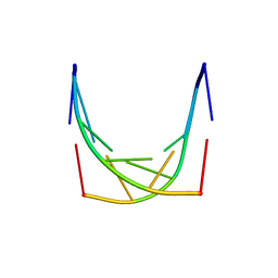
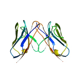
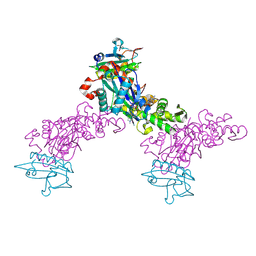
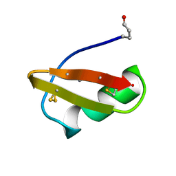
4GLK
 
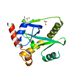 | | Structure and activity of AbiQ, a lactococcal anti-phage endoribonuclease belonging to the type-III toxin-antitoxin system | | 分子名称: | AbiQ, GLYCEROL | | 著者 | Samson, J, Spinelli, S, Cambillau, C, Moineau, S. | | 登録日 | 2012-08-14 | | 公開日 | 2013-01-16 | | 最終更新日 | 2023-09-13 | | 実験手法 | X-RAY DIFFRACTION (2.16 Å) | | 主引用文献 | Structure and activity of AbiQ, a lactococcal endoribonuclease belonging to the type III toxin-antitoxin system.
Mol.Microbiol., 87, 2013
|
|
1IH3
 | |
2M0O
 | |
2OTB
 | |
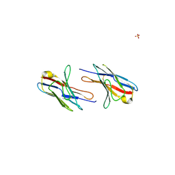
2OTE
 | | Crystal structure of a monomeric cyan fluorescent protein in the photobleached state | | 分子名称: | ACETATE ION, GFP-like fluorescent chromoprotein cFP484 | | 著者 | Henderson, J.N, Ai, H, Campbell, R.E, Remington, S.J. | | 登録日 | 2007-02-07 | | 公開日 | 2007-04-03 | | 最終更新日 | 2023-11-15 | | 実験手法 | X-RAY DIFFRACTION (1.47 Å) | | 主引用文献 | Structural basis for reversible photobleaching of a green fluorescent protein homologue.
Proc.Natl.Acad.Sci.Usa, 104, 2007
|
|
1IH4
 | |
1IH6
 | |
1T84
 | | Solution structure of the Wiskott-Aldrich Syndrome Protein (WASP) autoinhibited core domain complexed with (S)-wiskostatin, a small molecule inhibitor | | 分子名称: | (2S)-1-(3,6-DIBROMO-9H-CARBAZOL-9-YL)-3-(DIMETHYLAMINO)PROPAN-2-OL, Wiskott-Aldrich syndrome protein | | 著者 | Peterson, J.R, Bickford, L.C, Morgan, D, Kim, A.S, Ouerfelli, O, Kirschner, M.W, Rosen, M.K. | | 登録日 | 2004-05-11 | | 公開日 | 2004-07-13 | | 最終更新日 | 2024-05-22 | | 実験手法 | SOLUTION NMR | | 主引用文献 | Chemical inhibition of N-WASP by stabilization of a native autoinhibited conformation.
Nat.Struct.Mol.Biol., 11, 2004
|
|
1IH2
 | |
1K0E
 | | THE CRYSTAL STRUCTURE OF AMINODEOXYCHORISMATE SYNTHASE FROM FORMATE GROWN CRYSTALS | | 分子名称: | FORMIC ACID, TRYPTOPHAN, p-aminobenzoate synthase component I | | 著者 | Parsons, J.F, Jensen, P.Y, Pachikara, A.S, Howard, A.J, Eisenstein, E, Ladner, J.E. | | 登録日 | 2001-09-19 | | 公開日 | 2002-02-27 | | 最終更新日 | 2024-02-07 | | 実験手法 | X-RAY DIFFRACTION (2 Å) | | 主引用文献 | Structure of Escherichia coli aminodeoxychorismate synthase: architectural conservation and diversity in chorismate-utilizing enzymes.
Biochemistry, 41, 2002
|
|
2Q20
 | |
2Q1E
 | |
1IH1
 | |
1K0G
 | | THE CRYSTAL STRUCTURE OF AMINODEOXYCHORISMATE SYNTHASE FROM PHOSPHATE GROWN CRYSTALS | | 分子名称: | PHOSPHATE ION, TRYPTOPHAN, p-aminobenzoate synthase component I | | 著者 | Parsons, J.F, Jensen, P.Y, Pachikara, A.S, Howard, A.J, Eisenstein, E, Ladner, J.E. | | 登録日 | 2001-09-19 | | 公開日 | 2002-02-27 | | 最終更新日 | 2023-08-16 | | 実験手法 | X-RAY DIFFRACTION (2.05 Å) | | 主引用文献 | Structure of Escherichia coli aminodeoxychorismate synthase: architectural conservation and diversity in chorismate-utilizing enzymes.
Biochemistry, 41, 2002
|
|
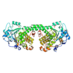
1MDU
 | | Crystal structure of the chicken actin trimer complexed with human gelsolin segment 1 (GS-1) | | 分子名称: | 2-AMINO-2-HYDROXYMETHYL-PROPANE-1,3-DIOL, ADENOSINE-5'-TRIPHOSPHATE, CALCIUM ION, ... | | 著者 | Dawson, J.F, Sablin, E.P, Spudich, J.A, Fletterick, R.J. | | 登録日 | 2002-08-07 | | 公開日 | 2003-01-07 | | 最終更新日 | 2011-07-13 | | 実験手法 | X-RAY DIFFRACTION (2.2 Å) | | 主引用文献 | Structure of an F-actin trimer disrupted by gelsolin and implications for the mechanism of severing
J.Biol.Chem., 278, 2003
|
|
1L6B
 | |
1LFO
 | | LIVER FATTY ACID BINDING PROTEIN-OLEATE COMPLEX | | 分子名称: | BUTENOIC ACID, LIVER FATTY ACID BINDING PROTEIN, OLEIC ACID, ... | | 著者 | Thompson, J, Winter, N, Terwey, D, Bratt, J, Banaszak, L. | | 登録日 | 1996-12-09 | | 公開日 | 1997-06-16 | | 最終更新日 | 2023-08-09 | | 実験手法 | X-RAY DIFFRACTION (2.3 Å) | | 主引用文献 | The crystal structure of the liver fatty acid-binding protein. A complex with two bound oleates.
J.Biol.Chem., 272, 1997
|
|
1LSH
 | |
1M8C
 | | SOLUTION STRUCTURE OF THE T State OF TURKEY OVOMUCOID AT PH 2.5 | | 分子名称: | Ovomucoid | | 著者 | Song, J, Laskowski Jr, M, Qasim, M.A, Markley, J.L. | | 登録日 | 2002-07-24 | | 公開日 | 2002-09-04 | | 最終更新日 | 2021-10-27 | | 実験手法 | SOLUTION NMR | | 主引用文献 | Two conformational states of Turkey ovomucoid third domain at low pH: three-dimensional structures, internal dynamics, and interconversion kinetics and thermodynamics.
Biochemistry, 42, 2003
|
|
1M8B
 | | Solution structure of the C State of turkey ovomucoid at pH 2.5 | | 分子名称: | Ovomucoid | | 著者 | Song, J, Laskowski Jr, M, Qasim, M.A, Markley, J.L. | | 登録日 | 2002-07-24 | | 公開日 | 2002-09-04 | | 最終更新日 | 2021-10-27 | | 実験手法 | SOLUTION NMR | | 主引用文献 | Two conformational states of Turkey ovomucoid third domain at low pH: three-dimensional structures, internal dynamics, and interconversion kinetics and thermodynamics.
Biochemistry, 42, 2003
|
|
1CBI
 | |
1E0F
 | | Crystal structure of the human alpha-thrombin-haemadin complex: an exosite II-binding inhibitor | | 分子名称: | HAEMADIN, THROMBIN | | 著者 | Richardson, J.L, Kroeger, B, Hoefken, W, Pereira, P, Huber, R, Bode, W, Fuentes-Prior, P. | | 登録日 | 2000-03-27 | | 公開日 | 2000-11-03 | | 最終更新日 | 2023-12-06 | | 実験手法 | X-RAY DIFFRACTION (3.1 Å) | | 主引用文献 | Crystal Structure of the Human Alpha-Thrombin-Haemadin Complex: An Exosite II-Binding Inhibitor
Embo J., 19, 2000
|
|
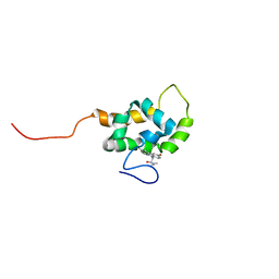
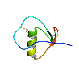
1IS0
 | | Crystal Structure of a Complex of the Src SH2 Domain with Conformationally Constrained Peptide Inhibitor | | 分子名称: | AY0 GLU GLU ILE peptide, Tyrosine-protein kinase transforming protein SRC | | 著者 | Davidson, J.P, Lubman, O, Rose, T, Waksman, G, Martin, S.F. | | 登録日 | 2001-11-02 | | 公開日 | 2002-02-06 | | 最終更新日 | 2023-12-27 | | 実験手法 | X-RAY DIFFRACTION (1.9 Å) | | 主引用文献 | Calorimetric and structural studies of 1,2,3-trisubstituted cyclopropanes as conformationally constrained peptide inhibitors of Src SH2 domain binding.
J.Am.Chem.Soc., 124, 2002
|
|
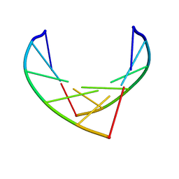
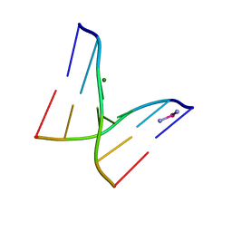
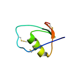
1BAH
 | | A TWO DISULFIDE DERIVATIVE OF CHARYBDOTOXIN WITH DISULFIDE 13-33 REPLACED BY TWO ALPHA-AMINOBUTYRIC ACIDS, NMR, 30 STRUCTURES | | 分子名称: | CHARYBDOTOXIN | | 著者 | Song, J, Gilquin, B, Jamin, N, Guenneugues, M, Dauplais, M, Vita, C, Menez, A. | | 登録日 | 1996-06-06 | | 公開日 | 1997-01-11 | | 最終更新日 | 2020-01-15 | | 実験手法 | SOLUTION NMR | | 主引用文献 | NMR solution structure of a two-disulfide derivative of charybdotoxin: structural evidence for conservation of scorpion toxin alpha/beta motif and its hydrophobic side chain packing.
Biochemistry, 36, 1997
|
|
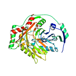
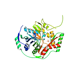
4Y7I
 | | Crystal Structure of MTMR8 | | 分子名称: | Myotubularin-related protein 8, PHOSPHATE ION | | 著者 | Yoo, K, Lee, J, Son, J, Shin, W, Im, D, Heo, Y.S. | | 登録日 | 2015-02-15 | | 公開日 | 2015-07-15 | | 最終更新日 | 2024-03-20 | | 実験手法 | X-RAY DIFFRACTION (2.802 Å) | | 主引用文献 | Structure of the catalytic phosphatase domain of MTMR8: implications for dimerization, membrane association and reversible oxidation.
Acta Crystallogr.,Sect.D, 71, 2015
|
|


