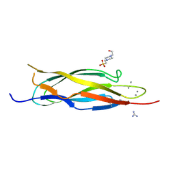
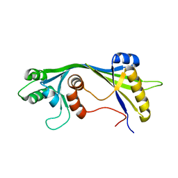
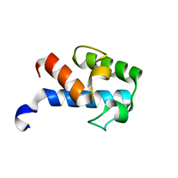
2HQK
 
 | | Crystal structure of a monomeric cyan fluorescent protein derived from Clavularia | | 分子名称: | ACETATE ION, CHLORIDE ION, Cyan fluorescent chromoprotein, ... | | 著者 | Henderson, J.N, Campbell, R.E, Ai, H, Remington, S.J. | | 登録日 | 2006-07-18 | | 公開日 | 2007-01-02 | | 最終更新日 | 2023-11-15 | | 実験手法 | X-RAY DIFFRACTION (1.19 Å) | | 主引用文献 | Directed evolution of a monomeric, bright and photostable version of Clavularia cyan fluorescent protein: structural characterization and applications in fluorescence imaging.
Biochem.J., 400, 2006
|
|
1TAD
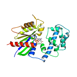 | | GTPASE MECHANISM OF GPROTEINS FROM THE 1.7-ANGSTROM CRYSTAL STRUCTURE OF TRANSDUCIN ALPHA-GDP-ALF4- | | 分子名称: | CACODYLATE ION, CALCIUM ION, GUANOSINE-5'-DIPHOSPHATE, ... | | 著者 | Sondek, J, Lambright, D.G, Noel, J.P, Hamm, H.E, Sigler, P.B. | | 登録日 | 1995-01-05 | | 公開日 | 1995-05-08 | | 最終更新日 | 2024-02-14 | | 実験手法 | X-RAY DIFFRACTION (1.7 Å) | | 主引用文献 | GTPase mechanism of Gproteins from the 1.7-A crystal structure of transducin alpha-GDP-AIF-4.
Nature, 372, 1994
|
|
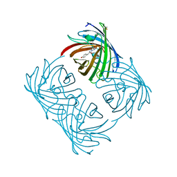
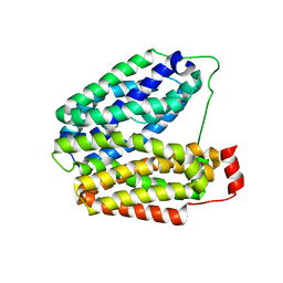
2A47
 | | Crystal structure of amFP486 H199T | | 分子名称: | BETA-MERCAPTOETHANOL, GFP-like fluorescent chromoprotein amFP486 | | 著者 | Henderson, J.N, Remington, S.J. | | 登録日 | 2005-06-28 | | 公開日 | 2005-08-16 | | 最終更新日 | 2023-11-15 | | 実験手法 | X-RAY DIFFRACTION (1.72 Å) | | 主引用文献 | Crystal structures and mutational analysis of amFP486, a cyan fluorescent protein from Anemonia majano
Proc.Natl.Acad.Sci.Usa, 102, 2005
|
|
2IAK
 | |
4FK9
 | |
2DPK
 | |
2A46
 | |
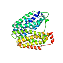
2A48
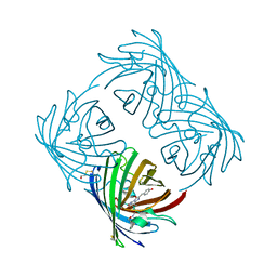 | | Crystal structure of amFP486 E150Q | | 分子名称: | BETA-MERCAPTOETHANOL, GFP-like fluorescent chromoprotein amFP486 | | 著者 | Henderson, J.N, Remington, S.J. | | 登録日 | 2005-06-28 | | 公開日 | 2005-08-16 | | 最終更新日 | 2023-11-15 | | 実験手法 | X-RAY DIFFRACTION (2 Å) | | 主引用文献 | Crystal structures and mutational analysis of amFP486, a cyan fluorescent protein from Anemonia majano
Proc.Natl.Acad.Sci.Usa, 102, 2005
|
|
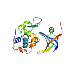
1T9M
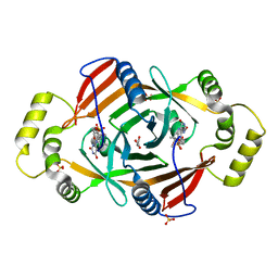 | | X-ray crystal structure of phzG from pseudomonas aeruginosa | | 分子名称: | ACETIC ACID, FLAVIN MONONUCLEOTIDE, SULFATE ION, ... | | 著者 | Parsons, J.F, Eisenstein, E, Ladner, J.E. | | 登録日 | 2004-05-18 | | 公開日 | 2004-11-02 | | 最終更新日 | 2023-08-23 | | 実験手法 | X-RAY DIFFRACTION (1.9 Å) | | 主引用文献 | Structure of the phenazine biosynthesis enzyme PhzG.
Acta Crystallogr.,Sect.D, 60, 2004
|
|
2FFT
 | | NMR structure of Spinach Thylakoid Soluble Phosphoprotein of 9 kDa in SDS Micelles | | 分子名称: | thylakoid soluble phosphoprotein | | 著者 | Song, J, Carlberg, I, Lee, M.S, Markley, J.L, Center for Eukaryotic Structural Genomics (CESG) | | 登録日 | 2005-12-20 | | 公開日 | 2006-01-17 | | 最終更新日 | 2024-05-29 | | 実験手法 | SOLUTION NMR | | 主引用文献 | Micelle-induced folding of spinach thylakoid soluble phosphoprotein of 9 kDa and its functional implications.
Biochemistry, 45, 2006
|
|
4GLA
 | | OBody NL8 bound to hen egg-white lysozyme | | 分子名称: | Lysozyme C, OBody NL8 | | 著者 | Steemson, J.D. | | 登録日 | 2012-08-14 | | 公開日 | 2013-08-14 | | 最終更新日 | 2014-02-12 | | 実験手法 | X-RAY DIFFRACTION (2.75 Å) | | 主引用文献 | Tracking Molecular Recognition at the Atomic Level with a New Protein Scaffold Based on the OB-Fold.
Plos One, 9, 2014
|
|
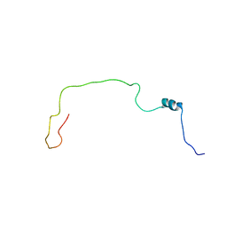
2ETT
 | | Solution Structure of Human Sorting Nexin 22 PX Domain | | 分子名称: | Sorting nexin-22 | | 著者 | Song, J, Zhao, Q, Tyler, R.C, Lee, M.S, Newman, C.L, Markley, J.L, Center for Eukaryotic Structural Genomics (CESG) | | 登録日 | 2005-10-27 | | 公開日 | 2005-11-15 | | 最終更新日 | 2024-05-22 | | 実験手法 | SOLUTION NMR | | 主引用文献 | Solution structure of human sorting nexin 22.
Protein Sci., 16, 2007
|
|
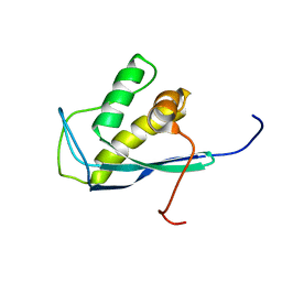
1TIZ
 | | Solution Structure of a Calmodulin-Like Calcium-Binding Domain from Arabidopsis thaliana | | 分子名称: | calmodulin-related protein, putative | | 著者 | Song, J, Zhao, Q, Thao, S, Frederick, R.O, Markley, J.L, Center for Eukaryotic Structural Genomics (CESG) | | 登録日 | 2004-06-02 | | 公開日 | 2004-08-10 | | 最終更新日 | 2024-05-22 | | 実験手法 | SOLUTION NMR | | 主引用文献 | Letter to the Editor: Solution structure of a calmodulin-like calcium-binding domain from Arabidopsis thaliana
J.Biomol.NMR, 30, 2004
|
|
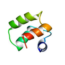
2JMU
 | |
2JU4
 | | NMR structure of the gamma subunit of cGMP phosphodiesterase | | 分子名称: | (3'R)-1'-oxyl-2',2',5',5'-tetramethyl-1,3'-bipyrrolidine-2,5-dione, Retinal rod rhodopsin-sensitive cGMP 3',5'-cyclic phosphodiesterase subunit gamma | | 著者 | Song, J, Guo, L.W, Ruoho, A.E, Markley, J.L, Center for Eukaryotic Structural Genomics (CESG) | | 登録日 | 2007-08-14 | | 公開日 | 2007-10-16 | | 最終更新日 | 2020-02-19 | | 実験手法 | SOLUTION NMR | | 主引用文献 | Intrinsically disordered gamma-subunit of cGMP phosphodiesterase encodes functionally relevant transient secondary and tertiary structure.
Proc.Natl.Acad.Sci.USA, 105, 2008
|
|
2ASQ
 | | Solution Structure of SUMO-1 in Complex with a SUMO-binding Motif (SBM) | | 分子名称: | Protein inhibitor of activated STAT2, Small ubiquitin-related modifier 1 | | 著者 | Song, J, Zhang, Z, Hu, W, Chen, Y. | | 登録日 | 2005-08-23 | | 公開日 | 2005-10-11 | | 最終更新日 | 2024-05-22 | | 実験手法 | SOLUTION NMR | | 主引用文献 | Small Ubiquitin-like Modifier (SUMO) Recognition of a SUMO Binding Motif: A reversal of the bound orientation
J.Biol.Chem., 280, 2005
|
|
1F6I
 | |
1F6J
 | |
1F69
 | |
1RPU
 | | Crystal Structure of CIRV p19 bound to siRNA | | 分子名称: | 19 kDa protein, 5'-R(P*CP*GP*UP*AP*CP*GP*CP*GP*UP*CP*AP*CP*GP*CP*GP*UP*AP*CP*GP*UP*U)-3' | | 著者 | Vargason, J.M, Szittya, G, Burgyan, J, Hall, T.M.T. | | 登録日 | 2003-12-03 | | 公開日 | 2004-01-13 | | 最終更新日 | 2024-02-14 | | 実験手法 | X-RAY DIFFRACTION (2.5 Å) | | 主引用文献 | Size selective recognition of siRNA by an RNA silencing suppressor
Cell(Cambridge,Mass.), 115, 2003
|
|
2KU7
 | |
2MF4
 | | 1H, 13C, 15N chemical shift assignments of Streptomyces virginiae VirA acp5a | | 分子名称: | Hybrid polyketide synthase-non ribosomal peptide synthetase | | 著者 | Davison, J, Dorival, J, Rabeharindranto, M.H, Mazon, H, Chagot, B, Gruez, A, Weissman, K.J. | | 登録日 | 2013-10-05 | | 公開日 | 2014-06-04 | | 最終更新日 | 2024-05-01 | | 実験手法 | SOLUTION NMR | | 主引用文献 | NMR assignements of ACP5a
To be Published
|
|
1PV6
 | | Crystal structure of lactose permease | | 分子名称: | Lactose permease | | 著者 | Abramson, J, Smirnova, I, Kasho, V, Verner, G, Kaback, H.R, Iwata, S. | | 登録日 | 2003-06-26 | | 公開日 | 2003-08-12 | | 最終更新日 | 2024-05-29 | | 実験手法 | X-RAY DIFFRACTION (3.5 Å) | | 主引用文献 | Structure and mechanism of the lactose permease of Escherichia coli
SCIENCE, 301, 2003
|
|
1PV7
 | | Crystal structure of lactose permease with TDG | | 分子名称: | Lactose permease, beta-D-galactopyranose-(1-1)-1-thio-beta-D-galactopyranose | | 著者 | Abramson, J, Smirnova, I, Kasho, V, Verner, G, Kaback, H.R, Iwata, S. | | 登録日 | 2003-06-26 | | 公開日 | 2003-08-12 | | 最終更新日 | 2024-05-29 | | 実験手法 | X-RAY DIFFRACTION (3.6 Å) | | 主引用文献 | Structure and mechanism of the lactose permease of Escherichia coli
SCIENCE, 301, 2003
|
|
1SPF
 | |


