1MNV
 
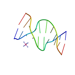
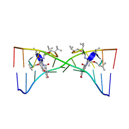 | | Actinomycin D binding to ATGCTGCAT | | 分子名称: | 5'-D(*AP*TP*GP*CP*TP*GP*CP*AP*T)-3', ACTINOMYCIN D | | 著者 | Hou, M.-H, Robinson, H, Gao, Y.-G, Wang, A.H.-J. | | 登録日 | 2002-09-06 | | 公開日 | 2002-11-22 | | 最終更新日 | 2024-07-10 | | 実験手法 | X-RAY DIFFRACTION (2.6 Å) | | 主引用文献 | Crystal Structure of Actinomycin D Bound to the Ctg Triplet Repeat Sequences Linked to Neurological Diseases
Nucleic Acids Res., 30, 2002
|
|
1D8X
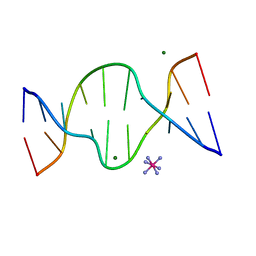 | | CRYSTAL STRUCTURE OF DNA SHEARED TANDEM G A BASE PAIRS | | 分子名称: | 5'-D(*CP*CP*GP*AP*AP*TP*GP*AP*GP*G)-3', COBALT HEXAMMINE(III), MAGNESIUM ION | | 著者 | Gao, Y.-G, Robinson, H, Sanishvili, R, Joachimiak, A, Wang, A.H.-J. | | 登録日 | 1999-10-26 | | 公開日 | 1999-11-05 | | 最終更新日 | 2024-02-07 | | 実験手法 | X-RAY DIFFRACTION (1.2 Å) | | 主引用文献 | Structure and recognition of sheared tandem G x A base pairs associated with human centromere DNA sequence at atomic resolution.
Biochemistry, 38, 1999
|
|
1R8U
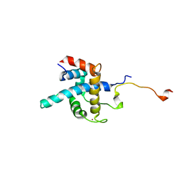 | | NMR structure of CBP TAZ1/CITED2 complex | | 分子名称: | CREB-binding protein, Cbp/p300-interacting transactivator 2, ZINC ION | | 著者 | De Guzman, R.N, Martinez-Yamout, M, Dyson, H.J, Wright, P.E. | | 登録日 | 2003-10-28 | | 公開日 | 2004-03-23 | | 最終更新日 | 2024-05-22 | | 実験手法 | SOLUTION NMR | | 主引用文献 | Interaction of the TAZ1 domain of the CREB-binding protein with the activation domain of CITED2: regulation by competition between intrinsically unstructured ligands for non-identical binding sites.
J.Biol.Chem., 279, 2004
|
|
1S4T
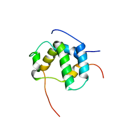
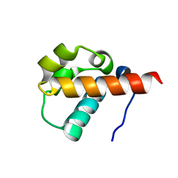
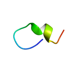 | | Solution structure of synthetic 21mer peptide spanning region 135-155 (in human numbering) of sheep prion protein | | 分子名称: | Major prion protein | | 著者 | Kozin, S.A, Lepage, C, Hui Bon Hoa, G, Rabesona, H, Mazur, A.K, Blond, A, Cheminant, M, Haertle, T, Debey, P, Rebuffat, S. | | 登録日 | 2004-01-18 | | 公開日 | 2004-01-27 | | 最終更新日 | 2024-05-22 | | 実験手法 | SOLUTION NMR | | 主引用文献 | Specific recognition between surface loop 2 (132-143) and helix 1 (144-154)
within sheep prion protein from in vitro studies of synthetic peptides
To be Published
|
|
1S2M
 | |
1PN9
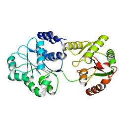
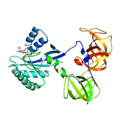
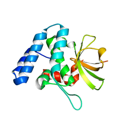
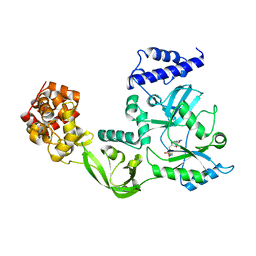
 | | Crystal structure of an insect delta-class glutathione S-transferase from a DDT-resistant strain of the malaria vector Anopheles gambiae | | 分子名称: | Glutathione S-transferase 1-6, S-HEXYLGLUTATHIONE | | 著者 | Chen, L, Hall, P.R, Zhou, X.E, Ranson, H, Hemingway, J, Meehan, E.J. | | 登録日 | 2003-06-12 | | 公開日 | 2003-12-09 | | 最終更新日 | 2024-04-03 | | 実験手法 | X-RAY DIFFRACTION (2 Å) | | 主引用文献 | Structure of an insect delta-class glutathione S-transferase from a DDT-resistant strain of the malaria vector Anopheles gambiae.
Acta Crystallogr.,Sect.D, 59, 2003
|
|
4FGO
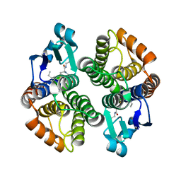
 | | Legionella pneumophila LapG (calcium-bound) | | 分子名称: | CALCIUM ION, Periplasmic protein | | 著者 | Chatterjee, D, Boyd, C.D, O'Toole, G.A, Sondermann, H. | | 登録日 | 2012-06-04 | | 公開日 | 2012-06-20 | | 最終更新日 | 2013-01-09 | | 実験手法 | X-RAY DIFFRACTION (1.903 Å) | | 主引用文献 | Structural characterization of a conserved, calcium-dependent periplasmic protease from Legionella pneumophila.
J.Bacteriol., 194, 2012
|
|
1RXR
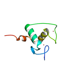 | | HIGH RESOLUTION SOLUTION STRUCTURE OF THE RETINOID X RECEPTOR DNA BINDING DOMAIN, NMR, 20 STRUCTURE | | 分子名称: | RETINOIC ACID RECEPTOR-ALPHA, ZINC ION | | 著者 | Holmbeck, S.M.A, Foster, M.P, Casimiro, D.R, Sem, D.S, Dyson, H.J, Wright, P.E. | | 登録日 | 1998-06-12 | | 公開日 | 1998-11-11 | | 最終更新日 | 2024-05-01 | | 実験手法 | SOLUTION NMR | | 主引用文献 | High-resolution solution structure of the retinoid X receptor DNA-binding domain.
J.Mol.Biol., 281, 1998
|
|
1DCR
 | | CRYSTAL STRUCTURE OF DNA SHEARED TANDEM G-A BASE PAIRS | | 分子名称: | 5'-D(*CP*CP*GP*AP*AP*(BRU)P*GP*AP*GP*G)-3', MAGNESIUM ION, SODIUM ION, ... | | 著者 | Gao, Y.-G, Robinson, H, Sanishvili, R, Joachimiak, A, Wang, A.H.-J. | | 登録日 | 1999-11-05 | | 公開日 | 1999-11-19 | | 最終更新日 | 2023-08-09 | | 実験手法 | X-RAY DIFFRACTION (1.6 Å) | | 主引用文献 | Structure and recognition of sheared tandem G x A base pairs associated with human centromere DNA sequence at atomic resolution.
Biochemistry, 38, 1999
|
|
4FGP
 | | Legionella pneumophila LapG (EGTA-treated) | | 分子名称: | Periplasmic protein | | 著者 | Chatterjee, D, Boyd, C.D, O'Toole, G.A, Sondermann, H. | | 登録日 | 2012-06-04 | | 公開日 | 2012-06-20 | | 最終更新日 | 2024-02-28 | | 実験手法 | X-RAY DIFFRACTION (1.73 Å) | | 主引用文献 | Structural characterization of a conserved, calcium-dependent periplasmic protease from Legionella pneumophila.
J.Bacteriol., 194, 2012
|
|
1CUR
 | | REDUCED RUSTICYANIN, NMR | | 分子名称: | COPPER (II) ION, CU(I) RUSTICYANIN | | 著者 | Botuyan, M.V, Dyson, H.J. | | 登録日 | 1996-04-19 | | 公開日 | 1996-11-08 | | 最終更新日 | 2024-05-22 | | 実験手法 | SOLUTION NMR | | 主引用文献 | NMR solution structure of Cu(I) rusticyanin from Thiobacillus ferrooxidans: structural basis for the extreme acid stability and redox potential.
J.Mol.Biol., 263, 1996
|
|
1S4U
 | |
1R5O
 | | crystal structure analysis of sup35 complexed with GMPPNP | | 分子名称: | Eukaryotic peptide chain release factor GTP-binding subunit, PHOSPHOAMINOPHOSPHONIC ACID-GUANYLATE ESTER | | 著者 | Kong, C, Song, H. | | 登録日 | 2003-10-11 | | 公開日 | 2004-05-25 | | 最終更新日 | 2023-10-25 | | 実験手法 | X-RAY DIFFRACTION (3.2 Å) | | 主引用文献 | Crystal structure and functional analysis of the eukaryotic class II release factor eRF3 from S. pombe
Mol.Cell, 14, 2004
|
|
1R5B
 | | Crystal structure analysis of sup35 | | 分子名称: | Eukaryotic peptide chain release factor GTP-binding subunit | | 著者 | Kong, C, Song, H. | | 登録日 | 2003-10-10 | | 公開日 | 2004-05-25 | | 最終更新日 | 2024-03-13 | | 実験手法 | X-RAY DIFFRACTION (2.35 Å) | | 主引用文献 | Crystal structure and functional analysis of the eukaryotic class II release factor eRF3 from S. pombe
Mol.Cell, 14, 2004
|
|
1R5N
 | | Crystal Structure Analysis of sup35 complexed with GDP | | 分子名称: | Eukaryotic peptide chain release factor GTP-binding subunit, GUANOSINE-5'-DIPHOSPHATE | | 著者 | Kong, C, Song, H. | | 登録日 | 2003-10-10 | | 公開日 | 2004-05-25 | | 最終更新日 | 2023-10-25 | | 実験手法 | X-RAY DIFFRACTION (2.9 Å) | | 主引用文献 | Crystal structure and functional analysis of the eukaryotic class II release factor eRF3 from S. pombe
Mol.Cell, 14, 2004
|
|
2L14
 | | Structure of CBP nuclear coactivator binding domain in complex with p53 TAD | | 分子名称: | CREB-binding protein, Cellular tumor antigen p53 | | 著者 | Lee, C, Martinez-Yamout, M.A, Dyson, H.J, Wright, P.E. | | 登録日 | 2010-07-22 | | 公開日 | 2010-11-03 | | 最終更新日 | 2024-05-01 | | 実験手法 | SOLUTION NMR | | 主引用文献 | Structure of the p53 transactivation domain in complex with the nuclear receptor coactivator binding domain of CREB binding protein.
Biochemistry, 49, 2010
|
|
4FGQ
 | | Legionella pneumophila LapG | | 分子名称: | Periplasmic protein | | 著者 | Chatterjee, D, Boyd, C.D, O'Toole, G.A, Sondermann, H. | | 登録日 | 2012-06-04 | | 公開日 | 2012-06-20 | | 最終更新日 | 2024-04-03 | | 実験手法 | X-RAY DIFFRACTION (1.645 Å) | | 主引用文献 | Structural characterization of a conserved, calcium-dependent periplasmic protease from Legionella pneumophila.
J.Bacteriol., 194, 2012
|
|
1D9R
 | | CRYSTAL STRUCTURE OF DNA SHEARED TANDEM G-A BASE PAIRS | | 分子名称: | 5'-D(*CP*CP*GP*AP*AP*(BRU)P*GP*AP*GP*G)-3', COBALT HEXAMMINE(III) | | 著者 | Gao, Y.-G, Robinson, H, Sanishvili, R, Joachimiak, A, Wang, A.H.-J. | | 登録日 | 1999-10-29 | | 公開日 | 1999-11-05 | | 最終更新日 | 2024-02-07 | | 実験手法 | X-RAY DIFFRACTION (1.5 Å) | | 主引用文献 | Structure and recognition of sheared tandem G x A base pairs associated with human centromere DNA sequence at atomic resolution.
Biochemistry, 38, 1999
|
|
1DGS
 | | CRYSTAL STRUCTURE OF NAD+-DEPENDENT DNA LIGASE FROM T. FILIFORMIS | | 分子名称: | ADENOSINE MONOPHOSPHATE, DNA LIGASE, ZINC ION | | 著者 | Lee, J.Y, Chang, C, Song, H.K, Kwon, S.T, Suh, S.W. | | 登録日 | 1999-11-25 | | 公開日 | 2000-11-27 | | 最終更新日 | 2011-07-13 | | 実験手法 | X-RAY DIFFRACTION (2.9 Å) | | 主引用文献 | Crystal structure of NAD(+)-dependent DNA ligase: modular architecture and functional implications.
EMBO J., 19, 2000
|
|
1SV4
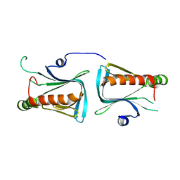
 | | Crystal Structure of Yan-SAM | | 分子名称: | Ets DNA-binding protein pokkuri | | 著者 | Qiao, F, Song, H, Kim, C.A, Sawaya, M.R, Hunter, J.B, Gingery, M, Rebay, I, Courey, A.J, Bowie, J.U. | | 登録日 | 2004-03-27 | | 公開日 | 2004-07-27 | | 最終更新日 | 2023-08-23 | | 実験手法 | X-RAY DIFFRACTION (2.15 Å) | | 主引用文献 | Derepression by depolymerization; structural insights into the regulation of yan by mae.
Cell(Cambridge,Mass.), 118, 2004
|
|
1Q67
 | | Crystal structure of Dcp1p | | 分子名称: | Decapping protein involved in mRNA degradation-Dcp1p | | 著者 | She, M, Decker, C.J, Liu, Y, Chen, N, Parker, R, Song, H. | | 登録日 | 2003-08-12 | | 公開日 | 2004-03-02 | | 最終更新日 | 2024-02-14 | | 実験手法 | X-RAY DIFFRACTION (2.3 Å) | | 主引用文献 | Crystal structure of Dcp1p and its functional implications in mRNA decapping
Nat.Struct.Mol.Biol., 11, 2004
|
|
1SB0
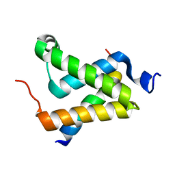 | | Solution structure of the KIX domain of CBP bound to the transactivation domain of c-Myb | | 分子名称: | protein CBP, protein c-Myb | | 著者 | Zor, T, De Guzman, R.N, Dyson, H.J, Wright, P.E. | | 登録日 | 2004-02-09 | | 公開日 | 2004-04-13 | | 最終更新日 | 2024-05-22 | | 実験手法 | SOLUTION NMR | | 主引用文献 | Solution Structure of the KIX Domain of CBP Bound to the Transactivation
Domain of c-Myb
J.Mol.Biol., 337, 2004
|
|
1TK1
 | | YEAST OXYGEN-DEPENDENT COPROPORPHYRINOGEN OXIDASE | | 分子名称: | Coproporphyrinogen III oxidase | | 著者 | Phillips, J.D, Whitby, F.G, Warby, C.A, Labbe, P, Yang, C, Pflugrath, J.W, Ferrara, J.D, Robinson, H, Kushner, J.P, Hill, C.P. | | 登録日 | 2004-06-08 | | 公開日 | 2004-07-20 | | 最終更新日 | 2024-02-14 | | 実験手法 | X-RAY DIFFRACTION (1.9 Å) | | 主引用文献 | Crystal Structure of the Oxygen-dependant Coproporphyrinogen Oxidase (Hem13p) of Saccharomyces cerevisiae
J.Biol.Chem., 279, 2004
|
|
4J40
 | | Crystal structure of the dual-domain GGDEF-EAL module of FimX from Pseudomonas aeruginosa | | 分子名称: | FimX | | 著者 | Navarro, M.V, De, N, Bae, N, Wang, Q, Sondermann, H. | | 登録日 | 2013-02-06 | | 公開日 | 2013-02-20 | | 最終更新日 | 2024-02-28 | | 実験手法 | X-RAY DIFFRACTION (2.99 Å) | | 主引用文献 | Structural analysis of the GGDEF-EAL domain-containing c-di-GMP receptor FimX.
Structure, 17, 2009
|
|
2KA6
 | | NMR structure of the CBP-TAZ2/STAT1-TAD complex | | 分子名称: | CREB-binding protein, Signal transducer and activator of transcription 1-alpha/beta, ZINC ION | | 著者 | Wojciak, J.M, Martinez-Yamout, M.A, Dyson, H.J, Wright, P.E. | | 登録日 | 2008-10-30 | | 公開日 | 2009-04-07 | | 最終更新日 | 2024-05-08 | | 実験手法 | SOLUTION NMR | | 主引用文献 | Structural basis for recruitment of CBP/p300 coactivators by STAT1 and STAT2 transactivation domains.
Embo J., 28, 2009
|
|


