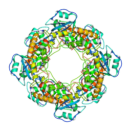
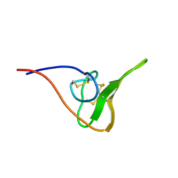
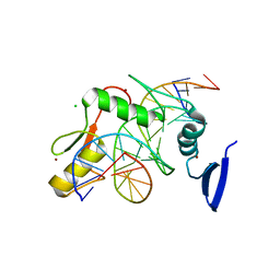
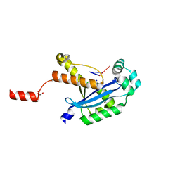
7MQG
 
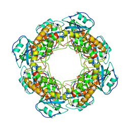 | | Bartonella henselae NrnC complexed with pAAAGG. C1 reconstruction. | | 分子名称: | NanoRNase C, RNA (5'-R(P*AP*AP*AP*GP*G)-3') | | 著者 | Lormand, J.D, Brownfield, B, Fromme, J.C, Sondermann, H. | | 登録日 | 2021-05-05 | | 公開日 | 2021-09-15 | | 最終更新日 | 2024-05-29 | | 実験手法 | ELECTRON MICROSCOPY (3.25 Å) | | 主引用文献 | Structural characterization of NrnC identifies unifying features of dinucleotidases.
Elife, 10, 2021
|
|
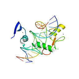
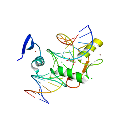
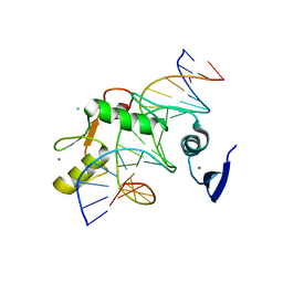
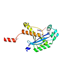
7MPM
 | |
7MPN
 | |
7MQI
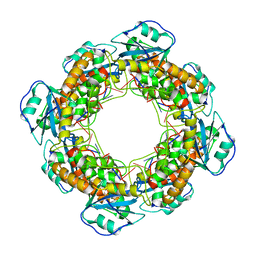 | | Bartonella henselae NrnC complexed with pAAAGG in the presence of Ca2+. C1 reconstruction. | | 分子名称: | NanoRNase C, RNA (5'-R(P*AP*AP*AP*GP*G)-3') | | 著者 | Lormand, J.D, Brownfield, B, Fromme, J.C, Sondermann, H. | | 登録日 | 2021-05-05 | | 公開日 | 2021-09-15 | | 最終更新日 | 2024-05-29 | | 実験手法 | ELECTRON MICROSCOPY (3.21 Å) | | 主引用文献 | Structural characterization of NrnC identifies unifying features of dinucleotidases.
Elife, 10, 2021
|
|
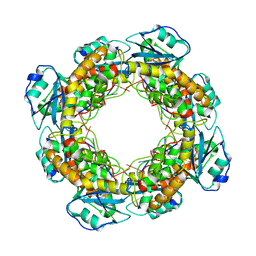
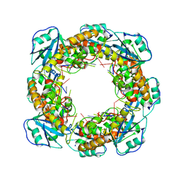
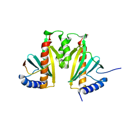
7MQH
 | | Bartonella henselae NrnC complexed with pAAAGG in the presence of Ca2+. D4 Symmetry. | | 分子名称: | NanoRNase C, RNA (5'-R(P*AP*AP*AP*GP*G)-3') | | 著者 | Lormand, J.D, Brownfield, B, Fromme, J.C, Sondermann, H. | | 登録日 | 2021-05-05 | | 公開日 | 2021-09-15 | | 最終更新日 | 2024-05-29 | | 実験手法 | ELECTRON MICROSCOPY (3.1 Å) | | 主引用文献 | Structural characterization of NrnC identifies unifying features of dinucleotidases.
Elife, 10, 2021
|
|
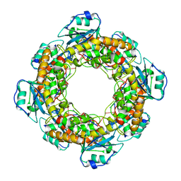
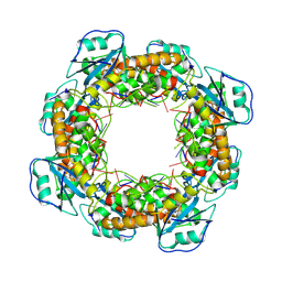
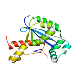
7MQF
 | | Bartonella henselae NrnC complexed with pAAAGG. D4 symmetry. | | 分子名称: | NanoRNase C, RNA (5'-R(P*AP*AP*AP*GP*G)-3') | | 著者 | Lormand, J.D, Brownfield, B, Fromme, J.C, Sondermann, H. | | 登録日 | 2021-05-05 | | 公開日 | 2021-09-15 | | 最終更新日 | 2024-05-29 | | 実験手法 | ELECTRON MICROSCOPY (2.88 Å) | | 主引用文献 | Structural characterization of NrnC identifies unifying features of dinucleotidases.
Elife, 10, 2021
|
|
7MQE
 | | Bartonella henselae NrnC complexed with pAGG. C1 reconstruction. | | 分子名称: | NanoRNase C, RNA (5'-R(P*AP*GP*G)-3') | | 著者 | Lormand, J.D, Brownfield, B, Fromme, J.C, Sondermann, H. | | 登録日 | 2021-05-05 | | 公開日 | 2021-09-15 | | 最終更新日 | 2024-05-29 | | 実験手法 | ELECTRON MICROSCOPY (3.69 Å) | | 主引用文献 | Structural characterization of NrnC identifies unifying features of dinucleotidases.
Elife, 10, 2021
|
|
3BUJ
 | | Crystal Structure of CalO2 | | 分子名称: | CalO2, PROTOPORPHYRIN IX CONTAINING FE | | 著者 | McCoy, J.G, Johnson, H.D, Singh, S, Bingman, C.A, Thorson, J.S, Phillips Jr, G.N. | | 登録日 | 2008-01-02 | | 公開日 | 2008-04-29 | | 最終更新日 | 2023-08-30 | | 実験手法 | X-RAY DIFFRACTION (2.47 Å) | | 主引用文献 | Structural characterization of CalO2: a putative orsellinic acid P450 oxidase in the calicheamicin biosynthetic pathway.
Proteins, 74, 2009
|
|
3K3H
 | | Crystal structure of the PDE9A catalytic domain in complex with (S)-BAY73-6691 | | 分子名称: | 1-(2-chlorophenyl)-6-[(2S)-3,3,3-trifluoro-2-methylpropyl]-1,7-dihydro-4H-pyrazolo[3,4-d]pyrimidin-4-one, High affinity cGMP-specific 3',5'-cyclic phosphodiesterase 9A, MAGNESIUM ION, ... | | 著者 | Wang, H, Luo, X, Ye, M, Hou, J, Robinson, H, Ke, H. | | 登録日 | 2009-10-02 | | 公開日 | 2010-02-16 | | 最終更新日 | 2023-09-06 | | 実験手法 | X-RAY DIFFRACTION (2.5 Å) | | 主引用文献 | Insight into Binding of Phosphodiesterase-9A Selective Inhibitors by Crystal Structures and Mutagenesis
J.Med.Chem., 53, 2010
|
|
2JOO
 | | The NMR Solution Structure of Recombinant RGD-hirudin | | 分子名称: | Hirudin variant-1 | | 著者 | Song, X, Mo, W, Liu, X, Yan, X, Song, H, Dai, L. | | 登録日 | 2007-03-14 | | 公開日 | 2008-03-18 | | 最終更新日 | 2023-12-20 | | 実験手法 | SOLUTION NMR | | 主引用文献 | The NMR solution structure of recombinant RGD-hirudin
Biochem.Biophys.Res.Commun., 360, 2007
|
|
2A9A
 | | Crystal structure of recombinant chicken sulfite oxidase with the bound product, sulfate, at the active site | | 分子名称: | MOLYBDENUM ATOM, PHOSPHONIC ACIDMONO-(2-AMINO-5,6-DIMERCAPTO-4-OXO-3,7,8A,9,10,10A-HEXAHYDRO-4H-8-OXA-1,3,9,10-TETRAAZA-ANTHRACEN-7-YLMETHYL)ESTER, SULFATE ION, ... | | 著者 | Karakas, E, Wilson, H.L, Graf, T.N, Xiang, S, Jaramillo-Busquets, S, Rajagopalan, K.V, Kisker, C. | | 登録日 | 2005-07-11 | | 公開日 | 2005-08-02 | | 最終更新日 | 2023-08-23 | | 実験手法 | X-RAY DIFFRACTION (2.003 Å) | | 主引用文献 | Structural insights into sulfite oxidase deficiency
J.Biol.Chem., 280, 2005
|
|
5VMU
 | | Kaiso (ZBTB33) zinc finger DNA binding domain in complex with a double CpG-methylated DNA resembling the specific Kaiso binding sequence (KBS) | | 分子名称: | CHLORIDE ION, DNA (5'-D(*CP*GP*TP*TP*AP*TP*TP*(5CM)P*GP*(5CM)P*GP*GP*GP*AP*AP*GP*CP*A)-3'), DNA (5'-D(*TP*GP*CP*TP*TP*CP*CP*(5CM)P*GP*(5CM)P*GP*AP*AP*TP*AP*AP*CP*G)-3'), ... | | 著者 | Nikolova, E.N, Stanfield, R.L, Martinez-Yamout, M.A, Dyson, H.J, Wright, P.E. | | 登録日 | 2017-04-28 | | 公開日 | 2018-04-04 | | 最終更新日 | 2023-10-04 | | 実験手法 | X-RAY DIFFRACTION (2.346 Å) | | 主引用文献 | CH···O Hydrogen Bonds Mediate Highly Specific Recognition of Methylated CpG Sites by the Zinc Finger Protein Kaiso.
Biochemistry, 57, 2018
|
|
5VMZ
 | | Kaiso (ZBTB33) E535Q mutant zinc finger DNA binding domain in complex with a double CpG-methylated DNA resembling the specific Kaiso binding sequence (KBS) | | 分子名称: | CHLORIDE ION, DNA (5'-D(*CP*GP*TP*TP*AP*TP*TP*(5CM)P*GP*(5CM)P*GP*GP*GP*AP*AP*GP*CP*A)-3'), DNA (5'-D(*TP*GP*CP*TP*TP*CP*CP*(5CM)P*GP*(5CM)P*GP*AP*AP*TP*AP*AP*CP*G)-3'), ... | | 著者 | Nikolova, E.N, Stanfield, R.L, Martinez-Yamout, M.A, Dyson, H.J, Wright, P.E. | | 登録日 | 2017-04-28 | | 公開日 | 2018-04-04 | | 最終更新日 | 2023-10-04 | | 実験手法 | X-RAY DIFFRACTION (2.319 Å) | | 主引用文献 | CH···O Hydrogen Bonds Mediate Highly Specific Recognition of Methylated CpG Sites by the Zinc Finger Protein Kaiso.
Biochemistry, 57, 2018
|
|
5VMV
 | | Kaiso (ZBTB33) zinc finger DNA binding domain in complex with its double CpG-methylated DNA consensus binding site | | 分子名称: | CHLORIDE ION, DNA (5'-D(*TP*GP*CP*TP*TP*CP*TP*(5CM)P*GP*(5CM)P*GP*AP*GP*AP*AP*GP*CP*A)-3'), Transcriptional regulator Kaiso, ... | | 著者 | Nikolova, E.N, Stanfield, R.L, Martinez-Yamout, M.A, Dyson, H.J, Wright, P.E. | | 登録日 | 2017-04-28 | | 公開日 | 2018-04-04 | | 最終更新日 | 2023-10-04 | | 実験手法 | X-RAY DIFFRACTION (2.313 Å) | | 主引用文献 | CH···O Hydrogen Bonds Mediate Highly Specific Recognition of Methylated CpG Sites by the Zinc Finger Protein Kaiso.
Biochemistry, 57, 2018
|
|
5VMX
 | | Kaiso (ZBTB33) zinc finger DNA binding domain in complex with a hemi CpG-methylated DNA resembling the specific Kaiso binding sequence (KBS) | | 分子名称: | CHLORIDE ION, DNA (5'-D(*CP*GP*TP*TP*AP*TP*TP*CP*GP*CP*GP*GP*GP*AP*AP*GP*CP*A)-3'), DNA (5'-D(*TP*GP*CP*TP*TP*CP*CP*(5CM)P*GP*(5CM)P*GP*AP*AP*TP*AP*AP*CP*G)-3'), ... | | 著者 | Nikolova, E.N, Stanfield, R.L, Martinez-Yamout, M.A, Dyson, H.J, Wright, P.E. | | 登録日 | 2017-04-28 | | 公開日 | 2018-04-04 | | 最終更新日 | 2023-10-04 | | 実験手法 | X-RAY DIFFRACTION (2.05 Å) | | 主引用文献 | CH···O Hydrogen Bonds Mediate Highly Specific Recognition of Methylated CpG Sites by the Zinc Finger Protein Kaiso.
Biochemistry, 57, 2018
|
|
5V8E
 | | Structure of Bacillus cereus PatB1 | | 分子名称: | Bacillus cereus PatB1, CITRIC ACID, DI(HYDROXYETHYL)ETHER, ... | | 著者 | Sychantha, D, Little, D.J, Chapman, R.N, Boons, G.J, Robinson, H, Howell, P.L, Clarke, A.J. | | 登録日 | 2017-03-21 | | 公開日 | 2017-10-18 | | 最終更新日 | 2017-12-20 | | 実験手法 | X-RAY DIFFRACTION (2.2 Å) | | 主引用文献 | PatB1 is an O-acetyltransferase that decorates secondary cell wall polysaccharides.
Nat. Chem. Biol., 14, 2018
|
|
3MF0
 | | Crystal structure of PDE5A GAF domain (89-518) | | 分子名称: | cGMP-specific 3',5'-cyclic phosphodiesterase | | 著者 | Wang, H, Robinson, H, Ke, H. | | 登録日 | 2010-04-01 | | 公開日 | 2010-09-22 | | 最終更新日 | 2024-02-21 | | 実験手法 | X-RAY DIFFRACTION (3.1 Å) | | 主引用文献 | Conformation changes, N-terminal involvement, and cGMP signal relay in the phosphodiesterase-5 GAF domain.
J.Biol.Chem., 285, 2010
|
|
3KXY
 | | Crystal Structure of the ExsC-ExsE Complex | | 分子名称: | Exoenzyme S synthesis protein C, ExsE | | 著者 | Vogelaar, N.J, Robinson, H.H, Schubot, F.D. | | 登録日 | 2009-12-04 | | 公開日 | 2010-06-30 | | 最終更新日 | 2024-02-21 | | 実験手法 | X-RAY DIFFRACTION (2.804 Å) | | 主引用文献 | Analysis of the Crystal Structure of the ExsC.ExsE Complex Reveals Distinctive Binding Interactions of the Pseudomonas aeruginosa Type III Secretion Chaperone ExsC with ExsE and ExsD.
Biochemistry, 49, 2010
|
|
4LYC
 | | Cd ions within a lysoyzme single crystal | | 分子名称: | CADMIUM ION, Lysozyme C | | 著者 | Wei, H, House, S, Wu, J, Zhang, J, Wang, Z, He, Y, Gao, Y.-G, Robinson, H, Li, W, Zuo, J.-M, Robertson, I.M, Lu, Y. | | 登録日 | 2013-07-30 | | 公開日 | 2015-02-25 | | 実験手法 | X-RAY DIFFRACTION (1.35 Å) | | 主引用文献 | Enhanced and tunable fluorescent quantum dots within a single crystal of protein
TO BE PUBLISHED
|
|
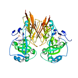
6N6D
 | |
6N6K
 | | Human REXO2 bound to pAG | | 分子名称: | MALONATE ION, RNA (5'-R(P*AP*G)-3'), RNA exonuclease 2 homolog,Small fragment nuclease, ... | | 著者 | Lormand, J.D, Sondermann, H. | | 登録日 | 2018-11-26 | | 公開日 | 2019-06-12 | | 最終更新日 | 2023-10-11 | | 実験手法 | X-RAY DIFFRACTION (1.418 Å) | | 主引用文献 | A dedicated diribonucleotidase resolves a key bottleneck for the terminal step of RNA degradation.
Elife, 8, 2019
|
|
3DYH
 | | T. Brucei Farnesyl Diphosphate Synthase Complexed with Bisphosphonate BPH-721 | | 分子名称: | 3-butoxy-1-(2,2-diphosphonoethyl)pyridinium, Farnesyl pyrophosphate synthase, MAGNESIUM ION | | 著者 | Cao, R, Gao, Y, Robinson, H, Goddard, A, Oldfield, E. | | 登録日 | 2008-07-27 | | 公開日 | 2009-05-05 | | 最終更新日 | 2024-02-21 | | 実験手法 | X-RAY DIFFRACTION (1.94 Å) | | 主引用文献 | Lipophilic bisphosphonates as dual farnesyl/geranylgeranyl diphosphate synthase inhibitors: an X-ray and NMR investigation.
J.Am.Chem.Soc., 131, 2009
|
|
3DYF
 | | T. Brucei Farnesyl Diphosphate Synthase Complexed with Bisphosphonate BPH-461 and Isopentyl Diphosphate | | 分子名称: | (4S)-2-METHYL-2,4-PENTANEDIOL, 3-FLUORO-1-(2-HYDROXY-2,2-DIPHOSPHONOETHYL)PYRIDINIUM, ACETATE ION, ... | | 著者 | Cao, R, Gao, Y, Robinson, H, Goddard, A, Oldfield, E. | | 登録日 | 2008-07-27 | | 公開日 | 2009-05-05 | | 最終更新日 | 2024-02-21 | | 実験手法 | X-RAY DIFFRACTION (2.65 Å) | | 主引用文献 | Lipophilic bisphosphonates as dual farnesyl/geranylgeranyl diphosphate synthase inhibitors: an X-ray and NMR investigation.
J.Am.Chem.Soc., 131, 2009
|
|
6N6F
 | |
6N6J
 | | Human REXO2 bound to pAA | | 分子名称: | 1,2-ETHANEDIOL, GLYCEROL, MALONATE ION, ... | | 著者 | Lormand, J.D, Sondermann, H. | | 登録日 | 2018-11-26 | | 公開日 | 2019-06-12 | | 最終更新日 | 2023-10-11 | | 実験手法 | X-RAY DIFFRACTION (1.317 Å) | | 主引用文献 | A dedicated diribonucleotidase resolves a key bottleneck for the terminal step of RNA degradation.
Elife, 8, 2019
|
|


