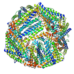
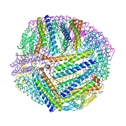
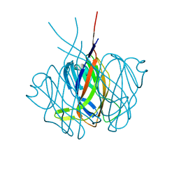
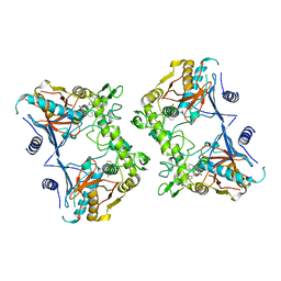
7EQX
 
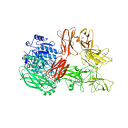
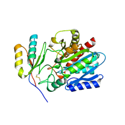 | | Crystal structure of an Aedes aegypti procarboxypeptidase B1 | | 分子名称: | Carboxypeptidase B, ZINC ION | | 著者 | Choong, Y.K, Gavor, E, Jobichen, C, Sivaraman, J. | | 登録日 | 2021-05-05 | | 公開日 | 2021-11-10 | | 最終更新日 | 2023-11-29 | | 実験手法 | X-RAY DIFFRACTION (2.08 Å) | | 主引用文献 | Structure of Aedes aegypti procarboxypeptidase B1 and its binding with Dengue virus for controlling infection.
Life Sci Alliance, 5, 2022
|
|
5H7R
 | |
5H7S
 | |
2LUS
 | |
6K3O
 | |
6K4M
 | |
6K43
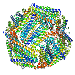 | |
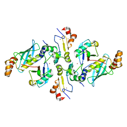
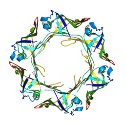
8H9D
 | | Crystal structure of Cas12a protein | | 分子名称: | Cas12A, MAGNESIUM ION, RNA (5'-R(P*AP*AP*UP*UP*UP*CP*UP*AP*CP*UP*AP*AP*GP*UP*GP*UP*AP*GP*AP*UP*C)-3'), ... | | 著者 | Jianwei, L, Jobichen, C, Sivaraman, J. | | 登録日 | 2022-10-25 | | 公開日 | 2023-02-08 | | 最終更新日 | 2023-11-29 | | 実験手法 | X-RAY DIFFRACTION (3.1 Å) | | 主引用文献 | Structures of apo Cas12a and its complex with crRNA and DNA reveal the dynamics of ternary complex formation and target DNA cleavage.
Plos Biol., 21, 2023
|
|
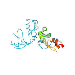
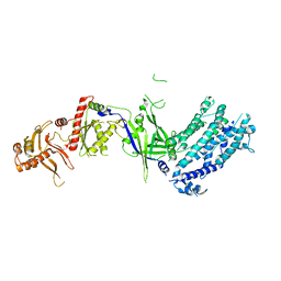
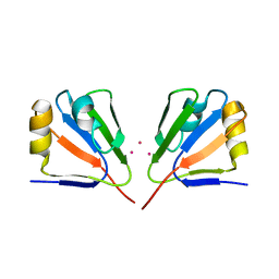
5XWR
 | | Crystal Structure of RBBP4-peptide complex | | 分子名称: | Histone-binding protein RBBP4, MET-SER-ARG-ARG-LYS-GLN-ALA-LYS-PRO-GLN-HIS-ILE | | 著者 | Jobichen, C, Lui, B.H, Daniel, G.T, Sivaraman, J. | | 登録日 | 2017-06-30 | | 公開日 | 2018-07-11 | | 最終更新日 | 2023-11-22 | | 実験手法 | X-RAY DIFFRACTION (2.69 Å) | | 主引用文献 | Targeting cancer addiction for SALL4 by shifting its transcriptome with a pharmacologic peptide.
Proc. Natl. Acad. Sci. U.S.A., 115, 2018
|
|
5XX9
 | |
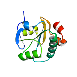
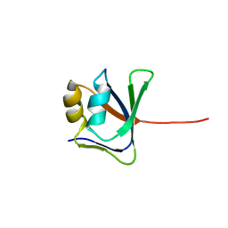
2PY2
 | | Structure of Herring Type II Antifreeze Protein | | 分子名称: | Antifreeze protein type II, CALCIUM ION | | 著者 | Liu, Y, Li, Z, Lin, Q, Seetharaman, J, Sivaraman, J, Hew, C.-L. | | 登録日 | 2007-05-15 | | 公開日 | 2007-06-26 | | 最終更新日 | 2011-07-13 | | 実験手法 | X-RAY DIFFRACTION (1.7 Å) | | 主引用文献 | Structure and Evolutionary Origin of Ca-Dependent Herring Type II Antifreeze Protein.
PLoS ONE, 2, 2007
|
|
2QCY
 | |
2MQ1
 | |
2EDM
 | |
6A2V
 | | Crystal structure of Hcp protein | | 分子名称: | Type VI secretion system tube protein Hcp | | 著者 | Jobichen, C, Sivaraman, J. | | 登録日 | 2018-06-13 | | 公開日 | 2018-09-19 | | 最終更新日 | 2023-11-22 | | 実験手法 | X-RAY DIFFRACTION (2.588 Å) | | 主引用文献 | Structural basis for the pathogenesis of Campylobacter jejuni Hcp1, a structural and effector protein of the Type VI Secretion System.
FEBS J., 285, 2018
|
|
2GJ2
 | | Crystal Structure of VP9 from White Spot Syndrome Virus | | 分子名称: | CADMIUM ION, wsv230 | | 著者 | Liu, Y, Wu, J.L, Song, J.X, Sivaraman, J, Hew, C.L. | | 登録日 | 2006-03-30 | | 公開日 | 2006-09-19 | | 最終更新日 | 2024-03-13 | | 実験手法 | X-RAY DIFFRACTION (2.35 Å) | | 主引用文献 | Identification of a Novel Nonstructural Protein, VP9, from White Spot Syndrome Virus: Its Structure Reveals a Ferredoxin Fold with Specific Metal Binding Sites
J.Virol., 80, 2006
|
|
2GJI
 | | NMR solution structure of VP9 from White Spot Syndrome Virus | | 分子名称: | wsv230 | | 著者 | Liu, Y, Wu, J.L, Song, J.X, Sivaraman, J, Hew, C.L. | | 登録日 | 2006-03-30 | | 公開日 | 2006-09-19 | | 最終更新日 | 2022-03-09 | | 実験手法 | SOLUTION NMR | | 主引用文献 | Identification of a Novel Nonstructural Protein, VP9, from White Spot Syndrome Virus: Its Structure Reveals a Ferredoxin Fold with Specific Metal Binding Sites
J.Virol., 80, 2006
|
|
1PVJ
 | | Crystal structure of the Streptococcal pyrogenic exotoxin B (SpeB)- inhibitor complex | | 分子名称: | (3R)-3-{[(BENZYLOXY)CARBONYL]AMINO}-2-OXO-4-PHENYLBUTANE-1-DIAZONIUM, pyrogenic exotoxin B | | 著者 | Ziomek, E, Sivaraman, J, Doran, J, Menard, R, Cygler, M. | | 登録日 | 2003-06-27 | | 公開日 | 2004-09-28 | | 最終更新日 | 2017-10-11 | | 実験手法 | X-RAY DIFFRACTION (3 Å) | | 主引用文献 | Inhibition of autoprocessing of the streptococcal pyrogenic exotoxin B (speB). Crystal structure of the proenzyme-inhibitor complex
To be published
|
|
3GGQ
 | | Dimerization of Hepatitis E Virus Capsid Protein E2s Domain is Essential for Virus-Host Interaction | | 分子名称: | BROMIDE ION, Capsid protein | | 著者 | Li, S.W, Tang, X.H, Seetharaman, J, Yang, C.Y, Gu, Y, Zhang, J, Du, H.L, Shih, J.W.K, Hew, C.L, Sivaraman, J, Xia, N.S. | | 登録日 | 2009-03-02 | | 公開日 | 2009-08-25 | | 最終更新日 | 2024-03-20 | | 実験手法 | X-RAY DIFFRACTION (2 Å) | | 主引用文献 | Dimerization of hepatitis E virus capsid protein E2s domain is essential for virus-host interaction
Plos Pathog., 5, 2009
|
|
2PJD
 | | Crystal structure of 16S rRNA methyltransferase RsmC | | 分子名称: | Ribosomal RNA small subunit methyltransferase C | | 著者 | Sunita, S, Purta, E, Durawa, M, Tkaczuk, K.L, Bujnicki, J.M, Sivaraman, J. | | 登録日 | 2007-04-16 | | 公開日 | 2007-07-03 | | 最終更新日 | 2011-07-13 | | 実験手法 | X-RAY DIFFRACTION (2.1 Å) | | 主引用文献 | Functional specialization of domains tandemly duplicated within 16S rRNA methyltransferase RsmC
Nucleic Acids Res., 35, 2007
|
|
1FC4
 | | 2-AMINO-3-KETOBUTYRATE COA LIGASE | | 分子名称: | 2-AMINO-3-KETOBUTYRATE CONENZYME A LIGASE, 2-AMINO-3-KETOBUTYRIC ACID, PYRIDOXAL-5'-PHOSPHATE | | 著者 | Schmidt, A, Matte, A, Li, Y, Sivaraman, J, Larocque, R, Schrag, J.D, Smith, C, Sauve, V, Cygler, M, Montreal-Kingston Bacterial Structural Genomics Initiative (BSGI) | | 登録日 | 2000-07-17 | | 公開日 | 2001-05-02 | | 最終更新日 | 2018-01-31 | | 実験手法 | X-RAY DIFFRACTION (2 Å) | | 主引用文献 | Three-dimensional structure of 2-amino-3-ketobutyrate CoA ligase from Escherichia coli complexed with a PLP-substrate intermediate: inferred reaction mechanism.
Biochemistry, 40, 2001
|
|
5XXZ
 | |
5XYA
 | | Crystal structure of a serine protease from Streptococcus species | | 分子名称: | 4-(2-AMINOETHYL)BENZENESULFONYL FLUORIDE, CALCIUM ION, Chemokine protease C, ... | | 著者 | Jobichen, C, Sivaraman, J. | | 登録日 | 2017-07-06 | | 公開日 | 2018-08-08 | | 最終更新日 | 2023-11-22 | | 実験手法 | X-RAY DIFFRACTION (3 Å) | | 主引用文献 | Structure of ScpC, a virulence protease fromStreptococcus pyogenes, reveals the functional domains and maturation mechanism.
Biochem. J., 475, 2018
|
|
5XYR
 | | Crystal structure of a serine protease from Streptococcus species | | 分子名称: | CALCIUM ION, CHLORIDE ION, Chemokine protease C, ... | | 著者 | Jobichen, C, Sivaraman, J. | | 登録日 | 2017-07-10 | | 公開日 | 2018-08-08 | | 最終更新日 | 2023-11-22 | | 実験手法 | X-RAY DIFFRACTION (2.8 Å) | | 主引用文献 | Structure of ScpC, a virulence protease fromStreptococcus pyogenes, reveals the functional domains and maturation mechanism.
Biochem. J., 475, 2018
|
|
3BUN
 | | Crystal structure of c-Cbl-TKB domain complexed with its binding motif in Sprouty4 | | 分子名称: | 13-meric peptide from Protein sprouty homolog 4, E3 ubiquitin-protein ligase CBL | | 著者 | Ng, C, Jackson, R.A, Buschdorf, J.P, Sun, Q, Guy, G.R, Sivaraman, J. | | 登録日 | 2008-01-03 | | 公開日 | 2008-02-26 | | 最終更新日 | 2023-11-15 | | 実験手法 | X-RAY DIFFRACTION (2 Å) | | 主引用文献 | Structural basis for a novel intrapeptidyl H-bond and reverse binding of c-Cbl-TKB domain substrates
Embo J., 27, 2008
|
|


