Loading
PDBj
MenuPDBj@FacebookPDBj@TwitterPDBj@YouTubewwPDB FoundationwwPDB
RCSB PDBPDBeBMRBAdv. SearchSearch help
PDB: 106 results

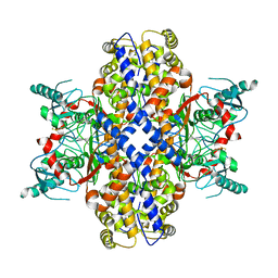
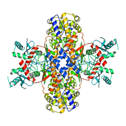
6VYF
DownloadVisualize
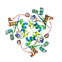
BU of 6vyf by Molmil
Cryo-EM structure of Plasmodium vivax hexokinase (Open state)
Descriptor: Phosphotransferase
Authors:Srivastava, S.S, Darling, J.E, Suryadi, J, Morris, J.C, Drew, M.E, Subramaniam, S.
Deposit date:2020-02-26
Release date:2020-05-06
Last modified:2024-03-06
Method:ELECTRON MICROSCOPY (3.3 Å)
Cite:Plasmodium vivax and human hexokinases share similar active sites but display distinct quaternary architectures
Iucrj, 7, 2020
6VYG
DownloadVisualize
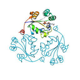
BU of 6vyg by Molmil
Cryo-EM structure of Plasmodium vivax hexokinase (Closed state)
Descriptor: Phosphotransferase
Authors:Srivastava, S.S, Darling, J.E, Suryadi, J, Morris, J.C, Drew, M.E, Subramaniam, S.
Deposit date:2020-02-26
Release date:2020-05-06
Method:ELECTRON MICROSCOPY (3.5 Å)
Cite:Plasmodium vivax and human hexokinases share similar active sites but display distinct quaternary architectures
Iucrj, 7, 2020
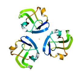
5HT7
DownloadVisualize
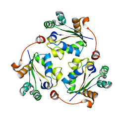
BU of 5ht7 by Molmil
Crystal structure of a transition-metal-ion-binding betagamma-crystallin from Methanosaeta thermophila
Descriptor: FE (II) ION, Uncharacterized protein
Authors:Srivastava, S.S, Sankaranarayanan, R.
Deposit date:2016-01-26
Release date:2017-02-01
Last modified:2024-03-20
Method:X-RAY DIFFRACTION (1.862 Å)
Cite:A Transition Metal-Binding, Trimeric beta gamma-Crystallin from Methane-Producing Thermophilic Archaea, Methanosaeta thermophila
Biochemistry, 56, 2017
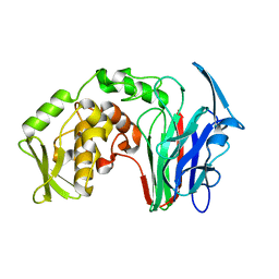
5H4E
DownloadVisualize
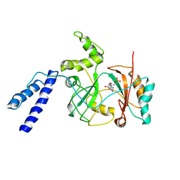
BU of 5h4e by Molmil
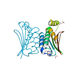
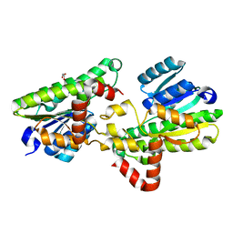
Crystal structure of a beta-1,3-glucanase domain (GH64) from Clostridium beijerinckii
Descriptor: beta 1-3 glucanase
Authors:Srivastava, S.S, Sankaranarayanan, R.
Deposit date:2016-10-31
Release date:2017-11-08
Last modified:2023-11-08
Method:X-RAY DIFFRACTION (1.863 Å)
Cite:Crystal structure of a beta-1,3-glucanase domain (GH64) from Clostridium beijerinckii
To Be Published
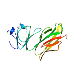
5Z6D
DownloadVisualize
BU of 5z6d by Molmil
Crystal structure of Abundant Perithecial Protein (APP) from Neurospora crassa
Descriptor: DUF1881 domain-containing protein
Authors:Srivastava, S.S, Sankaranarayanan, R.
Deposit date:2018-01-22
Release date:2019-01-23
Last modified:2024-03-27
Method:X-RAY DIFFRACTION (1.6 Å)
Cite:Interface interactions between beta gamma-crystallin domain and Ig-like domain render Ca2+-binding site inoperative in abundant perithecial protein of Neurospora crassa.
Mol.Microbiol., 110, 2018
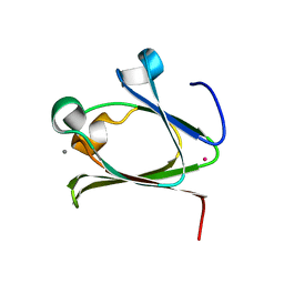
5Z6E
DownloadVisualize
BU of 5z6e by Molmil
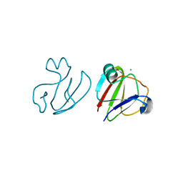
Crystal structure of a beta gamma-crystallin domain of Abundant Perithecial Protein (APP) from Neurospora crassa in the Ca2+-bound form
Descriptor: CALCIUM ION, DUF1881 domain-containing protein, POTASSIUM ION
Authors:Srivastava, S.S, Sankaranarayanan, R.
Deposit date:2018-01-22
Release date:2019-01-23
Last modified:2023-11-22
Method:X-RAY DIFFRACTION (1.864 Å)
Cite:Interface interactions between beta gamma-crystallin domain and Ig-like domain render Ca2+-binding site inoperative in abundant perithecial protein of Neurospora crassa.
Mol.Microbiol., 110, 2018
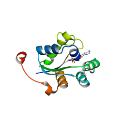
3Q8Y
DownloadVisualize
BU of 3q8y by Molmil
Crystal structure of Staphylococcus aureus nucleoside diphosphate kinase complexed with ADP and Vanadate
Descriptor: ADENOSINE-5'-DIPHOSPHATE, MAGNESIUM ION, Nucleoside diphosphate kinase, ...
Authors:Srivastava, S.K, Rajasree, K, Gopal, B.
Deposit date:2011-01-07
Release date:2011-07-27
Last modified:2023-11-01
Method:X-RAY DIFFRACTION (2.7 Å)
Cite:Conformational basis for substrate recognition and regulation of catalytic activity in Staphylococcus aureus nucleoside di-phosphate kinase.
Biochim.Biophys.Acta, 2011
3Q8V
DownloadVisualize
BU of 3q8v by Molmil
Crystal structure of Staphylococcus aureus nucleoside diphosphate kinase complexed with UDP
Descriptor: MAGNESIUM ION, Nucleoside diphosphate kinase, URIDINE-5'-DIPHOSPHATE
Authors:Srivastava, S.K, Rajasree, K, Gopal, B.
Deposit date:2011-01-07
Release date:2011-07-27
Last modified:2023-11-01
Method:X-RAY DIFFRACTION (2.5 Å)
Cite:Conformational basis for substrate recognition and regulation of catalytic activity in Staphylococcus aureus nucleoside di-phosphate kinase.
Biochim.Biophys.Acta, 2011
3Q89
DownloadVisualize
BU of 3q89 by Molmil
Crystal structure of Staphylococcus aureus nucleoside diphosphate kinase complexed with CDP
Descriptor: CYTIDINE-5'-DIPHOSPHATE, MAGNESIUM ION, Nucleoside diphosphate kinase
Authors:Srivastava, S.K, Rajasree, K, Gopal, B.
Deposit date:2011-01-06
Release date:2011-07-27
Last modified:2023-11-01
Method:X-RAY DIFFRACTION (2.9 Å)
Cite:Conformational basis for substrate recognition and regulation of catalytic activity in Staphylococcus aureus nucleoside di-phosphate kinase.
Biochim.Biophys.Acta, 2011
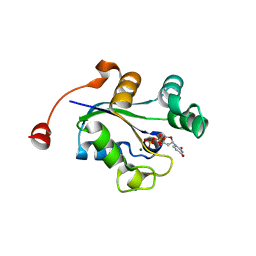
3Q8U
DownloadVisualize
BU of 3q8u by Molmil
Crystal structure of Staphylococcus aureus nucleoside diphosphate kinase complexed with ADP
Descriptor: ADENOSINE-5'-DIPHOSPHATE, MAGNESIUM ION, Nucleoside diphosphate kinase
Authors:Srivastava, S.K, Rajasree, K, Gopal, B.
Deposit date:2011-01-07
Release date:2011-07-27
Last modified:2023-11-01
Method:X-RAY DIFFRACTION (2.22 Å)
Cite:Conformational basis for substrate recognition and regulation of catalytic activity in Staphylococcus aureus nucleoside di-phosphate kinase.
Biochim.Biophys.Acta, 1814, 2011
3Q86
DownloadVisualize
BU of 3q86 by Molmil
Crystal structure of Staphylococcus aureus nucleoside diphosphate kinase complexed with GTP
Descriptor: GUANOSINE-5'-TRIPHOSPHATE, MAGNESIUM ION, Nucleoside diphosphate kinase
Authors:Srivastava, S.K, Rajasree, K, Gopal, B.
Deposit date:2011-01-06
Release date:2011-07-27
Last modified:2023-11-01
Method:X-RAY DIFFRACTION (2.38 Å)
Cite:Conformational basis for substrate recognition and regulation of catalytic activity in Staphylococcus aureus nucleoside di-phosphate kinase.
Biochim.Biophys.Acta, 1814, 2011
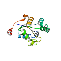
3Q83
DownloadVisualize
BU of 3q83 by Molmil
Crystal structure of Staphylococcus aureus nucleoside diphosphate kinase
Descriptor: Nucleoside diphosphate kinase
Authors:Srivastava, S.K, Rajasree, K, Gopal, B.
Deposit date:2011-01-06
Release date:2011-07-27
Last modified:2023-11-01
Method:X-RAY DIFFRACTION (2.5 Å)
Cite:Conformational basis for substrate recognition and regulation of catalytic activity in Staphylococcus aureus nucleoside di-phosphate kinase.
Biochim.Biophys.Acta, 1814, 2011
1ZAU
DownloadVisualize
BU of 1zau by Molmil
Adenylation domain of NAD+ dependent DNA ligase from M.tuberculosis
Descriptor: ADENOSINE MONOPHOSPHATE, DNA ligase
Authors:Srivastava, S.K, Ramachandran, R.
Deposit date:2005-04-07
Release date:2005-07-05
Last modified:2024-02-14
Method:X-RAY DIFFRACTION (3.15 Å)
Cite:NAD+-dependent DNA Ligase (Rv3014c) from Mycobacterium tuberculosis: CRYSTAL STRUCTURE OF THE ADENYLATION DOMAIN AND IDENTIFICATION OF NOVEL INHIBITORS
J.Biol.Chem., 280, 2005
4BXI
DownloadVisualize
BU of 4bxi by Molmil
Crystal structure of ATP binding domain of AgrC from Staphylococcus aureus
Descriptor: (4S)-2-METHYL-2,4-PENTANEDIOL, ACCESSORY GENE REGULATOR PROTEIN C, ACETATE ION, ...
Authors:Srivastava, S.K, Rajasree, K, Gopal, B.
Deposit date:2013-07-12
Release date:2014-06-04
Last modified:2023-12-20
Method:X-RAY DIFFRACTION (2.2 Å)
Cite:Influence of the Agrc-Agra Complex in the Response Time of Staphylococcus Aureus Quorum Sensing
J.Bacteriol., 196, 2014
3SNY
DownloadVisualize
BU of 3sny by Molmil
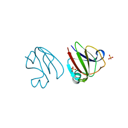
Crystal structure of a mutant T82R of a betagamma-crystallin domain from Clostridium beijerinckii
Descriptor: CALCIUM ION, Clostrillin, SULFATE ION
Authors:Srivastava, S.S, Sankaranarayanan, R.
Deposit date:2011-06-29
Release date:2011-11-16
Last modified:2023-11-01
Method:X-RAY DIFFRACTION (1.85 Å)
Cite:Decoding the molecular design principles underlying Ca(2+) binding to beta gamma-crystallin motifs
J.Mol.Biol., 415, 2012
3SO1
DownloadVisualize
BU of 3so1 by Molmil
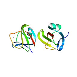
Crystal structure of a double mutant T41S T82S of a betagamma-crystallin domain from Clostridium beijerinckii
Descriptor: CALCIUM ION, Clostrillin, SULFATE ION
Authors:Srivastava, S.S, Sankaranarayanan, R.
Deposit date:2011-06-29
Release date:2011-11-16
Last modified:2023-11-01
Method:X-RAY DIFFRACTION (1.85 Å)
Cite:Decoding the molecular design principles underlying Ca(2+) binding to beta gamma-crystallin motifs
J.Mol.Biol., 415, 2012
3SO0
DownloadVisualize
BU of 3so0 by Molmil
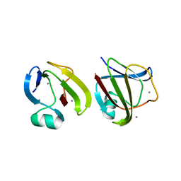
Crystal structure of a mutant T41S of a betagamma-crystallin domain from Clostridium beijerinckii
Descriptor: CALCIUM ION, Clostrillin
Authors:Srivastava, S.S, Sankaranarayanan, R.
Deposit date:2011-06-29
Release date:2011-11-16
Last modified:2023-11-01
Method:X-RAY DIFFRACTION (1.93 Å)
Cite:Decoding the molecular design principles underlying Ca(2+) binding to beta gamma-crystallin motifs
J.Mol.Biol., 415, 2012
3SNZ
DownloadVisualize
BU of 3snz by Molmil
Crystal structure of a mutant W39D of a betagamma-crystallin domain from Clostridium beijerinckii
Descriptor: CALCIUM ION, Clostrillin
Authors:Srivastava, S.S, Sankaranarayanan, R.
Deposit date:2011-06-29
Release date:2011-11-16
Last modified:2023-11-01
Method:X-RAY DIFFRACTION (2 Å)
Cite:Decoding the molecular design principles underlying Ca(2+) binding to beta gamma-crystallin motifs
J.Mol.Biol., 415, 2012
5FYD
DownloadVisualize
BU of 5fyd by Molmil
Structural and biochemical insights into 7beta-hydroxysteroid dehydrogenase stereoselectivity
Descriptor: GLYCEROL, OXIDOREDUCTASE, SHORT CHAIN DEHYDROGENASE/REDUCTASE FAMILY PROTEIN
Authors:Savino, S, Ferrandi, E, Forneris, F, Rovida, S, Riva, S, Monti, D, Mattevi, A.
Deposit date:2016-03-07
Release date:2016-04-06
Last modified:2024-01-10
Method:X-RAY DIFFRACTION (1.6 Å)
Cite:Structural and Biochemical Insights Into 7Beta-Hydroxysteroid Dehydrogenase Stereoselectivity.
Proteins, 84, 2016
7SY4
DownloadVisualize
BU of 7sy4 by Molmil
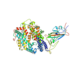
Cryo-EM structure of the SARS-CoV-2 D614G,N501Y,E484K mutant spike protein ectodomain bound to human ACE2 ectodomain (focused refinement of RBD and ACE2)
Descriptor: 2-acetamido-2-deoxy-beta-D-glucopyranose, Processed angiotensin-converting enzyme 2, Spike glycoprotein
Authors:Zhu, X, Mannar, D, Saville, J.W, Srivastava, S.S, Berezuk, A.M, Zhou, S, Tuttle, K.S, Kim, A, Li, W, Dimitrov, D.S, Subramaniam, S.
Deposit date:2021-11-24
Release date:2021-12-29
Last modified:2022-01-05
Method:ELECTRON MICROSCOPY (3.35 Å)
Cite:Structural analysis of receptor binding domain mutations in SARS-CoV-2 variants of concern that modulate ACE2 and antibody binding.
Cell Rep, 37, 2021
7SY6
DownloadVisualize
BU of 7sy6 by Molmil
Cryo-EM structure of the SARS-CoV-2 D614G,N501Y,E484K,K417N mutant spike protein ectodomain bound to human ACE2 ectodomain (focused refinement of RBD and ACE2)
Descriptor: 2-acetamido-2-deoxy-beta-D-glucopyranose, Processed angiotensin-converting enzyme 2, Spike glycoprotein
Authors:Zhu, X, Mannar, D, Saville, J.W, Srivastava, S.S, Berezuk, A.M, Zhou, S, Tuttle, K.S, Kim, A, Li, W, Dimitrov, D.S, Subramaniam, S.
Deposit date:2021-11-24
Release date:2021-12-29
Method:ELECTRON MICROSCOPY (2.81 Å)
Cite:Structural analysis of receptor binding domain mutations in SARS-CoV-2 variants of concern that modulate ACE2 and antibody binding.
Cell Rep, 37, 2021
7SXT
DownloadVisualize
BU of 7sxt by Molmil
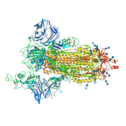
Cryo-EM structure of the SARS-CoV-2 D614G,N501Y mutant spike protein ectodomain
Descriptor: 2-acetamido-2-deoxy-beta-D-glucopyranose, 2-acetamido-2-deoxy-beta-D-glucopyranose-(1-4)-2-acetamido-2-deoxy-beta-D-glucopyranose, Spike glycoprotein
Authors:Zhu, X, Mannar, D, Saville, J.W, Srivastava, S.S, Berezuk, A.M, Zhou, S, Tuttle, K.S, Kim, A, Li, W, Dimitrov, D.S, Subramaniam, S.
Deposit date:2021-11-24
Release date:2021-12-29
Last modified:2022-01-05
Method:ELECTRON MICROSCOPY (2.31 Å)
Cite:Structural analysis of receptor binding domain mutations in SARS-CoV-2 variants of concern that modulate ACE2 and antibody binding.
Cell Rep, 37, 2021
7SY7
DownloadVisualize
BU of 7sy7 by Molmil
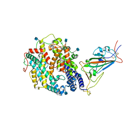
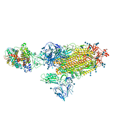
Cryo-EM structure of the SARS-CoV-2 D614G,N501Y,E484K,K417T mutant spike protein ectodomain bound to human ACE2 ectodomain (global refinement)
Descriptor: 2-acetamido-2-deoxy-beta-D-glucopyranose, 2-acetamido-2-deoxy-beta-D-glucopyranose-(1-4)-2-acetamido-2-deoxy-beta-D-glucopyranose, Processed angiotensin-converting enzyme 2, ...
Authors:Zhu, X, Mannar, D, Saville, J.W, Srivastava, S.S, Berezuk, A.M, Zhou, S, Tuttle, K.S, Kim, A, Li, W, Dimitrov, D.S, Subramaniam, S.
Deposit date:2021-11-24
Release date:2021-12-29
Method:ELECTRON MICROSCOPY (2.81 Å)
Cite:Structural analysis of receptor binding domain mutations in SARS-CoV-2 variants of concern that modulate ACE2 and antibody binding.
Cell Rep, 37, 2021
7SXS
DownloadVisualize
BU of 7sxs by Molmil
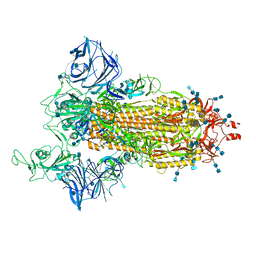
Cryo-EM structure of the SARS-CoV-2 D614G,L452R mutant spike protein ectodomain
Descriptor: 2-acetamido-2-deoxy-beta-D-glucopyranose, 2-acetamido-2-deoxy-beta-D-glucopyranose-(1-4)-2-acetamido-2-deoxy-beta-D-glucopyranose, Spike glycoprotein
Authors:Zhu, X, Mannar, D, Saville, J.W, Srivastava, S.S, Berezuk, A.M, Zhou, S, Tuttle, K.S, Kim, A, Li, W, Dimitrov, D.S, Subramaniam, S.
Deposit date:2021-11-24
Release date:2021-12-29
Last modified:2022-01-05
Method:ELECTRON MICROSCOPY (2.64 Å)
Cite:Structural analysis of receptor binding domain mutations in SARS-CoV-2 variants of concern that modulate ACE2 and antibody binding.
Cell Rep, 37, 2021
7SY5
DownloadVisualize
BU of 7sy5 by Molmil
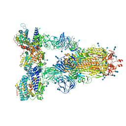
Cryo-EM structure of the SARS-CoV-2 D614G,N501Y,E484K,K417N mutant spike protein ectodomain bound to human ACE2 ectodomain (global refinement)
Descriptor: 2-acetamido-2-deoxy-beta-D-glucopyranose, 2-acetamido-2-deoxy-beta-D-glucopyranose-(1-4)-2-acetamido-2-deoxy-beta-D-glucopyranose, Processed angiotensin-converting enzyme 2, ...
Authors:Zhu, X, Mannar, D, Saville, J.W, Srivastava, S.S, Berezuk, A.M, Zhou, S, Tuttle, K.S, Kim, A, Li, W, Dimitrov, D.S, Subramaniam, S.
Deposit date:2021-11-24
Release date:2021-12-29
Last modified:2022-01-05
Method:ELECTRON MICROSCOPY (2.59 Å)
Cite:Structural analysis of receptor binding domain mutations in SARS-CoV-2 variants of concern that modulate ACE2 and antibody binding.
Cell Rep, 37, 2021

222926

數據於2024-07-24公開中

PDB statisticsPDBj update infoContact PDBjnumon