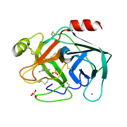
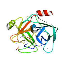
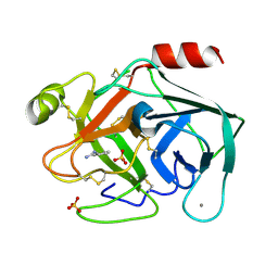
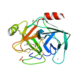
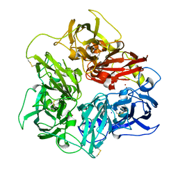
6SV6
 
 | | Non-terahertz irradiated structure of bovine trypsin (even frames of crystal x38) | | 分子名称: | BENZAMIDINE, CALCIUM ION, Cationic trypsin, ... | | 著者 | Ahlberg Gagner, V, Lundholm, I, Garcia-Bonete, M.J, Rodilla, H, Friedman, R, Zhaunerchyk, V, Bourenkov, G, Schneider, T, Stake, J, Katona, G. | | 登録日 | 2019-09-17 | | 公開日 | 2020-01-22 | | 最終更新日 | 2020-01-29 | | 実験手法 | X-RAY DIFFRACTION (1.15 Å) | | 主引用文献 | Clustering of atomic displacement parameters in bovine trypsin reveals a distributed lattice of atoms with shared chemical properties.
Sci Rep, 9, 2019
|
|
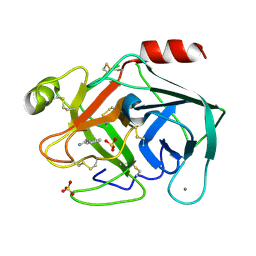
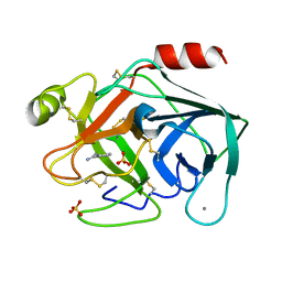
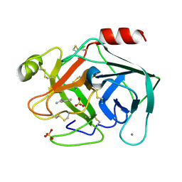
6SVN
 | | Reference structure of bovine trypsin (even frames of crystal x28) | | 分子名称: | BENZAMIDINE, CALCIUM ION, Cationic trypsin, ... | | 著者 | Ahlberg Gagner, V, Lundholm, I, Garcia-Bonete, M.J, Rodilla, H, Friedman, R, Zhaunerchyk, V, Bourenkov, G, Schneider, T, Stake, J, Katona, G. | | 登録日 | 2019-09-18 | | 公開日 | 2020-01-22 | | 最終更新日 | 2020-01-29 | | 実験手法 | X-RAY DIFFRACTION (1.16 Å) | | 主引用文献 | Clustering of atomic displacement parameters in bovine trypsin reveals a distributed lattice of atoms with shared chemical properties.
Sci Rep, 9, 2019
|
|
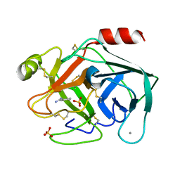
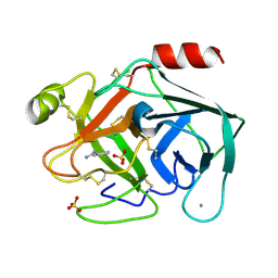
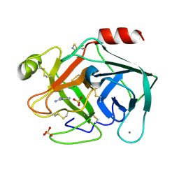
6SUX
 | | Terahertz irradiated structure of bovine trypsin (even frames of crystal x37) | | 分子名称: | BENZAMIDINE, CALCIUM ION, Cationic trypsin, ... | | 著者 | Ahlberg Gagner, V, Lundholm, I, Jose-Garcia, M.J, Rodilla, H, Friedman, R, Zhaunerchyk, V, Bourenkov, G, Schneider, T, Stake, J, Katona, G. | | 登録日 | 2019-09-17 | | 公開日 | 2020-01-22 | | 最終更新日 | 2020-01-29 | | 実験手法 | X-RAY DIFFRACTION (1.16 Å) | | 主引用文献 | Clustering of atomic displacement parameters in bovine trypsin reveals a distributed lattice of atoms with shared chemical properties.
Sci Rep, 9, 2019
|
|
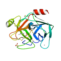
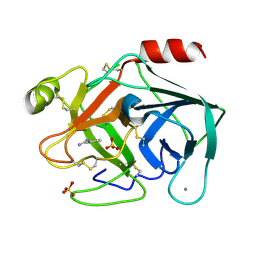
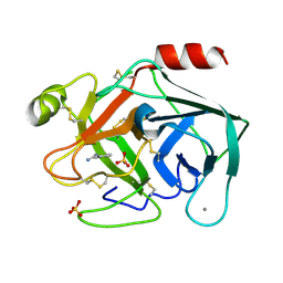
6SV0
 | | Non-terahertz irradiated structure of bovine trypsin (odd frames of crystal x37) | | 分子名称: | BENZAMIDINE, CALCIUM ION, Cationic trypsin, ... | | 著者 | Ahlberg Gagner, V, Lundholm, I, Garcia-Bonete, M.J, Rodilla, H, Friedman, R, Zhaunerchyk, V, Bourenkov, G, Schneider, T, Stake, J, Katona, G. | | 登録日 | 2019-09-17 | | 公開日 | 2020-01-22 | | 最終更新日 | 2020-01-29 | | 実験手法 | X-RAY DIFFRACTION (1.16 Å) | | 主引用文献 | Clustering of atomic displacement parameters in bovine trypsin reveals a distributed lattice of atoms with shared chemical properties.
Sci Rep, 9, 2019
|
|
6SV8
 | | Terahertz irradiated structure of bovine trypsin (odd frames of crystal x38) | | 分子名称: | BENZAMIDINE, CALCIUM ION, Cationic trypsin, ... | | 著者 | Ahlberg Gagner, V, Lundholm, I, Garcia-Bonete, M.J, Rodilla, H, Friedman, R, Zhaunerchyk, V, Bourenkov, G, Schneider, T, Stake, J, Katona, G. | | 登録日 | 2019-09-18 | | 公開日 | 2020-01-22 | | 最終更新日 | 2020-01-29 | | 実験手法 | X-RAY DIFFRACTION (1.15 Å) | | 主引用文献 | Clustering of atomic displacement parameters in bovine trypsin reveals a distributed lattice of atoms with shared chemical properties.
Sci Rep, 9, 2019
|
|
6SVD
 | | Non-terahertz irradiated structure of bovine trypsin (even frames of crystal x41) | | 分子名称: | BENZAMIDINE, CALCIUM ION, Cationic trypsin, ... | | 著者 | Ahlberg Gagner, V, Lundholm, I, Garcia-Bonete, M.J, Rodilla, H, Friedman, R, Zhaunerchyk, V, Bourenkov, G, Schneider, T, Stake, J, Katona, G. | | 登録日 | 2019-09-18 | | 公開日 | 2020-01-22 | | 最終更新日 | 2020-01-29 | | 実験手法 | X-RAY DIFFRACTION (1.15 Å) | | 主引用文献 | Clustering of atomic displacement parameters in bovine trypsin reveals a distributed lattice of atoms with shared chemical properties.
Sci Rep, 9, 2019
|
|
6SVW
 | | Reference structure of bovine trypsin (even frames of crystal x33) | | 分子名称: | BENZAMIDINE, CALCIUM ION, Cationic trypsin, ... | | 著者 | Ahlberg Gagner, V, Lundholm, I, Garcia-Bonete, M.J, Rodilla, H, Friedman, R, Zhaunerchyk, V, Bourenkov, G, Schneider, T, Stake, J, Katona, G. | | 登録日 | 2019-09-19 | | 公開日 | 2020-01-22 | | 最終更新日 | 2020-01-29 | | 実験手法 | X-RAY DIFFRACTION (1.16 Å) | | 主引用文献 | Clustering of atomic displacement parameters in bovine trypsin reveals a distributed lattice of atoms with shared chemical properties.
Sci Rep, 9, 2019
|
|
6SW0
 | | Reference structure of bovine trypsin (odd frames of crystal x34) | | 分子名称: | BENZAMIDINE, CALCIUM ION, Cationic trypsin, ... | | 著者 | Ahlberg Gagner, V, Lundholm, I, Garcia-Bonete, M.J, Rodilla, H, Friedman, R, Zhaunerchyk, V, Bourenkov, G, Schneider, T, Stake, J, Katona, G. | | 登録日 | 2019-09-19 | | 公開日 | 2020-01-22 | | 最終更新日 | 2020-01-29 | | 実験手法 | X-RAY DIFFRACTION (1.15 Å) | | 主引用文献 | Clustering of atomic displacement parameters in bovine trypsin reveals a distributed lattice of atoms with shared chemical properties.
Sci Rep, 9, 2019
|
|
6SVV
 | | Reference structure of bovine trypsin (odd frames of crystal x30) | | 分子名称: | BENZAMIDINE, CALCIUM ION, Cationic trypsin, ... | | 著者 | Ahlberg Gagner, V, Lundholm, I, Garcia-Bonete, M.J, Rodilla, H, Friedman, R, Zhaunerchyk, V, Bourenkov, G, Schneider, T, Stake, J, Katona, G. | | 登録日 | 2019-09-19 | | 公開日 | 2020-01-22 | | 最終更新日 | 2020-01-29 | | 実験手法 | X-RAY DIFFRACTION (1.15 Å) | | 主引用文献 | Clustering of atomic displacement parameters in bovine trypsin reveals a distributed lattice of atoms with shared chemical properties.
Sci Rep, 9, 2019
|
|
6SVG
 | | Terahertz irradiated structure of bovine trypsin (odd frames of crystal x41) | | 分子名称: | BENZAMIDINE, CALCIUM ION, Cationic trypsin, ... | | 著者 | Ahlberg Gagner, V, Lundholm, I, Garcia-Bonete, M.J, Rodilla, H, Friedman, R, Zhaunerchyk, V, Bourenkov, G, Schneider, T, Stake, J, Katona, G. | | 登録日 | 2019-09-18 | | 公開日 | 2020-01-22 | | 最終更新日 | 2020-01-29 | | 実験手法 | X-RAY DIFFRACTION (1.16 Å) | | 主引用文献 | Clustering of atomic displacement parameters in bovine trypsin reveals a distributed lattice of atoms with shared chemical properties.
Sci Rep, 9, 2019
|
|
6SVU
 | | Reference structure of bovine trypsin (even frames of crystal x30) | | 分子名称: | BENZAMIDINE, CALCIUM ION, Cationic trypsin, ... | | 著者 | Ahlberg Gagner, V, Lundholm, I, Garcia-Bonete, M.J, Rodilla, H, Friedman, R, Zhaunerchyk, V, Bourenkov, G, Schneider, T, Stake, J, Katona, G. | | 登録日 | 2019-09-19 | | 公開日 | 2020-01-22 | | 最終更新日 | 2020-01-29 | | 実験手法 | X-RAY DIFFRACTION (1.15 Å) | | 主引用文献 | Clustering of atomic displacement parameters in bovine trypsin reveals a distributed lattice of atoms with shared chemical properties.
Sci Rep, 9, 2019
|
|
6SVZ
 | | Reference structure of bovine trypsin (even frames of crystal x34) | | 分子名称: | BENZAMIDINE, CALCIUM ION, Cationic trypsin, ... | | 著者 | Ahlberg Gagner, V, Lundholm, I, Garcia-Bonete, M.J, Rodilla, H, Friedman, R, Zhaunerchyk, V, Bourenkov, G, Schneider, T, Stake, J, Katona, G. | | 登録日 | 2019-09-19 | | 公開日 | 2020-01-22 | | 最終更新日 | 2020-01-29 | | 実験手法 | X-RAY DIFFRACTION (1.15 Å) | | 主引用文献 | Clustering of atomic displacement parameters in bovine trypsin reveals a distributed lattice of atoms with shared chemical properties.
Sci Rep, 9, 2019
|
|
6SVB
 | | Terahertz irradiated structure of bovine trypsin (odd frames of crystal x40) | | 分子名称: | BENZAMIDINE, CALCIUM ION, Cationic trypsin, ... | | 著者 | Ahlberg Gagner, V, Lundholm, I, Garcia-Bonete, M.J, Rodilla, H, Friedman, R, Zhaunerchyk, V, Bourenkov, G, Schneider, T, Stake, J, Katona, G. | | 登録日 | 2019-09-18 | | 公開日 | 2020-01-22 | | 最終更新日 | 2020-01-29 | | 実験手法 | X-RAY DIFFRACTION (1.15 Å) | | 主引用文献 | Clustering of atomic displacement parameters in bovine trypsin reveals a distributed lattice of atoms with shared chemical properties.
Sci Rep, 9, 2019
|
|
6SVJ
 | | Terahertz irradiated structure of bovine trypsin (odd frames of crystal x42) | | 分子名称: | BENZAMIDINE, CALCIUM ION, Cationic trypsin, ... | | 著者 | Ahlberg Gagner, V, Lundholm, I, Garcia-Bonete, M.J, Rodilla, H, Friedman, R, Zhaunerchyk, V, Bourenkov, G, Schneider, T, Stake, J, Katona, G. | | 登録日 | 2019-09-18 | | 公開日 | 2020-01-22 | | 最終更新日 | 2020-01-29 | | 実験手法 | X-RAY DIFFRACTION (1.16 Å) | | 主引用文献 | Clustering of atomic displacement parameters in bovine trypsin reveals a distributed lattice of atoms with shared chemical properties.
Sci Rep, 9, 2019
|
|
6SVR
 | | Reference structure of bovine trypsin (odd frames of crystal x28) | | 分子名称: | BENZAMIDINE, CALCIUM ION, Cationic trypsin, ... | | 著者 | Ahlberg Gagner, V, Lundholm, I, Garcia-Bonete, M.J, Rodilla, H, Friedman, R, Zhaunerchyk, V, Bourenkov, G, Schneider, T, Stake, J, Katona, G. | | 登録日 | 2019-09-18 | | 公開日 | 2020-01-22 | | 最終更新日 | 2020-01-29 | | 実験手法 | X-RAY DIFFRACTION (1.16 Å) | | 主引用文献 | Clustering of atomic displacement parameters in bovine trypsin reveals a distributed lattice of atoms with shared chemical properties.
Sci Rep, 9, 2019
|
|
6SVI
 | | Non-terahertz irradiated structure of bovine trypsin (even frames of crystal x42) | | 分子名称: | BENZAMIDINE, CALCIUM ION, Cationic trypsin, ... | | 著者 | Ahlberg Gagner, V, Lundholm, I, Garcia-Bonete, M.J, Rodilla, H, Friedman, R, Zhaunerchyk, V, Bourenkov, G, Schneider, T, Stake, J, Katona, G. | | 登録日 | 2019-09-18 | | 公開日 | 2020-01-22 | | 最終更新日 | 2020-01-29 | | 実験手法 | X-RAY DIFFRACTION (1.16 Å) | | 主引用文献 | Clustering of atomic displacement parameters in bovine trypsin reveals a distributed lattice of atoms with shared chemical properties.
Sci Rep, 9, 2019
|
|
6SVX
 | | Reference structure of bovine trypsin (odd frames of crystal x33) | | 分子名称: | BENZAMIDINE, CALCIUM ION, Cationic trypsin, ... | | 著者 | Ahlberg Gagner, V, Lundholm, I, Garcia-Bonete, M.J, Rodilla, H, Friedman, R, Zhaunerchyk, V, Bourenkov, G, Schneider, T, Stake, J, Katona, G. | | 登録日 | 2019-09-19 | | 公開日 | 2020-01-22 | | 最終更新日 | 2020-01-29 | | 実験手法 | X-RAY DIFFRACTION (1.16 Å) | | 主引用文献 | Clustering of atomic displacement parameters in bovine trypsin reveals a distributed lattice of atoms with shared chemical properties.
Sci Rep, 9, 2019
|
|
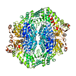
8CGY
 | | Trypanosoma brucei IMP dehydrogenase (ori) crystallized in High Five cells reveals native ligands ATP, GDP and phosphate. Diffraction data collection at 100 K in cellulo; XDS processing | | 分子名称: | ADENOSINE-5'-TRIPHOSPHATE, GUANOSINE-5'-DIPHOSPHATE, Inosine-5'-monophosphate dehydrogenase, ... | | 著者 | Boger, J, Schoenherr, R, Lahey-Rudolph, J.M, Harms, M, Kaiser, J, Nachtschatt, S, Wobbe, M, Duden, R, Bourenkov, G, Schneider, T, Redecke, L. | | 登録日 | 2023-02-06 | | 公開日 | 2024-02-21 | | 最終更新日 | 2024-03-06 | | 実験手法 | X-RAY DIFFRACTION (3 Å) | | 主引用文献 | A streamlined approach to structure elucidation using in cellulo crystallized recombinant proteins, InCellCryst.
Nat Commun, 15, 2024
|
|
7YXA
 | | XFEL crystal structure of the human sphingosine 1 phosphate receptor 5 in complex with ONO-5430608 | | 分子名称: | (2R)-2,3-dihydroxypropyl (9Z)-octadec-9-enoate, 2-acetamido-2-deoxy-beta-D-glucopyranose, 4-[6-(2-naphthalen-1-ylethoxy)-2,3,4,5-tetrahydro-1H-3-benzazepin-3-ium-3-yl]butanoic acid, ... | | 著者 | Lyapina, E, Marin, E, Gusach, A, Orekhov, P, Gerasimov, A, Luginina, A, Vakhrameev, D, Ergasheva, M, Kovaleva, M, Khusainov, G, Khorn, P, Shevtsov, M, Kovalev, K, Okhrimenko, I, Bukhdruker, S, Popov, P, Hu, H, Weierstall, U, Liu, W, Cho, Y, Gushchin, I, Rogachev, A, Bourenkov, G, Park, S, Park, G, Huyn, H.J, Park, J, Gordeliy, V, Borshchevskiy, V, Mishin, A, Cherezov, V. | | 登録日 | 2022-02-15 | | 公開日 | 2022-08-10 | | 最終更新日 | 2024-02-07 | | 実験手法 | X-RAY DIFFRACTION (2.2 Å) | | 主引用文献 | Structural basis for receptor selectivity and inverse agonism in S1P 5 receptors.
Nat Commun, 13, 2022
|
|
5N4L
 | | Rat ceruloplasmin trigonal form | | 分子名称: | CALCIUM ION, COPPER (II) ION, Ceruloplasmin, ... | | 著者 | Samygina, V.R, Sokolov, A.V, Bourenkov, G, Vasilyev, V.B. | | 登録日 | 2017-02-11 | | 公開日 | 2017-12-13 | | 最終更新日 | 2024-01-17 | | 実験手法 | X-RAY DIFFRACTION (3.2 Å) | | 主引用文献 | Rat ceruloplasmin: a new labile copper binding site and zinc/copper mosaic.
Metallomics, 9, 2017
|
|
5N0K
 | | Rat ceruloplasmin orthorhombic form | | 分子名称: | 2-acetamido-2-deoxy-beta-D-glucopyranose, CALCIUM ION, COPPER (II) ION, ... | | 著者 | Samygina, V.R, Sokolov, A.V, Bourenkov, G, Vasilyev, V.B. | | 登録日 | 2017-02-03 | | 公開日 | 2017-12-13 | | 最終更新日 | 2024-01-17 | | 実験手法 | X-RAY DIFFRACTION (2.3 Å) | | 主引用文献 | Rat ceruloplasmin: a new labile copper binding site and zinc/copper mosaic.
Metallomics, 9, 2017
|
|
4RJI
 | | Acetolactate synthase from Bacillus subtilis bound to ThDP - crystal form I | | 分子名称: | Acetolactate synthase, MAGNESIUM ION, TETRAETHYLENE GLYCOL, ... | | 著者 | Sommer, B, von Moeller, H, Haack, M, Qoura, F, Langner, C, Bourenkov, G, Garbe, D, Brueck, T, Loll, B. | | 登録日 | 2014-10-09 | | 公開日 | 2014-10-22 | | 最終更新日 | 2023-09-20 | | 実験手法 | X-RAY DIFFRACTION (3.2 Å) | | 主引用文献 | Detailed Structure-Function Correlations of Bacillus subtilis Acetolactate Synthase.
Chembiochem, 16, 2015
|
|
7UI5
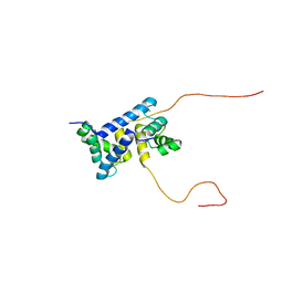 | | Evolution avoids a pathological stabilizing interaction in the immune protein S100A9 | | 分子名称: | CALCIUM ION, Protein S100-A9 | | 著者 | Reardon, P.N, Harman, J.L, Costello, S.M, Warren, G.D, Phillips, S.R, Connor, P.J, Marqusee, S, Harms, M.J. | | 登録日 | 2022-03-28 | | 公開日 | 2022-10-26 | | 最終更新日 | 2024-05-15 | | 実験手法 | SOLUTION NMR | | 主引用文献 | Evolution avoids a pathological stabilizing interaction in the immune protein S100A9.
Proc.Natl.Acad.Sci.USA, 119, 2022
|
|
8C5K
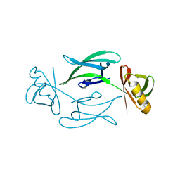 | | HEX-1 (in cellulo, in situ) crystallized and diffracted in High Five cells. Growth and SX data collection at 296 K on CrystalDirect plates | | 分子名称: | Woronin body major protein | | 著者 | Lahey-Rudolph, J.M, Schoenherr, R, Boger, J, Harms, M, Kaiser, J, Nachtschatt, S, Wobbe, M, Duden, R, Koenig, P, Bourenkov, G, Schneider, T, Redecke, L. | | 登録日 | 2023-01-09 | | 公開日 | 2024-01-17 | | 最終更新日 | 2024-03-06 | | 実験手法 | X-RAY DIFFRACTION (2.16 Å) | | 主引用文献 | A streamlined approach to structure elucidation using in cellulo crystallized recombinant proteins, InCellCryst.
Nat Commun, 15, 2024
|
|
8CD4
 | | structure of HEX-1 from N. crassa crystallized in cellulo (cytosol), diffracted at 100K and resolved using CrystFEL | | 分子名称: | Woronin body major protein | | 著者 | Boger, J, Schoenherr, R, Lahey-Rudolph, J.M, Harms, M, Kaiser, J, Nachtschatt, S, Wobbe, M, Koenig, P, Bourenkov, G, Schneider, T, Redecke, L. | | 登録日 | 2023-01-30 | | 公開日 | 2024-02-21 | | 最終更新日 | 2024-03-06 | | 実験手法 | X-RAY DIFFRACTION (1.83 Å) | | 主引用文献 | A streamlined approach to structure elucidation using in cellulo crystallized recombinant proteins, InCellCryst.
Nat Commun, 15, 2024
|
|


