8ATD
 
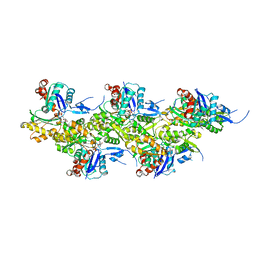
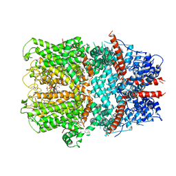
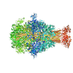
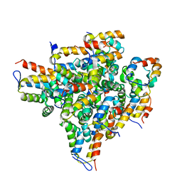
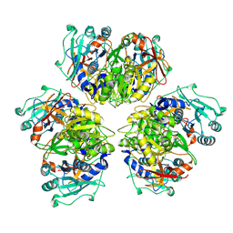 | | Wild type hexamer oxalyl-CoA synthetase (OCS) | | 分子名称: | Oxalate--CoA ligase | | 著者 | Lill, P, Burgi, J, Raunser, S, Wilmanns, M, Gatsogiannis, C. | | 登録日 | 2022-08-23 | | 公開日 | 2023-02-08 | | 最終更新日 | 2023-02-22 | | 実験手法 | ELECTRON MICROSCOPY (3.1 Å) | | 主引用文献 | Asymmetric horseshoe-like assembly of peroxisomal yeast oxalyl-CoA synthetase.
Biol.Chem., 404, 2023
|
|
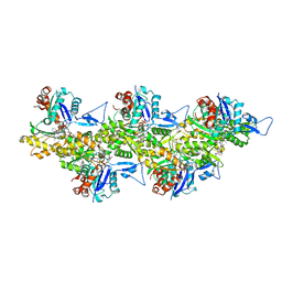
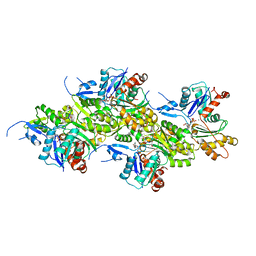
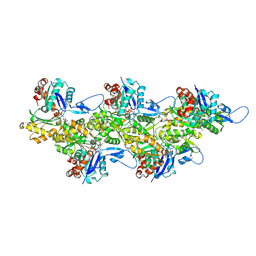
4UWE
 | |
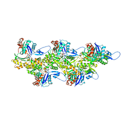
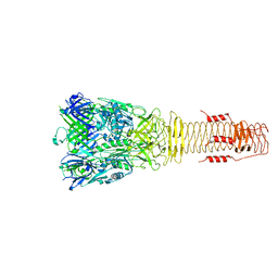
6T23
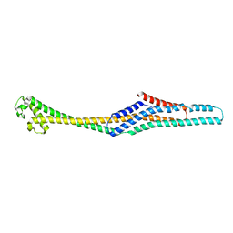
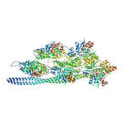
 | | Cryo-EM structure of jasplakinolide-stabilized F-actin (aged) | | 分子名称: | (4~{R},7~{R},10~{S},13~{S},15~{E},19~{S})-10-(4-azanylbutyl)-4-(4-hydroxyphenyl)-7-(1~{H}-indol-3-ylmethyl)-8,13,15,19-tetramethyl-1-oxa-5,8,11-triazacyclononadec-15-ene-2,6,9,12-tetrone, ADENOSINE-5'-DIPHOSPHATE, Actin, ... | | 著者 | Pospich, S, Merino, F, Raunser, S. | | 登録日 | 2019-10-07 | | 公開日 | 2020-03-04 | | 最終更新日 | 2020-04-15 | | 実験手法 | ELECTRON MICROSCOPY (3.1 Å) | | 主引用文献 | Structural Effects and Functional Implications of Phalloidin and Jasplakinolide Binding to Actin Filaments.
Structure, 28, 2020
|
|
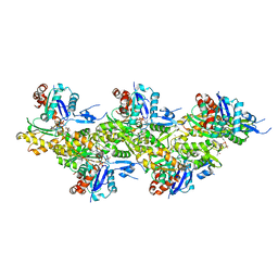
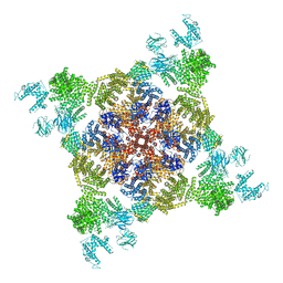
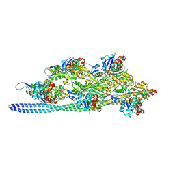
8OID
 | | Cryo-EM structure of ADP-bound, filamentous beta-actin harboring the N111S mutation | | 分子名称: | ADENOSINE-5'-DIPHOSPHATE, Actin, cytoplasmic 1, ... | | 著者 | Oosterheert, W, Blanc, F.E.C, Roy, A, Belyy, A, Hofnagel, O, Hummer, G, Bieling, P, Raunser, S. | | 登録日 | 2023-03-22 | | 公開日 | 2023-08-09 | | 最終更新日 | 2023-11-22 | | 実験手法 | ELECTRON MICROSCOPY (2.3 Å) | | 主引用文献 | Molecular mechanisms of inorganic-phosphate release from the core and barbed end of actin filaments.
Nat.Struct.Mol.Biol., 30, 2023
|
|
6T25
 | | Cryo-EM structure of phalloidin-Alexa Flour-546-stabilized F-actin (copolymerized) | | 分子名称: | ADENOSINE-5'-DIPHOSPHATE, Actin, alpha skeletal muscle, ... | | 著者 | Pospich, S, Merino, F, Raunser, S. | | 登録日 | 2019-10-07 | | 公開日 | 2020-03-04 | | 最終更新日 | 2020-04-15 | | 実験手法 | ELECTRON MICROSCOPY (3.6 Å) | | 主引用文献 | Structural Effects and Functional Implications of Phalloidin and Jasplakinolide Binding to Actin Filaments.
Structure, 28, 2020
|
|
8OI8
 | | Cryo-EM structure of ADP-bound, filamentous beta-actin harboring the R183W mutation | | 分子名称: | ADENOSINE-5'-DIPHOSPHATE, Actin, cytoplasmic 1, ... | | 著者 | Oosterheert, W, Blanc, F.E.C, Roy, A, Belyy, A, Hofnagel, O, Hummer, G, Bieling, P, Raunser, S. | | 登録日 | 2023-03-22 | | 公開日 | 2023-08-16 | | 最終更新日 | 2023-11-22 | | 実験手法 | ELECTRON MICROSCOPY (2.28 Å) | | 主引用文献 | Molecular mechanisms of inorganic-phosphate release from the core and barbed end of actin filaments.
Nat.Struct.Mol.Biol., 30, 2023
|
|
8OI6
 | | Cryo-EM structure of the undecorated barbed end of filamentous beta/gamma actin | | 分子名称: | ADENOSINE-5'-DIPHOSPHATE, Actin, cytoplasmic 1, ... | | 著者 | Oosterheert, W, Blanc, F.E.C, Roy, A, Belyy, A, Hofnagel, O, Hummer, G, Bieling, P, Raunser, S. | | 登録日 | 2023-03-22 | | 公開日 | 2023-08-09 | | 最終更新日 | 2023-11-22 | | 実験手法 | ELECTRON MICROSCOPY (3.59 Å) | | 主引用文献 | Molecular mechanisms of inorganic-phosphate release from the core and barbed end of actin filaments.
Nat.Struct.Mol.Biol., 30, 2023
|
|
6H3L
 | |
4UWA
 | |
6G1K
 | | Electron cryo-microscopy structure of the canonical TRPC4 ion channel | | 分子名称: | (2R)-3-(phosphonooxy)propane-1,2-diyl dihexanoate, CHOLESTEROL HEMISUCCINATE, Transient receptor potential cation channel subfamily c member 4a | | 著者 | Vinayagam, D, Mager, T, Apelbaum, A, Bothe, A, Merino, F, Hofnagel, O, Gatsogiannis, C, Raunser, S. | | 登録日 | 2018-03-21 | | 公開日 | 2018-05-02 | | 最終更新日 | 2018-08-01 | | 実験手法 | ELECTRON MICROSCOPY (3.6 Å) | | 主引用文献 | Electron cryo-microscopy structure of the canonical TRPC4 ion channel.
Elife, 7, 2018
|
|
6FHL
 | | Cryo-EM structure of F-actin in complex with ADP-Pi | | 分子名称: | ADENOSINE-5'-DIPHOSPHATE, Actin, alpha skeletal muscle, ... | | 著者 | Merino, F, Pospich, S, Funk, J, Wagner, T, Kuellmer, F, Arndt, H.-D, Bieling, P, Raunser, S. | | 登録日 | 2018-01-15 | | 公開日 | 2018-06-13 | | 最終更新日 | 2018-08-29 | | 実験手法 | ELECTRON MICROSCOPY (3.3 Å) | | 主引用文献 | Structural transitions of F-actin upon ATP hydrolysis at near-atomic resolution revealed by cryo-EM.
Nat. Struct. Mol. Biol., 25, 2018
|
|
6GY7
 | | Crystal structure of XaxB from Xenorhabdus nematophil | | 分子名称: | XaxB | | 著者 | Schubert, E, Raunser, S, Vetter, I.R, Prumbaum, D, Penczek, P.A. | | 登録日 | 2018-06-28 | | 公開日 | 2018-07-25 | | 実験手法 | X-RAY DIFFRACTION (3.4 Å) | | 主引用文献 | Membrane insertion of alpha-xenorhabdolysin in near-atomic detail.
Elife, 7, 2018
|
|
5JLF
 | |
8CPZ
 | | Photorhabdus luminescens TcdA1 prepore-to-pore intermediate, K1179W mutant | | 分子名称: | TcdA1 | | 著者 | Nganga, P.N, Roderer, D, Belyy, A, Prumbaum, D, Raunser, S. | | 登録日 | 2023-03-03 | | 公開日 | 2024-03-13 | | 実験手法 | ELECTRON MICROSCOPY (2.9 Å) | | 主引用文献 | Kinetics of the syringe-like injection mechanism of Tc toxins
to be published
|
|
8CQ0
 | | Photorhabdus luminescens TcdA1 prepore-to-pore intermediate, K567W K2008W mutant | | 分子名称: | TcdA1 | | 著者 | Nganga, P.N, Roderer, D, Belyy, A, Prumbaum, D, Raunser, S. | | 登録日 | 2023-03-03 | | 公開日 | 2024-03-13 | | 実験手法 | ELECTRON MICROSCOPY (3.2 Å) | | 主引用文献 | Kinetics of the syringe-like injection mechanism of Tc toxins
to be published
|
|
8CQ2
 | | Photorhabdus luminescens TcdA1 prepore-to-pore intermediate, C16S, C20S, C870S, T1279C mutant | | 分子名称: | TcdA1 | | 著者 | Nganga, P.N, Roderer, D, Belyy, A, Prumbaum, D, Raunser, S. | | 登録日 | 2023-03-03 | | 公開日 | 2024-03-13 | | 実験手法 | ELECTRON MICROSCOPY (3.6 Å) | | 主引用文献 | Kinetics of the syringe-like injection mechanism of Tc toxins
to be published
|
|
3J8A
 | | Structure of the F-actin-tropomyosin complex | | 分子名称: | ADENOSINE-5'-DIPHOSPHATE, Actin, alpha skeletal muscle, ... | | 著者 | von der Ecken, J, Mueller, M, Lehman, W, Manstein, J.M, Penczek, A.P, Raunser, S. | | 登録日 | 2014-10-08 | | 公開日 | 2014-12-10 | | 最終更新日 | 2018-07-18 | | 実験手法 | ELECTRON MICROSCOPY (3.7 Å) | | 主引用文献 | Structure of the F-actin--tropomyosin complex.
Nature, 519, 2015
|
|
2OF5
 | | Oligomeric Death Domain complex | | 分子名称: | Death domain-containing protein CRADD, Leucine-rich repeat and death domain-containing protein | | 著者 | Park, H.H, Logette, E, Raunser, S, Cuenin, S, Walz, T, Tschopp, J, Wu, H. | | 登録日 | 2007-01-02 | | 公開日 | 2007-04-17 | | 最終更新日 | 2023-12-27 | | 実験手法 | X-RAY DIFFRACTION (3.2 Å) | | 主引用文献 | Death domain assembly mechanism revealed by crystal structure of the oligomeric PIDDosome core complex.
Cell(Cambridge,Mass.), 128, 2007
|
|
5JLH
 | | Cryo-EM structure of a human cytoplasmic actomyosin complex at near-atomic resolution | | 分子名称: | ADENOSINE-5'-DIPHOSPHATE, Actin, cytoplasmic 2, ... | | 著者 | von der Ecken, J, Heissler, S.M, Pathan-Chhatbar, S, Manstein, D.J, Raunser, S. | | 登録日 | 2016-04-27 | | 公開日 | 2016-06-15 | | 最終更新日 | 2022-11-09 | | 実験手法 | ELECTRON MICROSCOPY (3.9 Å) | | 主引用文献 | Cryo-EM structure of a human cytoplasmic actomyosin complex at near-atomic resolution.
Nature, 534, 2016
|
|
4A7H
 | | Structure of the Actin-Tropomyosin-Myosin Complex (rigor ATM 2) | | 分子名称: | ACTIN, ALPHA SKELETAL MUSCLE, ADENOSINE-5'-DIPHOSPHATE, ... | | 著者 | Behrmann, E, Mueller, M, Penczek, P.A, Mannherz, H.G, Manstein, D.J, Raunser, S. | | 登録日 | 2011-11-14 | | 公開日 | 2012-08-01 | | 最終更新日 | 2017-08-30 | | 実験手法 | ELECTRON MICROSCOPY (7.8 Å) | | 主引用文献 | Structure of the Rigor Actin-Tropomyosin-Myosin Complex.
Cell(Cambridge,Mass.), 150, 2012
|
|
4A7F
 | | Structure of the Actin-Tropomyosin-Myosin Complex (rigor ATM 3) | | 分子名称: | ACTIN, ALPHA SKELETAL MUSCLE, ADENOSINE-5'-DIPHOSPHATE, ... | | 著者 | Behrmann, E, Mueller, M, Penczek, P.A, Mannherz, H.G, Manstein, D.J, Raunser, S. | | 登録日 | 2011-11-14 | | 公開日 | 2012-08-01 | | 最終更新日 | 2017-08-30 | | 実験手法 | ELECTRON MICROSCOPY (7.7 Å) | | 主引用文献 | Structure of the Rigor Actin-Tropomyosin-Myosin Complex.
Cell(Cambridge,Mass.), 150, 2012
|
|
4A7L
 | | Structure of the Actin-Tropomyosin-Myosin Complex (rigor ATM 1) | | 分子名称: | ACTIN, ALPHA SKELETON MUSCLE, ADENOSINE-5'-DIPHOSPHATE, ... | | 著者 | Behrmann, E, Mueller, M, Penczek, P.A, Mannherz, H.G, Manstein, D.J, Raunser, S. | | 登録日 | 2011-11-14 | | 公開日 | 2012-08-01 | | 最終更新日 | 2019-10-23 | | 実験手法 | ELECTRON MICROSCOPY (8.1 Å) | | 主引用文献 | Structure of the Rigor Actin-Tropomyosin-Myosin Complex.
Cell(Cambridge,Mass.), 150, 2012
|
|
4A7N
 | | Structure of bare F-actin filaments obtained from the same sample as the Actin-Tropomyosin-Myosin Complex | | 分子名称: | ADENOSINE-5'-DIPHOSPHATE, CALCIUM ION, F-ACTIN | | 著者 | Behrmann, E, Mueller, M, Penczek, P.A, Mannherz, H.G, Manstein, D.J, Raunser, S. | | 登録日 | 2011-11-14 | | 公開日 | 2012-08-01 | | 最終更新日 | 2017-08-30 | | 実験手法 | ELECTRON MICROSCOPY (8.9 Å) | | 主引用文献 | Structure of the Rigor Actin-Tropomyosin-Myosin Complex.
Cell(Cambridge,Mass.), 150, 2012
|
|
7NEP
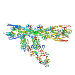 | | Homology model of the in situ actomyosin complex from the A-band of mouse psoas muscle sarcomere in the rigor state | | 分子名称: | Actin, alpha skeletal muscle, Myosin light chain 1/3, ... | | 著者 | Wang, Z, Grange, M, Wagner, T, Kho, A.L, Gautel, M, Raunser, S. | | 登録日 | 2021-02-04 | | 公開日 | 2021-04-07 | | 最終更新日 | 2024-05-01 | | 実験手法 | ELECTRON MICROSCOPY (10.2 Å) | | 主引用文献 | The molecular basis for sarcomere organization in vertebrate skeletal muscle.
Cell, 184, 2021
|
|
7ND2
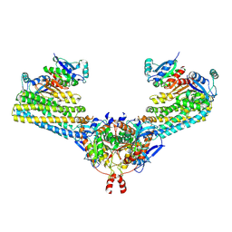 | | Cryo-EM structure of the human FERRY complex | | 分子名称: | Glutamine amidotransferase-like class 1 domain-containing protein 1, Protein phosphatase 1 regulatory subunit 21, Quinone oxidoreductase-like protein 1 | | 著者 | Quentin, D, Klink, B.U, Raunser, S. | | 登録日 | 2021-01-29 | | 公開日 | 2022-03-02 | | 最終更新日 | 2023-06-14 | | 実験手法 | ELECTRON MICROSCOPY (4 Å) | | 主引用文献 | Structural basis of mRNA binding by the human FERRY Rab5 effector complex.
Mol.Cell, 83, 2023
|
|


