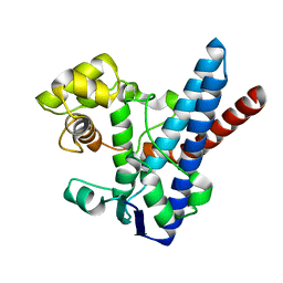
3M7G
 
 | |
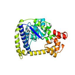
3M6K
 | |
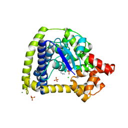
3M6Z
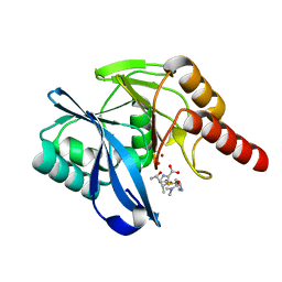
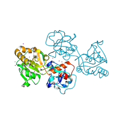
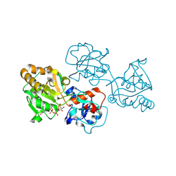
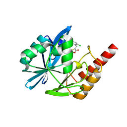
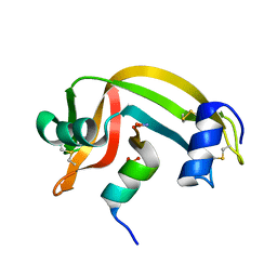
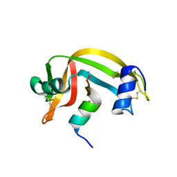
 | | Crystal structure of an N-terminal 44 kDa fragment of topoisomerase V in the presence of guanidium hydrochloride | | 分子名称: | CHLORIDE ION, GUANIDINE, MAGNESIUM ION, ... | | 著者 | Rajan, R, Taneja, B, Mondragon, A. | | 登録日 | 2010-03-16 | | 公開日 | 2010-08-04 | | 最終更新日 | 2011-07-13 | | 実験手法 | X-RAY DIFFRACTION (1.4 Å) | | 主引用文献 | Structures of minimal catalytic fragments of topoisomerase v reveals conformational changes relevant for DNA binding.
Structure, 18, 2010
|
|
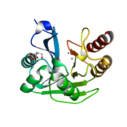
5N5I
 | | Crystal Structure of VIM-1 metallo-beta-lactamase in complex with hydrolysed meropenem | | 分子名称: | (2~{S},3~{R},4~{S})-2-[(2~{S},3~{R})-1,3-bis(oxidanyl)-1-oxidanylidene-butan-2-yl]-4-[(3~{S},5~{S})-5-(dimethylcarbamoy l)pyrrolidin-3-yl]sulfanyl-3-methyl-3,4-dihydro-2~{H}-pyrrole-5-carboxylic acid, Beta-lactamase VIM-1, ZINC ION | | 著者 | Salimraj, R, Hinchliffe, P, Spencer, J. | | 登録日 | 2017-02-14 | | 公開日 | 2018-03-07 | | 最終更新日 | 2024-01-17 | | 実験手法 | X-RAY DIFFRACTION (2.2 Å) | | 主引用文献 | Crystal structures of VIM-1 complexes explain active site heterogeneity in VIM-class metallo-beta-lactamases.
FEBS J., 286, 2019
|
|
8D0O
 | | Human alpha1,3-fucosyltransferase FUT9, heavy atom derivative | | 分子名称: | 2-acetamido-2-deoxy-beta-D-glucopyranose-(1-4)-2-acetamido-2-deoxy-beta-D-glucopyranose, 4-galactosyl-N-acetylglucosaminide 3-alpha-L-fucosyltransferase 9, CESIUM ION, ... | | 著者 | Kadirvelraj, R, Wood, Z.A. | | 登録日 | 2022-05-26 | | 公開日 | 2023-05-24 | | 最終更新日 | 2023-08-09 | | 実験手法 | X-RAY DIFFRACTION (2.1 Å) | | 主引用文献 | Structural basis for Lewis antigen synthesis by the alpha 1,3-fucosyltransferase FUT9.
Nat.Chem.Biol., 19, 2023
|
|
8D34
 | |
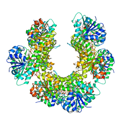
3D4K
 | | Concanavalin A Complexed to a Synthetic Analog of the Trimannoside | | 分子名称: | CALCIUM ION, Concanavalin-A, GLYCEROL, ... | | 著者 | Kadirvelraj, R, Foley, B.L, Dyekjaer, J.D, Woods, R.J. | | 登録日 | 2008-05-14 | | 公開日 | 2009-03-03 | | 最終更新日 | 2023-08-30 | | 実験手法 | X-RAY DIFFRACTION (1.8 Å) | | 主引用文献 | Involvement of water in carbohydrate-protein binding: concanavalin A revisited.
J.Am.Chem.Soc., 130, 2008
|
|
8D0Q
 | | Human FUT9 bound to GDP-CF3-Fucose and H-Type 2 | | 分子名称: | 2-acetamido-2-deoxy-beta-D-glucopyranose-(1-4)-2-acetamido-2-deoxy-beta-D-glucopyranose, 4-galactosyl-N-acetylglucosaminide 3-alpha-L-fucosyltransferase 9, GUANOSINE-5'-DIPHOSPHATE, ... | | 著者 | Kadirvelraj, R, Wood, Z.A. | | 登録日 | 2022-05-26 | | 公開日 | 2023-05-31 | | 最終更新日 | 2023-10-25 | | 実験手法 | X-RAY DIFFRACTION (1.39 Å) | | 主引用文献 | Structural basis for Lewis antigen synthesis by the alpha 1,3-fucosyltransferase FUT9.
Nat.Chem.Biol., 19, 2023
|
|
8D0W
 | | Human FUT9 bound to H-Type 2 | | 分子名称: | 2-acetamido-2-deoxy-beta-D-glucopyranose-(1-4)-2-acetamido-2-deoxy-beta-D-glucopyranose, 4-galactosyl-N-acetylglucosaminide 3-alpha-L-fucosyltransferase 9, GLYCEROL, ... | | 著者 | Kadirvelraj, R, Wood, Z.A. | | 登録日 | 2022-05-26 | | 公開日 | 2023-05-24 | | 最終更新日 | 2023-10-25 | | 実験手法 | X-RAY DIFFRACTION (1.332 Å) | | 主引用文献 | Structural basis for Lewis antigen synthesis by the alpha 1,3-fucosyltransferase FUT9.
Nat.Chem.Biol., 19, 2023
|
|
8D0X
 | | Human FUT9 bound to LNnT | | 分子名称: | 1,2-ETHANEDIOL, 4-galactosyl-N-acetylglucosaminide 3-alpha-L-fucosyltransferase 9, GLYCEROL, ... | | 著者 | Kadirvelraj, R, Wood, Z.A. | | 登録日 | 2022-05-26 | | 公開日 | 2023-05-24 | | 最終更新日 | 2023-10-25 | | 実験手法 | X-RAY DIFFRACTION (1.33 Å) | | 主引用文献 | Structural basis for Lewis antigen synthesis by the alpha 1,3-fucosyltransferase FUT9.
Nat.Chem.Biol., 19, 2023
|
|
8D0P
 | | Human FUT9, unliganded | | 分子名称: | 1,2-ETHANEDIOL, 4-galactosyl-N-acetylglucosaminide 3-alpha-L-fucosyltransferase 9, SULFATE ION, ... | | 著者 | Kadirvelraj, R, Wood, Z.A. | | 登録日 | 2022-05-26 | | 公開日 | 2023-05-24 | | 最終更新日 | 2023-10-25 | | 実験手法 | X-RAY DIFFRACTION (1.09 Å) | | 主引用文献 | Structural basis for Lewis antigen synthesis by the alpha 1,3-fucosyltransferase FUT9.
Nat.Chem.Biol., 19, 2023
|
|
8D0S
 | | Human FUT9 bound to GDP and LNnT | | 分子名称: | 1,2-ETHANEDIOL, 4-galactosyl-N-acetylglucosaminide 3-alpha-L-fucosyltransferase 9, GUANOSINE-5'-DIPHOSPHATE, ... | | 著者 | Kadirvelraj, R, Wood, Z.A. | | 登録日 | 2022-05-26 | | 公開日 | 2023-05-24 | | 最終更新日 | 2023-10-25 | | 実験手法 | X-RAY DIFFRACTION (1.37 Å) | | 主引用文献 | Structural basis for Lewis antigen synthesis by the alpha 1,3-fucosyltransferase FUT9.
Nat.Chem.Biol., 19, 2023
|
|
8D0U
 | | Human FUT9 bound to GDP | | 分子名称: | 1,2-ETHANEDIOL, 4-galactosyl-N-acetylglucosaminide 3-alpha-L-fucosyltransferase 9, GUANOSINE-5'-DIPHOSPHATE, ... | | 著者 | Kadirvelraj, R, Wood, Z.A. | | 登録日 | 2022-05-26 | | 公開日 | 2023-05-24 | | 最終更新日 | 2023-10-25 | | 実験手法 | X-RAY DIFFRACTION (1.29 Å) | | 主引用文献 | Structural basis for Lewis antigen synthesis by the alpha 1,3-fucosyltransferase FUT9.
Nat.Chem.Biol., 19, 2023
|
|
8D0R
 | | Human FUT9 bound to GDP and H-Type 2 | | 分子名称: | 1,2-ETHANEDIOL, 4-galactosyl-N-acetylglucosaminide 3-alpha-L-fucosyltransferase 9, GUANOSINE-5'-DIPHOSPHATE, ... | | 著者 | Kadirvelraj, R, Wood, Z.A. | | 登録日 | 2022-05-26 | | 公開日 | 2023-05-24 | | 最終更新日 | 2023-10-25 | | 実験手法 | X-RAY DIFFRACTION (1.4 Å) | | 主引用文献 | Structural basis for Lewis antigen synthesis by the alpha 1,3-fucosyltransferase FUT9.
Nat.Chem.Biol., 19, 2023
|
|
5N5H
 | | Crystal structure of metallo-beta-lactamase VIM-1 in complex with ML302F inhibitor | | 分子名称: | (2Z)-2-sulfanyl-3-(2,3,6-trichlorophenyl)prop-2-enoic acid, Beta-lactamase VIM-1, ZINC ION | | 著者 | Salimraj, R, Hinchliffe, P, Spencer, J. | | 登録日 | 2017-02-14 | | 公開日 | 2018-03-07 | | 最終更新日 | 2024-01-17 | | 実験手法 | X-RAY DIFFRACTION (1.3 Å) | | 主引用文献 | Crystal structures of VIM-1 complexes explain active site heterogeneity in VIM-class metallo-beta-lactamases.
FEBS J., 286, 2019
|
|
5N5G
 | |
3PTZ
 | |
4GFJ
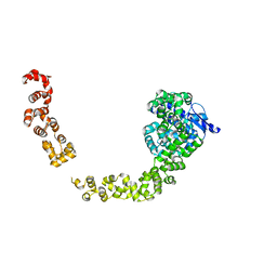
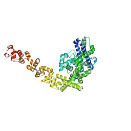 | | Crystal structure of Topo-78, an N-terminal 78kDa fragment of topoisomerase V | | 分子名称: | GLYCEROL, Topoisomerase V, ZINC ION | | 著者 | Rajan, R, Prasad, R, Taneja, B, Wilson, S.H, Mondragon, A. | | 登録日 | 2012-08-03 | | 公開日 | 2012-12-05 | | 最終更新日 | 2023-09-13 | | 実験手法 | X-RAY DIFFRACTION (2.91 Å) | | 主引用文献 | Identification of one of the apurinic/apyrimidinic lyase active sites of topoisomerase V by structural and functional studies.
Nucleic Acids Res., 41, 2013
|
|
3PHZ
 | |
3PRJ
 | |
1RBF
 | |
1RBI
 | |
1RBC
 | |
5HM5
 | |
1RBE
 | |


