3OR7
 
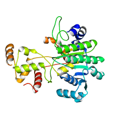
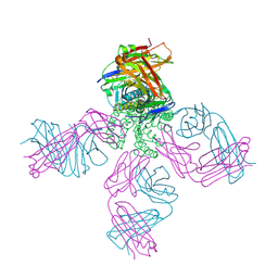 | | On the structural basis of modal gating behavior in K+channels - E71I | | 分子名称: | POTASSIUM ION, Voltage-gated potassium channel, antibody fab fragment heavy chain, ... | | 著者 | Chakrapani, S, Cordero-Morales, J.F, Jogini, V, Pan, A.C, Cortes, D.M, Roux, B, Perozo, E. | | 登録日 | 2010-09-06 | | 公開日 | 2011-01-05 | | 最終更新日 | 2018-08-29 | | 実験手法 | X-RAY DIFFRACTION (2.3 Å) | | 主引用文献 | On the structural basis of modal gating behavior in K(+) channels.
Nat.Struct.Mol.Biol., 18, 2011
|
|
5V23
 | |
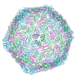
5KUA
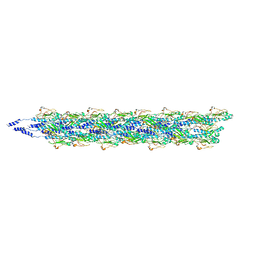 | | Cryo-EM reconstruction of Neisseria meningitidis Type IV pilus | | 分子名称: | pilin | | 著者 | Kolappan, S, Coureuil, M, Yu, X, Nassif, X, Craig, L, Egelman, E.H. | | 登録日 | 2016-07-13 | | 公開日 | 2016-10-12 | | 最終更新日 | 2016-11-30 | | 実験手法 | ELECTRON MICROSCOPY (6 Å) | | 主引用文献 | Structure of the Neisseria meningitidis Type IV pilus.
Nat Commun, 7, 2016
|
|
6VTJ
 | |
6VTZ
 | |
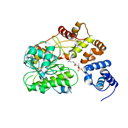
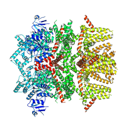
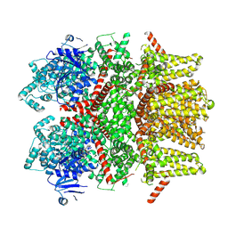
1JSC
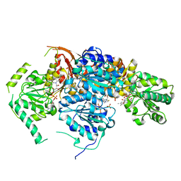 | | Crystal Structure of the Catalytic Subunit of Yeast Acetohydroxyacid Synthase: A target for Herbicidal Inhibitors | | 分子名称: | ACETOHYDROXY-ACID SYNTHASE, DIHYDROGENPHOSPHATE ION, FLAVIN-ADENINE DINUCLEOTIDE, ... | | 著者 | Pang, S.S, Duggleby, R.G, Guddat, L.W. | | 登録日 | 2001-08-17 | | 公開日 | 2002-01-16 | | 最終更新日 | 2023-08-16 | | 実験手法 | X-RAY DIFFRACTION (2.6 Å) | | 主引用文献 | Crystal structure of yeast acetohydroxyacid synthase: a target for herbicidal inhibitors.
J.Mol.Biol., 317, 2002
|
|
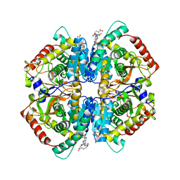
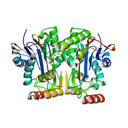
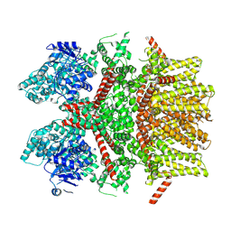
4QT0
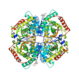 | | Crystal structure of human muscle L-lactate dehydrogenase in complex with inhibitor 1, 3-{[3-CARBAMOYL-7-(2,4-DIMETHOXYPYRIMIDIN-5-YL)QUINOLIN-4-YL]AMINO}BENZOIC ACID | | 分子名称: | 3-{[3-carbamoyl-7-(2,4-dimethoxypyrimidin-5-yl)quinolin-4-yl]amino}benzoic acid, L-lactate dehydrogenase A chain | | 著者 | Kolappan, S, Craig, L. | | 登録日 | 2014-07-06 | | 公開日 | 2014-12-03 | | 最終更新日 | 2024-02-28 | | 実験手法 | X-RAY DIFFRACTION (3.2 Å) | | 主引用文献 | Structures of lactate dehydrogenase A (LDHA) in apo, ternary and inhibitor-bound forms.
Acta Crystallogr.,Sect.D, 71, 2015
|
|
4QS4
 | |
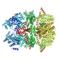
4QSM
 | | Crystal structure of human muscle L-lactate dehydrogenase in complex with inhibitor 2, 3-{[7-(2,4-dimethoxypyrimidin-5-yl)-3-sulfamoylquinolin-4-yl]amino}benzoic acid | | 分子名称: | 3-{[7-(2,4-dimethoxypyrimidin-5-yl)-3-sulfamoylquinolin-4-yl]amino}benzoic acid, L-lactate dehydrogenase A chain | | 著者 | Kolappan, S, Craig, L. | | 登録日 | 2014-07-06 | | 公開日 | 2014-12-03 | | 最終更新日 | 2024-02-28 | | 実験手法 | X-RAY DIFFRACTION (3 Å) | | 主引用文献 | Structures of lactate dehydrogenase A (LDHA) in apo, ternary and inhibitor-bound forms.
Acta Crystallogr.,Sect.D, 71, 2015
|
|
9FHP
 | |
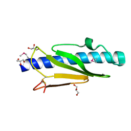
4RZJ
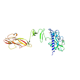
 | | Structure of the complex of type 1 ribosome inactivating protein from Momordica balsamina with N-acetylglucosamine at 1.98 Angstrom resolution using crystals grown in different conditions | | 分子名称: | 2-acetamido-2-deoxy-beta-D-glucopyranose, GLYCEROL, Ribosome inactivating protein | | 著者 | Pandey, S, Kushwaha, G.S, Kaur, P, Sharma, S, Singh, T.P. | | 登録日 | 2014-12-22 | | 公開日 | 2015-01-14 | | 最終更新日 | 2023-09-20 | | 実験手法 | X-RAY DIFFRACTION (1.98 Å) | | 主引用文献 | Structure of the complex of type 1 ribosome inactivating protein from Momordica balsamina with N-acetylglucosamine at 1.98 Angstrom resolution using crystals grown in different conditions
TO BE PUBLISHED
|
|
6P5G
 | |
6P4I
 | |
6P5F
 | |
6P5E
 | |
6P5D
 | |
6Y09
 | |
6ZAY
 | |
6NQ6
 | |
8SLE
 | | Cryo-EM structure of the rat TRPM5 channel in trace calcium, trace-3 | | 分子名称: | Transient receptor potential cation channel subfamily M member 5 | | 著者 | Karuppan, S, Schrag, L.G, Jara-Oseguera, A, Zubcevic, L. | | 登録日 | 2023-04-21 | | 公開日 | 2024-07-03 | | 最終更新日 | 2024-07-10 | | 実験手法 | ELECTRON MICROSCOPY (4.3 Å) | | 主引用文献 | Structural dynamics at cytosolic interprotomer interfaces control gating of a mammalian TRPM5 channel.
Proc.Natl.Acad.Sci.USA, 121, 2024
|
|
8SLI
 | | Cryo-EM structure of the rat TRPM5 channel in 2mM calcium, high-1 | | 分子名称: | CALCIUM ION, Transient receptor potential cation channel subfamily M member 5 | | 著者 | Karuppan, S, Schrag, L.G, Jara-Oseguera, A, Zubcevic, L. | | 登録日 | 2023-04-21 | | 公開日 | 2024-07-03 | | 最終更新日 | 2024-07-10 | | 実験手法 | ELECTRON MICROSCOPY (4.9 Å) | | 主引用文献 | Structural dynamics at cytosolic interprotomer interfaces control gating of a mammalian TRPM5 channel.
Proc.Natl.Acad.Sci.USA, 121, 2024
|
|
8SLQ
 | | Cryo-EM structure of the rat TRPM5 channel in 2mM calcium, high-3 | | 分子名称: | Transient receptor potential cation channel subfamily M member 5 | | 著者 | Karuppan, S, Schrag, L.G, Jara-Oseguera, A, Zubcevic, L. | | 登録日 | 2023-04-24 | | 公開日 | 2024-07-03 | | 最終更新日 | 2024-07-10 | | 実験手法 | ELECTRON MICROSCOPY (4.5 Å) | | 主引用文献 | Structural dynamics at cytosolic interprotomer interfaces control gating of a mammalian TRPM5 channel.
Proc.Natl.Acad.Sci.USA, 121, 2024
|
|
8SLW
 | | Cryo-EM structure of the rat TRPM5 channel in 2mM calcium, high-4 | | 分子名称: | CALCIUM ION, Transient receptor potential cation channel subfamily M member 5 | | 著者 | Karuppan, S, Schrag, L.G, Jara-Oseguera, A, Zubcevic, L. | | 登録日 | 2023-04-24 | | 公開日 | 2024-07-03 | | 最終更新日 | 2024-07-10 | | 実験手法 | ELECTRON MICROSCOPY (4.2 Å) | | 主引用文献 | Structural dynamics at cytosolic interprotomer interfaces control gating of a mammalian TRPM5 channel.
Proc.Natl.Acad.Sci.USA, 121, 2024
|
|
8SLP
 | | Cryo-EM structure of the rat TRPM5 channel in 2mM calcium, high-2 | | 分子名称: | CALCIUM ION, Transient receptor potential cation channel subfamily M member 5 | | 著者 | Karuppan, S, Schrag, L.G, Jara-Oseguera, A, Zubcevic, L. | | 登録日 | 2023-04-24 | | 公開日 | 2024-07-03 | | 最終更新日 | 2024-07-10 | | 実験手法 | ELECTRON MICROSCOPY (3.8 Å) | | 主引用文献 | Structural dynamics at cytosolic interprotomer interfaces control gating of a mammalian TRPM5 channel.
Proc.Natl.Acad.Sci.USA, 121, 2024
|
|
8SL8
 | | Cryo-EM structure of the rat TRPM5 channel in trace calcium, trace-1 | | 分子名称: | Transient receptor potential cation channel subfamily M member 5 | | 著者 | Karuppan, S, Schrag, L.G, Jara-Oseguera, A, Zubcevic, L. | | 登録日 | 2023-04-21 | | 公開日 | 2024-07-03 | | 最終更新日 | 2024-07-10 | | 実験手法 | ELECTRON MICROSCOPY (4.2 Å) | | 主引用文献 | Structural dynamics at cytosolic interprotomer interfaces control gating of a mammalian TRPM5 channel.
Proc.Natl.Acad.Sci.USA, 121, 2024
|
|


