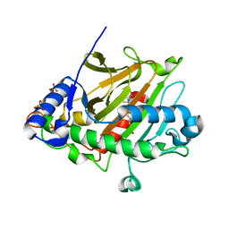
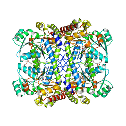
1IUS
 
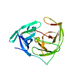
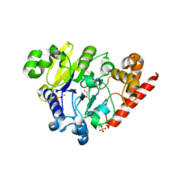
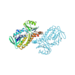 | | P-HYDROXYBENZOATE HYDROXYLASE COMPLEXED WITH 4-AMINOBENZOATE AT PH 5.0 | | 分子名称: | 4-AMINOBENZOIC ACID, FLAVIN-ADENINE DINUCLEOTIDE, P-HYDROXYBENZOATE HYDROXYLASE | | 著者 | Gatti, D.L, Entsch, B, Ballou, D.P, Ludwig, M.L. | | 登録日 | 1995-11-22 | | 公開日 | 1996-04-03 | | 最終更新日 | 2024-02-07 | | 実験手法 | X-RAY DIFFRACTION (2.2 Å) | | 主引用文献 | pH-dependent structural changes in the active site of p-hydroxybenzoate hydroxylase point to the importance of proton and water movements during catalysis.
Biochemistry, 35, 1996
|
|
8BOS
 | |
6INE
 | | Crystal Structure of human ASH1L-MRG15 complex | | 分子名称: | GLYCEROL, Histone-lysine N-methyltransferase ASH1L, Mortality factor 4-like protein 1, ... | | 著者 | Hou, P, Huang, C, Liu, C.P, Yu, T, Yin, Y, Zhu, B, Xu, R.M. | | 登録日 | 2018-10-25 | | 公開日 | 2019-03-20 | | 最終更新日 | 2023-11-22 | | 実験手法 | X-RAY DIFFRACTION (2.6 Å) | | 主引用文献 | Structural Insights into Stimulation of Ash1L's H3K36 Methyltransferase Activity through Mrg15 Binding.
Structure, 27, 2019
|
|
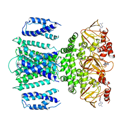
5DS6
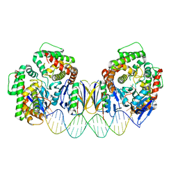 | | Crystal structure the Escherichia coli Cas1-Cas2 complex bound to protospacer DNA with splayed ends | | 分子名称: | CRISPR-associated endonuclease Cas1, CRISPR-associated endoribonuclease Cas2, DNA (28-MER), ... | | 著者 | Nunez, J.K, Harrington, L.B, Kranzusch, P.J, Engelman, A.N, Doudna, J.A. | | 登録日 | 2015-09-16 | | 公開日 | 2015-10-28 | | 最終更新日 | 2023-09-27 | | 実験手法 | X-RAY DIFFRACTION (3.352 Å) | | 主引用文献 | Foreign DNA capture during CRISPR-Cas adaptive immunity.
Nature, 527, 2015
|
|
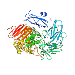
6L50
 | | Crystal structure of Zika NS2B-NS3 protease with compound 16 | | 分子名称: | 2-sulfanylidene-1,3-thiazolidin-4-one, NS3 protease, Serine protease subunit NS2B | | 著者 | Quek, J.P. | | 登録日 | 2019-10-21 | | 公開日 | 2020-07-15 | | 最終更新日 | 2023-11-22 | | 実験手法 | X-RAY DIFFRACTION (1.95 Å) | | 主引用文献 | Identification and structural characterization of small molecule fragments targeting Zika virus NS2B-NS3 protease.
Antiviral Res., 175, 2020
|
|
8OKU
 | | Salt-Inducible Kinase 3 in complex with an inhibitor | | 分子名称: | Serine/threonine-protein kinase SIK3, ~{N}-ethyl-4-[5-[1-(2-hydroxyethyl)pyrazol-4-yl]benzimidazol-1-yl]-2,6-dimethoxy-benzamide | | 著者 | Flower, T.G, Leonard, P.M, Lamers, M.B.A.C, Mollat, P. | | 登録日 | 2023-03-29 | | 公開日 | 2024-01-10 | | 最終更新日 | 2024-01-24 | | 実験手法 | X-RAY DIFFRACTION (3.1 Å) | | 主引用文献 | Optimization of Selectivity and Pharmacokinetic Properties of Salt-Inducible Kinase Inhibitors that Led to the Discovery of Pan-SIK Inhibitor GLPG3312.
J.Med.Chem., 67, 2024
|
|
4K9B
 | | Structure of Ternary Complex of cGAS with dsDNA and Bound c[G(2 ,5 )pA(3 ,5 )p] | | 分子名称: | ADENOSINE MONOPHOSPHATE, Cyclic GMP-AMP synthase, DNA-F, ... | | 著者 | Gao, P, Wu, Y, Patel, D.J. | | 登録日 | 2013-04-19 | | 公開日 | 2013-05-15 | | 最終更新日 | 2024-05-29 | | 実験手法 | X-RAY DIFFRACTION (2.26 Å) | | 主引用文献 | Cyclic [G(2',5')pA(3',5')p] is the metazoan second messenger produced by DNA-activated cyclic GMP-AMP synthase.
Cell(Cambridge,Mass.), 153, 2013
|
|
6WKL
 | |
6WKM
 | |
8BTL
 | | Crystal structure of a complex between the E2 conjugating enzyme UBE2A and the E3 ligase module from UBR4 | | 分子名称: | Ubiquitin conjugating enzyme E2 A, ZINC ION, cDNA FLJ12511 fis, ... | | 著者 | Virdee, S, Mabbitt, P.D, Barnsby-Greer, L. | | 登録日 | 2022-11-29 | | 公開日 | 2023-12-13 | | 最終更新日 | 2024-04-17 | | 実験手法 | X-RAY DIFFRACTION (3.2 Å) | | 主引用文献 | UBE2A and UBE2B are recruited by an atypical E3 ligase module in UBR4.
Nat.Struct.Mol.Biol., 31, 2024
|
|
8BSY
 | |
5DVP
 | | Crystal structure of Mycobacterium tuberculosis L,D-transpeptidase 2 with Doripenem adduct | | 分子名称: | (2S,3R,4S)-2-[(2S,3R)-3-hydroxy-1-oxobutan-2-yl]-3-methyl-4-({(3S,5S)-5-[(sulfamoylamino)methyl]pyrrolidin-3-yl}sulfanyl)-3,4-dihydro-2H-pyrrole-5-carboxylic acid, L,D-transpeptidase 2, PHOSPHONOACETALDEHYDE, ... | | 著者 | Kumar, P, Lamichhane, G. | | 登録日 | 2015-09-21 | | 公開日 | 2016-09-28 | | 最終更新日 | 2023-09-27 | | 実験手法 | X-RAY DIFFRACTION (2.18 Å) | | 主引用文献 | Non-classical transpeptidases yield insight into new antibacterials.
Nat. Chem. Biol., 13, 2017
|
|
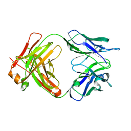
6WJL
 | | Crystal structure of Glypican-2 core protein in complex with D3 Fab | | 分子名称: | D3 Fab Heavy chain, D3 Fab Light Chain, Glypican-2, ... | | 著者 | Raman, S, Maris, J.M, Bosse, K.R, Julien, J.P. | | 登録日 | 2020-04-14 | | 公開日 | 2021-04-21 | | 最終更新日 | 2023-10-18 | | 実験手法 | X-RAY DIFFRACTION (3.3 Å) | | 主引用文献 | A GPC2 antibody-drug conjugate is efficacious against neuroblastoma and small-cell lung cancer via binding a conformational epitope.
Cell Rep Med, 2, 2021
|
|
8BSX
 | |
8BSW
 | |
7RU0
 | | SthK R120A Open State 1 | | 分子名称: | ADENOSINE-3',5'-CYCLIC-MONOPHOSPHATE, SthK | | 著者 | Gao, X, Nimigean, C, Schmidpeter, P. | | 登録日 | 2021-08-16 | | 公開日 | 2022-11-23 | | 最終更新日 | 2024-06-05 | | 実験手法 | ELECTRON MICROSCOPY (4.3 Å) | | 主引用文献 | Gating intermediates reveal inhibitory role of the voltage sensor in a cyclic nucleotide-modulated ion channel.
Nat Commun, 13, 2022
|
|
6I60
 | |
4PEF
 | | Dbr1 in complex with sulfate | | 分子名称: | GLYCEROL, MANGANESE (II) ION, RNA lariat debranching enzyme, ... | | 著者 | Montemayor, E.J, Katolik, A, Clark, N.E, Taylor, A.B, Schuermann, J.P, Combs, D.J, Johnsson, R, Holloway, S.P, Stevens, S.W, Damha, M.J, Hart, P.J. | | 登録日 | 2014-04-23 | | 公開日 | 2014-08-27 | | 最終更新日 | 2024-04-03 | | 実験手法 | X-RAY DIFFRACTION (1.96 Å) | | 主引用文献 | Structural basis of lariat RNA recognition by the intron debranching enzyme Dbr1.
Nucleic Acids Res., 42, 2014
|
|
8BSV
 | |
1E70
 | | 2-F-glucosylated MYROSINASE FROM SINAPIS ALBA | | 分子名称: | 2-acetamido-2-deoxy-beta-D-glucopyranose, 2-acetamido-2-deoxy-beta-D-glucopyranose-(1-4)-2-acetamido-2-deoxy-beta-D-glucopyranose, 2-deoxy-2-fluoro-alpha-D-glucopyranose, ... | | 著者 | Burmeister, W.P. | | 登録日 | 2000-08-23 | | 公開日 | 2001-01-05 | | 最終更新日 | 2023-12-13 | | 実験手法 | X-RAY DIFFRACTION (1.65 Å) | | 主引用文献 | High Resolution X-Ray Crystallography Shows that Ascorbate is a Cofactor for Myrosinase and Substitutes for the Function of the Catalytic Base
J.Biol.Chem., 275, 2000
|
|
4PG9
 | | MHC Class I in complex with Sendai virus nucleoprotein peptide FAPGNYPAL | | 分子名称: | Beta-2-microglobulin, GLYCEROL, H-2 class I histocompatibility antigen, ... | | 著者 | Celie, P, Joosten, R.P, Perrakis, A, Neefjes, J. | | 登録日 | 2014-05-01 | | 公開日 | 2015-01-07 | | 最終更新日 | 2023-09-27 | | 実験手法 | X-RAY DIFFRACTION (2.4 Å) | | 主引用文献 | The first step of peptide selection in antigen presentation by MHC class I molecules.
Proc.Natl.Acad.Sci.USA, 112, 2015
|
|
5E38
 | | Structural basis of mapping the spontaneous mutations with 5-flourouracil in uracil phosphoribosyltransferase from Mycobacterium tuberculosis | | 分子名称: | Uracil phosphoribosyltransferase | | 著者 | Ghode, P, Jobichen, C, Ramachandran, S, Bifani, P, Sivaraman, J. | | 登録日 | 2015-10-02 | | 公開日 | 2015-10-21 | | 最終更新日 | 2023-11-08 | | 実験手法 | X-RAY DIFFRACTION (3 Å) | | 主引用文献 | Structural basis of mapping the spontaneous mutations with 5-flurouracil in uracil phosphoribosyltransferase from Mycobacterium tuberculosis
Biochem.Biophys.Res.Commun., 467, 2015
|
|
6LD7
 | |
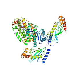
6E0E
 | | Crystal structure of Glucokinase in complex with compound 6 | | 分子名称: | 2-({2-[(4-methyl-1,3-thiazol-2-yl)amino]pyridin-3-yl}oxy)benzonitrile, Glucokinase, alpha-D-glucopyranose | | 著者 | Hinklin, R.J, Baer, B.R, Boyd, S.A, Chicarelli, M.D, Condroski, K.R, DeWolf, W.E, Fischer, J, Frank, M, Hingorani, G.P, Lee, P.A, Neitzel, N.A, Pratt, S.A, Singh, A, Sullivan, F.X, Turner, T, Voegtli, W.C, Wallace, E.M, Williams, L, Aicher, T.D. | | 登録日 | 2018-07-06 | | 公開日 | 2019-07-10 | | 最終更新日 | 2024-03-13 | | 実験手法 | X-RAY DIFFRACTION (2.7 Å) | | 主引用文献 | Discovery and preclinical development of AR453588 as an anti-diabetic glucokinase activator.
Bioorg.Med.Chem., 28, 2020
|
|
8BV8
 | | Crystal structure of the phage Mu protein Mom inactive mutant S124A | | 分子名称: | Methylcarbamoylase mom | | 著者 | Silva, R.M.B, Slyvka, A, Lee, Y.J, Guan, C, Lund, S.R, Raleigh, E.A, Skowronek, K, Bochtler, M, Weigele, P.R. | | 登録日 | 2022-12-08 | | 公開日 | 2023-12-20 | | 実験手法 | X-RAY DIFFRACTION (2.03 Å) | | 主引用文献 | Crystal structure of the phage Mu protein Mom catalytic mutant S124A
To Be Published
|
|


