6ROA
 
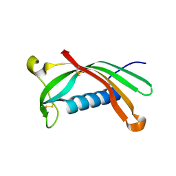 | | Crystal structure of V57G mutant of human cystatin C | | 分子名称: | Cystatin-C | | 著者 | Orlikowska, M, Behrendt, I, Borek, D, Otwinowski, Z, Skowron, P, Szymanska, A. | | 登録日 | 2019-05-10 | | 公開日 | 2019-08-07 | | 最終更新日 | 2024-10-16 | | 実験手法 | X-RAY DIFFRACTION (2.65 Å) | | 主引用文献 | NMR and crystallographic structural studies of the extremely stable monomeric variant of human cystatin C with single amino acid substitution.
Febs J., 287, 2020
|
|
7PQ8
 
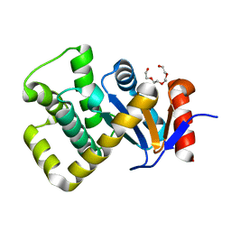 | | Crystal structure of Campylobacter jejuni DsbA1 | | 分子名称: | TETRAETHYLENE GLYCOL, Thiol:disulfide interchange protein DsbA | | 著者 | Orlikowska, M, Bocian-Ostrzycka, K.M, Banas, A.M, Jagusztyn-Krynicka, E.K. | | 登録日 | 2021-09-16 | | 公開日 | 2021-12-29 | | 最終更新日 | 2024-01-31 | | 実験手法 | X-RAY DIFFRACTION (1.329 Å) | | 主引用文献 | Interplay between DsbA1, DsbA2 and C8J_1298 Periplasmic Oxidoreductases of Campylobacter jejuni and Their Impact on Bacterial Physiology and Pathogenesis.
Int J Mol Sci, 22, 2021
|
|
3S67
 
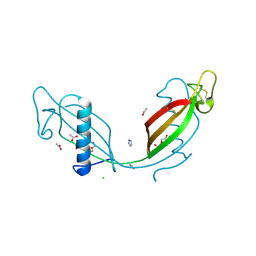 | | Crystal structure of V57P mutant of human cystatin C | | 分子名称: | ACETATE ION, CHLORIDE ION, Cystatin-C, ... | | 著者 | Orlikowska, M, Szymanska, A, Borek, D, Otwinowski, Z, Skowron, P, Jankowska, E. | | 登録日 | 2011-05-25 | | 公開日 | 2012-05-30 | | 最終更新日 | 2024-10-16 | | 実験手法 | X-RAY DIFFRACTION (2.26 Å) | | 主引用文献 | Structural characterization of V57D and V57P mutants of human cystatin C, an amyloidogenic protein.
Acta Crystallogr.,Sect.D, 69, 2013
|
|
3SVA
 
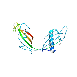 | | Crystal structure of V57D mutant of human cystatin C | | 分子名称: | ACETATE ION, Cystatin-C, DI(HYDROXYETHYL)ETHER | | 著者 | Orlikowska, M, Szymanska, A, Borek, D, Otwinowski, Z, Skowron, P, Jankowska, E. | | 登録日 | 2011-07-12 | | 公開日 | 2012-08-01 | | 最終更新日 | 2024-10-09 | | 実験手法 | X-RAY DIFFRACTION (3.02 Å) | | 主引用文献 | Structural characterization of V57D and V57P mutants of human cystatin C, an amyloidogenic protein.
Acta Crystallogr.,Sect.D, 69, 2013
|
|
5NQ7
 
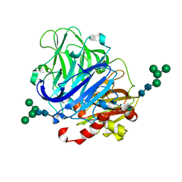 | | Crystal structure of laccases from Pycnoporus sanguineus, izoform I | | 分子名称: | COPPER (II) ION, Laccase, PEROXIDE ION, ... | | 著者 | Orlikowska, M, de J.Rostro-Alanis, M, Bujacz, A, Hernandez-Luna, C, Rubio, R, Parra, R, Bujacz, G. | | 登録日 | 2017-04-19 | | 公開日 | 2017-11-01 | | 最終更新日 | 2024-11-06 | | 実験手法 | X-RAY DIFFRACTION (2.75 Å) | | 主引用文献 | Structural studies of two thermostable laccases from the white-rot fungus Pycnoporus sanguineus.
Int. J. Biol. Macromol., 107, 2018
|
|
5NQ9
 
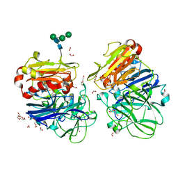 | | Crystal structure of laccases from Pycnoporus sanguineus, izoform II, monoclinic | | 分子名称: | 2-acetamido-2-deoxy-beta-D-glucopyranose, 2-acetamido-2-deoxy-beta-D-glucopyranose-(1-4)-2-acetamido-2-deoxy-beta-D-glucopyranose, COPPER (II) ION, ... | | 著者 | Orlikowska, M, de J.Rostro-Alanis, M, Bujacz, A, Hernandez-Luna, C, Rubio, R, Parra, R, Bujacz, G. | | 登録日 | 2017-04-19 | | 公開日 | 2017-11-01 | | 最終更新日 | 2024-11-06 | | 実験手法 | X-RAY DIFFRACTION (2.72 Å) | | 主引用文献 | Structural studies of two thermostable laccases from the white-rot fungus Pycnoporus sanguineus.
Int. J. Biol. Macromol., 107, 2018
|
|
5NQ8
 
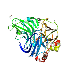 | | Crystal structure of laccases from Pycnoporus sanguineus, izoform II | | 分子名称: | 1-ETHOXY-2-(2-METHOXYETHOXY)ETHANE, COPPER (II) ION, Laccase, ... | | 著者 | Orlikowska, M, de J.Rostro-Alanis, M, Bujacz, A, Hernandez-Luna, C, Rubio, R, Parra, R, Bujacz, G. | | 登録日 | 2017-04-19 | | 公開日 | 2017-11-01 | | 最終更新日 | 2024-11-06 | | 実験手法 | X-RAY DIFFRACTION (2 Å) | | 主引用文献 | Structural studies of two thermostable laccases from the white-rot fungus Pycnoporus sanguineus.
Int. J. Biol. Macromol., 107, 2018
|
|
3QRD
 
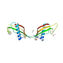 | | Crystal structure of L68V mutant of human cystatin C | | 分子名称: | Cystatin-C, DI(HYDROXYETHYL)ETHER | | 著者 | Orlikowska, M, Borek, D, Otwinowski, Z, Skowron, P, Szymanska, A. | | 登録日 | 2011-02-17 | | 公開日 | 2012-02-29 | | 最終更新日 | 2023-09-13 | | 実験手法 | X-RAY DIFFRACTION (2.19 Å) | | 主引用文献 | Crystal structure of L68V mutant of human cystatin C
To be Published
|
|
3NX0
 
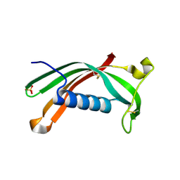 | | Hinge-loop mutation can be used to control 3D domain swapping and amyloidogenesis of human cystatin C | | 分子名称: | Cystatin-C, SULFATE ION | | 著者 | Orlikowska, M, Jankowska, E, Kolodziejczyk, R, Jaskolski, M, Szymanska, A. | | 登録日 | 2010-07-12 | | 公開日 | 2010-12-01 | | 最終更新日 | 2011-07-13 | | 実験手法 | X-RAY DIFFRACTION (2.04 Å) | | 主引用文献 | Hinge-loop mutation can be used to control 3D domain swapping and amyloidogenesis of human cystatin C.
J.Struct.Biol., 173, 2011
|
|
3PS8
 
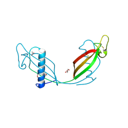 | | Crystal structure of L68V mutant of human cystatin C | | 分子名称: | ACETATE ION, Cystatin-C, DI(HYDROXYETHYL)ETHER | | 著者 | Orlikowska, M, Borek, D, Otwinowski, Z, Skowron, P, Szymanska, A. | | 登録日 | 2010-12-01 | | 公開日 | 2011-12-21 | | 最終更新日 | 2013-04-03 | | 実験手法 | X-RAY DIFFRACTION (2.55 Å) | | 主引用文献 | Crystal structure of L68V mutant of human cystatin C
To be Published
|
|
3KWM
 
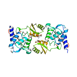 | | Crystal structure of ribose-5-isomerase A | | 分子名称: | D-Glyceraldehyde, DI(HYDROXYETHYL)ETHER, PHOSPHATE ION, ... | | 著者 | Orlikowska, M, Rostankowski, R, Nakka, C, Hattne, J, Grimshaw, S, Borek, D, Otwinowski, Z, Center for Structural Genomics of Infectious Diseases (CSGID) | | 登録日 | 2009-12-01 | | 公開日 | 2010-01-05 | | 最終更新日 | 2023-09-06 | | 実験手法 | X-RAY DIFFRACTION (2.32 Å) | | 主引用文献 |
|
|
4M8L
 
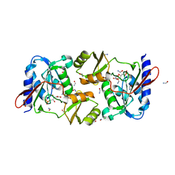 | | crystal structure of RpiA-R5P complex | | 分子名称: | CHLORIDE ION, FORMAMIDE, RIBULOSE-5-PHOSPHATE, ... | | 著者 | Rostankowski, R, Borek, D, Orlikowska, M, Nakka, C, Grimshaw, S, Otwinowski, Z, Center for Structural Genomics of Infectious Diseases (CSGID) | | 登録日 | 2013-08-13 | | 公開日 | 2013-10-02 | | 最終更新日 | 2023-09-20 | | 実験手法 | X-RAY DIFFRACTION (2.37 Å) | | 主引用文献 | crystal structure of RpiA-R5P complex
TO BE PUBLISHED
|
|
7PQ7
 
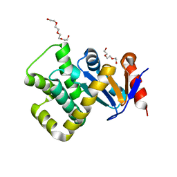 | | Crystal structure of Campylobacter jejuni DsbA1 | | 分子名称: | TETRAETHYLENE GLYCOL, TRIETHYLENE GLYCOL, Thiol:disulfide interchange protein DsbA | | 著者 | Wilk, P, Orlikowska, M, Banas, A.M, Bocian-Ostrzycka, K.M, Jagusztyn-Krynicka, E.K. | | 登録日 | 2021-09-16 | | 公開日 | 2021-12-29 | | 最終更新日 | 2024-01-31 | | 実験手法 | X-RAY DIFFRACTION (1.55 Å) | | 主引用文献 | Interplay between DsbA1, DsbA2 and C8J_1298 Periplasmic Oxidoreductases of Campylobacter jejuni and Their Impact on Bacterial Physiology and Pathogenesis.
Int J Mol Sci, 22, 2021
|
|
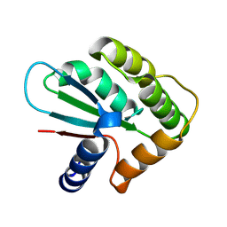
8PXD
 
 | |
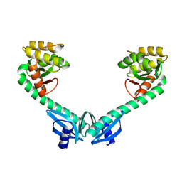
8PXF
 
 | |
