3KA9
 
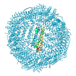 | | Frog M-ferritin, EEH mutant, with cobalt | | 分子名称: | CHLORIDE ION, COBALT (II) ION, Ferritin, ... | | 著者 | Tosha, T, Ng, H.L, Theil, E, Alber, T, Bhattasali, O. | | 登録日 | 2009-10-19 | | 公開日 | 2010-10-06 | | 最終更新日 | 2023-09-06 | | 実験手法 | X-RAY DIFFRACTION (1.45 Å) | | 主引用文献 | Frog M-ferritin, EEH mutant, with cobalt
To be Published
|
|
5WIF
 | | Crystal structure of spermidine/spermine N-acetyltransferase SpeG from Yersinia pestis | | 分子名称: | 1-METHOXY-2-[2-(2-METHOXY-ETHOXY]-ETHANE, BORIC ACID, DI(HYDROXYETHYL)ETHER, ... | | 著者 | Filippova, E.V, Wawrzak, Z, Kiryukhina, O, Shatsman, S, Anderson, W.F, Center for Structural Genomics of Infectious Diseases (CSGID) | | 登録日 | 2017-07-19 | | 公開日 | 2017-08-02 | | 最終更新日 | 2023-10-04 | | 実験手法 | X-RAY DIFFRACTION (2.5 Å) | | 主引用文献 | Crystal structure of spermidine/spermine N-acetyltransferase SpeG from Yersinia pestis
To Be Published
|
|
7NHQ
 | | Structure of PSII-I prime (PSII with Psb28, and Psb34) | | 分子名称: | 1,2-DIPALMITOYL-PHOSPHATIDYL-GLYCEROLE, 1,2-DISTEAROYL-MONOGALACTOSYL-DIGLYCERIDE, 2,3-DIMETHYL-5-(3,7,11,15,19,23,27,31,35-NONAMETHYL-2,6,10,14,18,22,26,30,34-HEXATRIACONTANONAENYL-2,5-CYCLOHEXADIENE-1,4-DIONE-2,3-DIMETHYL-5-SOLANESYL-1,4-BENZOQUINONE, ... | | 著者 | Zabret, J, Bohn, S, Schuller, S.K, Arnolds, O, Chan, A, Tajkhorshid, E, Stoll, R, Engel, B.D, Rudack, T, Schuller, J.M, Nowaczyk, M.M. | | 登録日 | 2021-02-11 | | 公開日 | 2021-05-05 | | 最終更新日 | 2024-05-01 | | 実験手法 | ELECTRON MICROSCOPY (2.68 Å) | | 主引用文献 | Structural insights into photosystem II assembly.
Nat.Plants, 7, 2021
|
|
5V36
 | | 1.88 Angstrom Resolution Crystal Structure of Glutathione Reductase from Streptococcus mutans UA159 in Complex with FAD | | 分子名称: | 4-(2-HYDROXYETHYL)-1-PIPERAZINE ETHANESULFONIC ACID, BETA-MERCAPTOETHANOL, CHLORIDE ION, ... | | 著者 | Minasov, G, Shuvalova, L, Dubrovska, I, Kiryukhina, O, Grimshaw, S, Kwon, K, Anderson, W.F, Center for Structural Genomics of Infectious Diseases (CSGID) | | 登録日 | 2017-03-06 | | 公開日 | 2017-03-22 | | 最終更新日 | 2023-10-04 | | 実験手法 | X-RAY DIFFRACTION (1.88 Å) | | 主引用文献 | 1.88 Angstrom Resolution Crystal Structure of Glutathione Reductase from Streptococcus mutans UA159 in Complex with FAD.
To Be Published
|
|
5V4D
 | | Crystal Structure of the Protein of Unknown Function of the Conserved Rid Protein Family YyfA from Yersinia pestis | | 分子名称: | ACETIC ACID, CALCIUM ION, GLYCEROL, ... | | 著者 | Kim, Y, Chhor, G, Endres, M, Krishnan, A, Babnigg, G, Schneewind, O, Anderson, W.F, Joachimiak, A, Center for Structural Genomics of Infectious Diseases (CSGID) | | 登録日 | 2017-03-09 | | 公開日 | 2017-04-05 | | 最終更新日 | 2023-10-04 | | 実験手法 | X-RAY DIFFRACTION (1.6 Å) | | 主引用文献 | Crystal Structure of the Protein of Unknown Function of the Conserved Rid Protein Family YyfA from Yersinia pestis
To Be Published
|
|
3JSY
 | | N-terminal fragment of ribosomal protein L10 from Methanococcus jannaschii | | 分子名称: | Acidic ribosomal protein P0 homolog | | 著者 | Kravchenko, O, Mitroshin, I, Nikulin, A.D, Piendl, W, Garber, M, Nikonov, S. | | 登録日 | 2009-09-11 | | 公開日 | 2010-05-26 | | 最終更新日 | 2024-03-20 | | 実験手法 | X-RAY DIFFRACTION (1.6 Å) | | 主引用文献 | Structure of a two-domain N-terminal fragment of ribosomal protein L10 from Methanococcus jannaschii reveals a specific piece of the archaeal ribosomal stalk
J.Mol.Biol., 399, 2010
|
|
1F3D
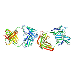 | | CATALYTIC ANTIBODY 4B2 IN COMPLEX WITH ITS AMIDINIUM HAPTEN. | | 分子名称: | 2-(4-AMINOBENZYLAMINO)-3,4,5,6-TETRAHYDROPYRIDINIUM, CATALYTIC ANTIBODY 4B2, SULFATE ION | | 著者 | Golinelli-Pimpaneau, B, Goncalves, O, Dintinger, T, Blanchard, D, Knossow, M, Tellier, C. | | 登録日 | 2000-06-02 | | 公開日 | 2000-09-13 | | 最終更新日 | 2012-07-04 | | 実験手法 | X-RAY DIFFRACTION (1.87 Å) | | 主引用文献 | Structural evidence for a programmed general base in the active site of a catalytic antibody.
Proc.Natl.Acad.Sci.USA, 97, 2000
|
|
3J25
 | | Structural basis for TetM-mediated tetracycline resistance | | 分子名称: | PHOSPHOMETHYLPHOSPHONIC ACID GUANYLATE ESTER, Tetracycline resistance protein tetM | | 著者 | Doenhoefer, A, Franckenberg, S, Wickles, S, Berninghausen, O, Beckmann, R, Wilson, D.N. | | 登録日 | 2012-08-22 | | 公開日 | 2012-10-17 | | 最終更新日 | 2024-02-21 | | 実験手法 | ELECTRON MICROSCOPY (7.2 Å) | | 主引用文献 | Structural basis for TetM-mediated tetracycline resistance.
Proc.Natl.Acad.Sci.USA, 109, 2012
|
|
3KA8
 | | Frog M-ferritin, EQH mutant, with cobalt | | 分子名称: | CHLORIDE ION, COBALT (II) ION, Ferritin, ... | | 著者 | Tosha, T, Ng, H.L, Theil, E, Alber, T, Bhattasali, O. | | 登録日 | 2009-10-19 | | 公開日 | 2010-10-06 | | 最終更新日 | 2023-09-06 | | 実験手法 | X-RAY DIFFRACTION (1.35 Å) | | 主引用文献 | Moving Metal Ions through Ferritin-Protein Nanocages from Three-Fold Pores to Catalytic Sites.
J.Am.Chem.Soc., 132, 2010
|
|
3K6Y
 | | Crystal structure of Rv3671c protease from M. tuberculosis, active form | | 分子名称: | POSSIBLE MEMBRANE-ASSOCIATED SERINE PROTEASE | | 著者 | Biswas, T, Small, J, Vandal, O, Ehrt, S, Tsodikov, O.V. | | 登録日 | 2009-10-10 | | 公開日 | 2010-10-13 | | 最終更新日 | 2023-09-06 | | 実験手法 | X-RAY DIFFRACTION (1.3 Å) | | 主引用文献 | Structural insight into serine protease Rv3671c that Protects M. tuberculosis from oxidative and acidic stress.
Structure, 18, 2010
|
|
3KA3
 | | Frog M-ferritin with magnesium | | 分子名称: | CHLORIDE ION, Ferritin, middle subunit, ... | | 著者 | Tosha, T, Ng, H.L, Theil, E, Alber, T, Bhattasali, O. | | 登録日 | 2009-10-18 | | 公開日 | 2010-10-06 | | 最終更新日 | 2024-02-21 | | 実験手法 | X-RAY DIFFRACTION (1.4 Å) | | 主引用文献 | Moving Metal Ions through Ferritin-Protein Nanocages from Three-Fold Pores to Catalytic Sites.
J.Am.Chem.Soc., 132, 2010
|
|
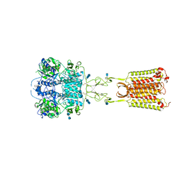
7M3E
 | | Asymmetric Activation of the Calcium Sensing Receptor Homodimer | | 分子名称: | 2-acetamido-2-deoxy-beta-D-glucopyranose, 2-acetamido-2-deoxy-beta-D-glucopyranose-(1-4)-2-acetamido-2-deoxy-beta-D-glucopyranose, 2-chloro-6-[(2R)-2-hydroxy-3-{[2-methyl-1-(naphthalen-2-yl)propan-2-yl]amino}propoxy]benzonitrile, ... | | 著者 | Gao, Y, Robertson, M.J, Zhang, C, Meyerowitz, J.G, Panova, O, Skiniotis, G. | | 登録日 | 2021-03-18 | | 公開日 | 2021-06-30 | | 最終更新日 | 2021-07-21 | | 実験手法 | ELECTRON MICROSCOPY (3.2 Å) | | 主引用文献 | Asymmetric activation of the calcium-sensing receptor homodimer.
Nature, 595, 2021
|
|
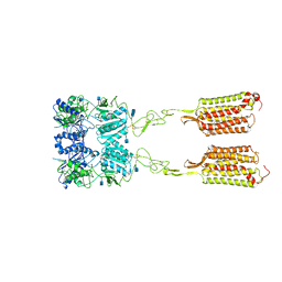
7M3G
 | | Asymmetric Activation of the Calcium Sensing Receptor Homodimer | | 分子名称: | 2-[4-[(3S)-3-[[(1R)-1-naphthalen-1-ylethyl]amino]pyrrolidin-1-yl]phenyl]ethanoic acid, 2-acetamido-2-deoxy-beta-D-glucopyranose, 2-acetamido-2-deoxy-beta-D-glucopyranose-(1-4)-2-acetamido-2-deoxy-beta-D-glucopyranose, ... | | 著者 | Gao, Y, Robertson, M.J, Zhang, C, Meyerowitz, J.G, Panova, O, Skiniotis, G. | | 登録日 | 2021-03-18 | | 公開日 | 2021-06-30 | | 最終更新日 | 2021-07-21 | | 実験手法 | ELECTRON MICROSCOPY (2.5 Å) | | 主引用文献 | Asymmetric activation of the calcium-sensing receptor homodimer.
Nature, 595, 2021
|
|
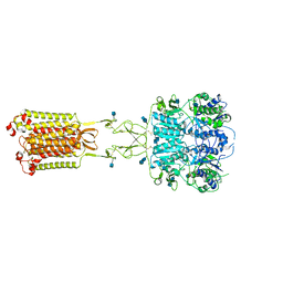
7M3J
 | | Asymmetric Activation of the Calcium Sensing Receptor Homodimer | | 分子名称: | 2-acetamido-2-deoxy-beta-D-glucopyranose, 2-acetamido-2-deoxy-beta-D-glucopyranose-(1-4)-2-acetamido-2-deoxy-beta-D-glucopyranose, 2-chloro-6-[(2R)-2-hydroxy-3-{[2-methyl-1-(naphthalen-2-yl)propan-2-yl]amino}propoxy]benzonitrile, ... | | 著者 | Gao, Y, Robertson, M.J, Zhang, C, Meyerowitz, J.G, Panova, O, Skiniotis, G. | | 登録日 | 2021-03-18 | | 公開日 | 2021-06-30 | | 最終更新日 | 2021-07-21 | | 実験手法 | ELECTRON MICROSCOPY (4.1 Å) | | 主引用文献 | Asymmetric activation of the calcium-sensing receptor homodimer.
Nature, 595, 2021
|
|
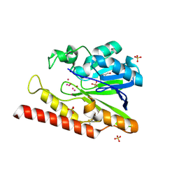
7M3F
 | | Asymmetric Activation of the Calcium Sensing Receptor Homodimer | | 分子名称: | 2-acetamido-2-deoxy-beta-D-glucopyranose, 2-acetamido-2-deoxy-beta-D-glucopyranose-(1-4)-2-acetamido-2-deoxy-beta-D-glucopyranose, CALCIUM ION, ... | | 著者 | Gao, Y, Robertson, M.J, Zhang, C, Meyerowitz, J.G, Panova, O, Skiniotis, G. | | 登録日 | 2021-03-18 | | 公開日 | 2021-06-30 | | 最終更新日 | 2021-07-21 | | 実験手法 | ELECTRON MICROSCOPY (2.8 Å) | | 主引用文献 | Asymmetric activation of the calcium-sensing receptor homodimer.
Nature, 595, 2021
|
|
5WCK
 | | Native FEZ-1 metallo-beta-lactamase from Legionella gormanii | | 分子名称: | FEZ-1 protein, GLYCEROL, SULFATE ION, ... | | 著者 | Garcia-Saez, I, Mercuri, P.S, Kahn, R, Papamicael, C, Shabalin, I.G, Raczynska, J.E, Jaskolski, M, Minor, W, Frere, J.M, Galleni, M, Dideberg, O. | | 登録日 | 2017-06-30 | | 公開日 | 2018-06-20 | | 最終更新日 | 2023-10-04 | | 実験手法 | X-RAY DIFFRACTION (1.65 Å) | | 主引用文献 | Three-dimensional structure of FEZ-1, a monomeric subclass B3 metallo-beta-lactamase from Fluoribacter gormanii, in native form and in complex with D-captopril.
J. Mol. Biol., 325, 2003
|
|
3MM0
 | | Crystal structure of chimeric avidin | | 分子名称: | Avidin, Avidin-related protein 4/5 | | 著者 | Livnah, O, Eisenberg-Domovich, Y, Maatta, J.A.E, Kulomaa, M.S, Hytonen, V.P, Nordlund, H.R. | | 登録日 | 2010-04-19 | | 公開日 | 2010-10-27 | | 最終更新日 | 2023-09-06 | | 実験手法 | X-RAY DIFFRACTION (2.7 Å) | | 主引用文献 | Chimeric avidin shows stability against harsh chemical conditions-biochemical analysis and 3D structure.
Biotechnol.Bioeng., 108, 2011
|
|
3M8O
 | | Human IgA1 Fab fragment | | 分子名称: | CHLORIDE ION, GLYCEROL, IMMUNOGLOBULIN A1 HEAVY CHAIN, ... | | 著者 | Buschiazzo, A, Trajtenberg, F, Correa, A, Oppezzo, P, Pritsch, O, Dighiero, G. | | 登録日 | 2010-03-18 | | 公開日 | 2011-03-30 | | 最終更新日 | 2023-11-01 | | 実験手法 | X-RAY DIFFRACTION (1.55 Å) | | 主引用文献 | Structure of a human IgA1 Fab fragment at 1.55 angstrom resolution: potential effect of the constant domains on antigen-affinity modulation
Acta Crystallogr.,Sect.D, 69, 2013
|
|
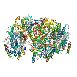
5VFB
 | | 1.36 Angstrom Resolution Crystal Structure of Malate Synthase G from Pseudomonas aeruginosa in Complex with Glycolic Acid. | | 分子名称: | CHLORIDE ION, GLYCOLIC ACID, Malate synthase G, ... | | 著者 | Minasov, G, Shuvalova, L, Dubrovska, I, Kiryukhina, O, Grimshaw, S, Kwon, K, Anderson, W.F, Center for Structural Genomics of Infectious Diseases (CSGID) | | 登録日 | 2017-04-07 | | 公開日 | 2017-04-19 | | 最終更新日 | 2023-11-15 | | 実験手法 | X-RAY DIFFRACTION (1.36 Å) | | 主引用文献 | 1.36 Angstrom Resolution Crystal Structure of Malate Synthase G from Pseudomonas aeruginosa in Complex with Glycolic Acid.
To Be Published
|
|
3KXR
 | | Structure of the cystathionine beta-synthase pair domain of the putative Mg2+ transporter SO5017 from Shewanella oneidensis MR-1. | | 分子名称: | CHLORIDE ION, Magnesium transporter, putative | | 著者 | Fratczak, Z, Zimmerman, M.D, Chruszcz, M, Cymborowski, M, Kagan, O, Savchenko, A, Edwards, A, Joachimiak, A, Minor, W, Midwest Center for Structural Genomics (MCSG) | | 登録日 | 2009-12-03 | | 公開日 | 2009-12-15 | | 最終更新日 | 2022-04-13 | | 実験手法 | X-RAY DIFFRACTION (2.41 Å) | | 主引用文献 | Structure of the cystathionine beta-synthase pair domain of the putative Mg2+ transporter SO5017 from Shewanella oneidensis MR-1.
To be Published
|
|
7MPV
 | | CmcC from Type II Cut MCP | | 分子名称: | BMC domain-containing protein | | 著者 | Ochoa, J.M, Mijares, O, Sawaya, M.R, Yeates, T.O. | | 登録日 | 2021-05-05 | | 公開日 | 2021-09-08 | | 最終更新日 | 2024-04-03 | | 実験手法 | X-RAY DIFFRACTION (2.292 Å) | | 主引用文献 | Structural characterization of hexameric shell proteins from two types of choline-utilization bacterial microcompartments
Acta Crystallogr.,Sect.F, 77, 2021
|
|
7MN4
 | | CmcB E35G mutant from Type II Cut MCP | | 分子名称: | BMC domain-containing protein | | 著者 | Ochoa, J.M, Mijares, O, Sawaya, M.R, Yeates, T.O. | | 登録日 | 2021-04-30 | | 公開日 | 2021-09-08 | | 最終更新日 | 2024-04-03 | | 実験手法 | X-RAY DIFFRACTION (1.8 Å) | | 主引用文献 | Structural characterization of hexameric shell proteins from two types of choline-utilization bacterial microcompartments
Acta Crystallogr.,Sect.F, 77, 2021
|
|
5UU1
 | | Crystal Structure of Human Vaccinia-related kinase 2 (VRK-2) bound to BI-D1870 | | 分子名称: | (7S)-2-[(3,5-difluoro-4-hydroxyphenyl)amino]-5,7-dimethyl-8-(3-methylbutyl)-7,8-dihydropteridin-6(5H)-one, Serine/threonine-protein kinase VRK2 | | 著者 | Counago, R.M, Bountra, C, Arruda, P, Edwards, A.M, Gileadi, O, Structural Genomics Consortium (SGC) | | 登録日 | 2017-02-15 | | 公開日 | 2017-03-01 | | 最終更新日 | 2023-10-04 | | 実験手法 | X-RAY DIFFRACTION (2 Å) | | 主引用文献 | Structural characterization of human Vaccinia-Related Kinases (VRK) bound to small-molecule inhibitors identifies different P-loop conformations.
Sci Rep, 7, 2017
|
|
3LSI
 | | Pyranose 2-oxidase T169A, tetragonal | | 分子名称: | FLAVIN-ADENINE DINUCLEOTIDE, Pyranose 2-oxidase | | 著者 | Tan, T.C, Spadiut, O, Divne, C. | | 登録日 | 2010-02-12 | | 公開日 | 2010-08-25 | | 最終更新日 | 2024-02-21 | | 実験手法 | X-RAY DIFFRACTION (1.9 Å) | | 主引用文献 | H-bonding and positive charge at the N5/O4 locus are critical for covalent flavin attachment in trametes pyranose 2-oxidase.
J.Mol.Biol., 402, 2010
|
|
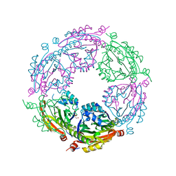
3LQK
 | | Crystal structure of dipicolinate synthase subunit B from Bacillus halodurans C | | 分子名称: | Dipicolinate synthase subunit B, PHOSPHATE ION | | 著者 | Nocek, B, Kagan, O, Savchenko, A, Edwards, A, Joachimiak, A, Midwest Center for Structural Genomics (MCSG) | | 登録日 | 2010-02-09 | | 公開日 | 2010-03-23 | | 最終更新日 | 2017-11-01 | | 実験手法 | X-RAY DIFFRACTION (2.1 Å) | | 主引用文献 | Crystal structure of dipicolinate synthase subunit B from Bacillus halodurans C
To be Published
|
|


