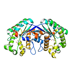
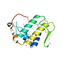
4KEG
 
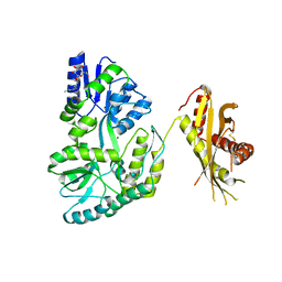 | | Crystal Structure of MBP Fused Human SPLUNC1 | | 分子名称: | MAGNESIUM ION, Maltose-binding periplasmic/Palate lung and nasal epithelium clone fusion protein, octyl beta-D-glucopyranoside | | 著者 | Ning, F, Wang, C, Niu, L, Chu, H.W, Zhang, G. | | 登録日 | 2013-04-25 | | 公開日 | 2014-04-30 | | 最終更新日 | 2020-07-29 | | 実験手法 | X-RAY DIFFRACTION (2.5 Å) | | 主引用文献 | The Lipid Ligands of the SPLUNC1 Protein
To be Published
|
|
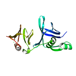
4HG9
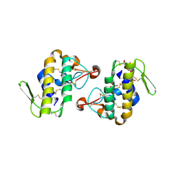 | | Crystal structure of AhV_bPA, a basic PLA2 from Agkistrodon halys pallas venom | | 分子名称: | Basic phospholipase A2 B, CALCIUM ION, CITRIC ACID, ... | | 著者 | Zeng, F, Niu, L, Li, X, Teng, M. | | 登録日 | 2012-10-07 | | 公開日 | 2012-10-17 | | 最終更新日 | 2023-09-20 | | 実験手法 | X-RAY DIFFRACTION (1.6 Å) | | 主引用文献 | Crystal structure of AhV_bPA, a basic PLA2 from Agkistrodon halys pallas venom
TO BE PUBLISHED
|
|
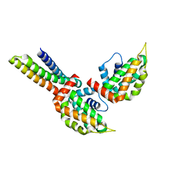
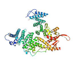
4FMM
 | | Dimeric Sec14 family homolog 3 from Saccharomyces cerevisiae presents some novel features of structure that lead to a surprising "dimer-monomer" state change induced by substrate binding | | 分子名称: | GLYCEROL, MAGNESIUM ION, Phosphatidylinositol transfer protein PDR16 | | 著者 | Yuan, Y, Zhao, W, Wang, X, Gao, Y, Niu, L, Teng, M. | | 登録日 | 2012-06-18 | | 公開日 | 2013-02-27 | | 最終更新日 | 2024-02-28 | | 実験手法 | X-RAY DIFFRACTION (2.34 Å) | | 主引用文献 | Dimeric Sfh3 has structural changes in its binding pocket that are associated with a dimer-monomer state transformation induced by substrate binding.
Acta Crystallogr.,Sect.D, 69, 2013
|
|
3LKX
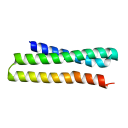
 | | Human nac dimerization domain | | 分子名称: | Nascent polypeptide-associated complex subunit alpha, Transcription factor BTF3 | | 著者 | Liu, Y, Hu, Y, Li, X, Niu, L, Teng, M. | | 登録日 | 2010-01-28 | | 公開日 | 2010-03-23 | | 最終更新日 | 2023-11-01 | | 実験手法 | X-RAY DIFFRACTION (2.5 Å) | | 主引用文献 | The crystal structure of the human nascent polypeptide-associated complex domain reveals a nucleic acid-binding region on the NACA subunit
Biochemistry, 49, 2010
|
|
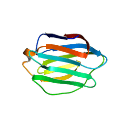
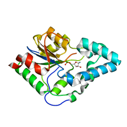
3MD1
 | | Crystal Structure of the Second RRM Domain of Yeast Poly(U)-Binding Protein (Pub1) | | 分子名称: | GLYCEROL, Nuclear and cytoplasmic polyadenylated RNA-binding protein PUB1 | | 著者 | Li, H, Shi, H, Li, Y, Cui, Y, Niu, L, Teng, M. | | 登録日 | 2010-03-29 | | 公開日 | 2010-05-05 | | 最終更新日 | 2023-11-01 | | 実験手法 | X-RAY DIFFRACTION (1.6 Å) | | 主引用文献 | Crystal Structure of the Second RRM Domain of Yeast Poly(U)-Binding Protein (Pub1)
To be published
|
|
3OJB
 | |
3ONL
 | | yeast Ent3_ENTH-Vti1p_Habc complex structure | | 分子名称: | Epsin-3, t-SNARE VTI1 | | 著者 | Wang, J, Fang, P, Niu, L, Teng, M. | | 登録日 | 2010-08-29 | | 公開日 | 2011-07-20 | | 最終更新日 | 2023-11-01 | | 実験手法 | X-RAY DIFFRACTION (2.2 Å) | | 主引用文献 | Epsin N-terminal homology domains bind on opposite sides of two SNAREs
Proc.Natl.Acad.Sci.USA, 108, 2011
|
|
3MIL
 | | Crystal structure of isoamyl acetate-hydrolyzing esterase from Saccharomyces cerevisiae | | 分子名称: | GLYCEROL, Isoamyl acetate-hydrolyzing esterase | | 著者 | Ma, J, Lu, Q, Yuan, Y, Li, K, Ge, H, Go, Y, Niu, L, Teng, M. | | 登録日 | 2010-04-11 | | 公開日 | 2010-11-24 | | 最終更新日 | 2024-03-20 | | 実験手法 | X-RAY DIFFRACTION (1.6 Å) | | 主引用文献 | Crystal structure of isoamyl acetate-hydrolyzing esterase from Saccharomyces cerevisiae reveals a novel active site architecture and the basis of substrate specificity
Proteins, 79, 2011
|
|
3ONJ
 | |
3OPX
 | |
3ONK
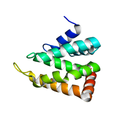 | | yeast Ent3_ENTH domain | | 分子名称: | Epsin-3 | | 著者 | Wang, J, Fang, P, Niu, L, Teng, M. | | 登録日 | 2010-08-29 | | 公開日 | 2011-07-20 | | 最終更新日 | 2023-11-01 | | 実験手法 | X-RAY DIFFRACTION (2.09 Å) | | 主引用文献 | Epsin N-terminal homology domains bind on opposite sides of two SNAREs
Proc.Natl.Acad.Sci.USA, 108, 2011
|
|
4H0S
 | |
4IFS
 | | Crystal structure of the hSSRP1 Middle domain | | 分子名称: | CHLORIDE ION, FACT complex subunit SSRP1 | | 著者 | Zhang, W.J, Zeng, F.X, Shao, C, Liu, Y.W, Niu, L.W, Li, X, Teng, M.K. | | 登録日 | 2012-12-15 | | 公開日 | 2014-01-29 | | 最終更新日 | 2023-11-08 | | 実験手法 | X-RAY DIFFRACTION (1.93 Å) | | 主引用文献 | Crystal structure of the hSSRP1 Middle domain
To be Published
|
|
7EWF
 | |
7EWM
 | |
7YDS
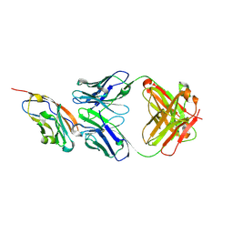 | | The structure of the bispecific antibody targeted PD-L1 and 4-1BB | | 分子名称: | Anti-PDL1-VH-CH1, Anti-PDL1-VL-CL, Programmed cell death 1 ligand 1 | | 著者 | Gao, Y, Zhu, M, Liu, W.T, Cheng, L.S, Zhu, Z.L, Niu, L.W. | | 登録日 | 2022-07-04 | | 公開日 | 2023-07-19 | | 最終更新日 | 2023-11-29 | | 実験手法 | X-RAY DIFFRACTION (2.3 Å) | | 主引用文献 | A bispecific antibody targeted PD-L1 and 4-1BB induces a potent antitumor immune activity in colorectal cancer without systemic toxicity
To Be Published
|
|
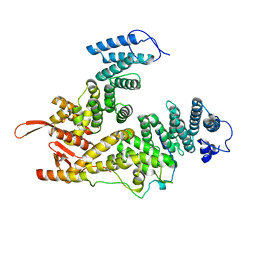
4IQ8
 | | Crystal structure of glyceraldehyde-3-phosphate dehydrogenase 3 from Saccharomyces cerevisiae | | 分子名称: | Glyceraldehyde-3-phosphate dehydrogenase 3 | | 著者 | Wang, H, Liu, Q, Niu, L, Teng, M, Li, X. | | 登録日 | 2013-01-11 | | 公開日 | 2013-02-06 | | 最終更新日 | 2023-09-20 | | 実験手法 | X-RAY DIFFRACTION (2.49 Å) | | 主引用文献 | Preliminary crystallographic analysis of glyceraldehyde-3-phosphate dehydrogenase 3 from Saccharomyces cerevisiae.
Acta Crystallogr.,Sect.F, 68, 2012
|
|


