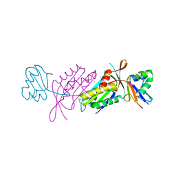
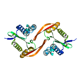
1V02
 
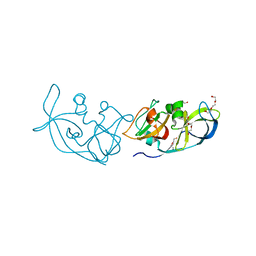
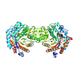 | | Crystal structure of the Sorghum bicolor dhurrinase 1 | | 分子名称: | DHURRINASE | | 著者 | Moriniere, J, Verdoucq, L, Bevan, D.R, Esen, A, Henrissat, B, Czjzek, M. | | 登録日 | 2004-03-22 | | 公開日 | 2004-05-20 | | 最終更新日 | 2023-12-13 | | 実験手法 | X-RAY DIFFRACTION (1.8 Å) | | 主引用文献 | Structural Determinants of Substrate Specificity in Family 1 Beta-Glucosidases: Novel Insights from the Crystal Structure of Sorghum Dhurrinase-1, a Plant Beta-Glucosidase with Strict Specificity, in Complex with its Natural Substrate
J.Biol.Chem., 279, 2004
|
|
4L8H
 | |
3N6W
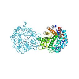
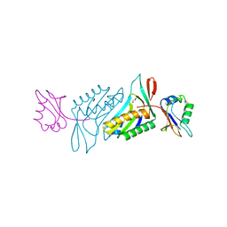
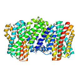
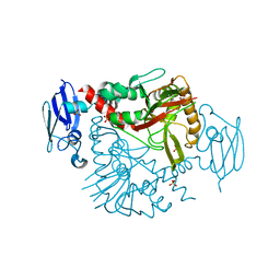 | | Crystal structure of human gamma-butyrobetaine hydroxylase | | 分子名称: | Gamma-butyrobetaine dioxygenase, SULFATE ION, ZINC ION | | 著者 | Rumnieks, J, Zeltins, A, Leonchiks, A, Kazaks, A, Kotelovica, S, Tars, K. | | 登録日 | 2010-05-26 | | 公開日 | 2010-07-21 | | 最終更新日 | 2024-02-21 | | 実験手法 | X-RAY DIFFRACTION (2 Å) | | 主引用文献 | Crystal structure of human gamma-butyrobetaine hydroxylase.
Biochem.Biophys.Res.Commun., 398, 2010
|
|
1KNO
 | |
6TKT
 | |
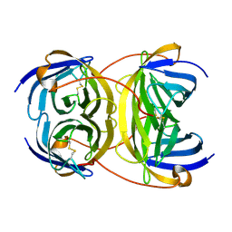
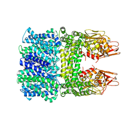
1JBO
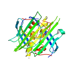
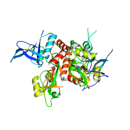
 | | The 1.45A Three-Dimensional Structure of c-Phycocyanin from the Thermophylic Cyanobacterium Synechococcus elongatus | | 分子名称: | C-Phycocyanin alpha chain, C-Phycocyanin beta chain, PHYCOCYANOBILIN | | 著者 | Nield, J, Rizkallah, P.J, Barber, J, Chayen, N.E. | | 登録日 | 2002-05-02 | | 公開日 | 2003-03-18 | | 最終更新日 | 2023-08-16 | | 実験手法 | X-RAY DIFFRACTION (1.45 Å) | | 主引用文献 | The 1.45A three-dimensional structure of C-phycocyanin from the
thermophilic cyanobacterium Synechococcus elongatus
J.STRUCT.BIOL., 141, 2003
|
|
1BQ7
 | |
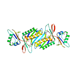
1V08
 | | Crystal structure of the Zea maze beta-glucosidase-1 in complex with gluco-tetrazole | | 分子名称: | BETA-GLUCOSIDASE, NOJIRIMYCINE TETRAZOLE | | 著者 | Moriniere, J, Verdoucq, L, Bevan, D.R, Esen, A, Henrissat, B, Czjzek, M. | | 登録日 | 2004-03-25 | | 公開日 | 2004-05-20 | | 最終更新日 | 2023-12-13 | | 実験手法 | X-RAY DIFFRACTION (1.9 Å) | | 主引用文献 | Structural Determinants of Substrate Specificity in Family 1 Beta-Glucosidases: Novel Insights from the Crystal Structure of Sorghum Dhurrinase-1, a Plant Beta-Glucosidase with Strict Specificity, in Complex with its Natural Substrate
J.Biol.Chem., 279, 2004
|
|
3BII
 | | Crystal Structure of Activated MPT Synthase | | 分子名称: | CHLORIDE ION, Molybdopterin-converting factor subunit 1, Molybdopterin-converting factor subunit 2 | | 著者 | Daniels, J.N, Schindelin, H. | | 登録日 | 2007-11-30 | | 公開日 | 2008-02-19 | | 最終更新日 | 2017-10-25 | | 実験手法 | X-RAY DIFFRACTION (2.5 Å) | | 主引用文献 | Crystal structure of a molybdopterin synthase-precursor Z complex: insight into its sulfur transfer mechanism and its role in molybdenum cofactor deficiency.
Biochemistry, 47, 2008
|
|
1V03
 | | Crystal structure of the Sorghum bicolor dhurrinase 1 | | 分子名称: | ACETONITRILE, DHURRINASE, PHENOL, ... | | 著者 | Moriniere, J, Verdoucq, L, Bevan, D.R, Esen, A, Henrissat, B, Czjzek, M. | | 登録日 | 2004-03-22 | | 公開日 | 2004-05-20 | | 最終更新日 | 2023-12-13 | | 実験手法 | X-RAY DIFFRACTION (2 Å) | | 主引用文献 | Structural Determinants of Substrate Specificity in Family 1 Beta-Glucosidases: Novel Insights from the Crystal Structure of Sorghum Dhurrinase-1, a Plant Beta-Glucosidase with Strict Specificity, in Complex with its Natural Substrate
J.Biol.Chem., 279, 2004
|
|
2Y32
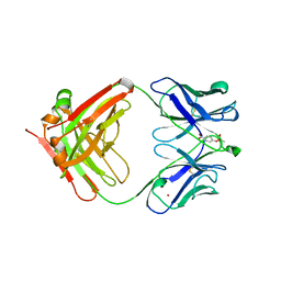
 | | Crystal structure of bradavidin | | 分子名称: | BLR5658 PROTEIN | | 著者 | Leppiniemi, J, Gronroos, T, Johnson, M.S, Kulomaa, M.S, Hytonen, V.P, Airenne, T.T. | | 登録日 | 2010-12-17 | | 公開日 | 2011-12-28 | | 最終更新日 | 2023-12-20 | | 実験手法 | X-RAY DIFFRACTION (1.78 Å) | | 主引用文献 | Structure of Bradavidin - C-Terminal Residues Act as Intrinsic Ligands.
Plos One, 7, 2012
|
|
2QIE
 | | Staphylococcus aureus molybdopterin synthase in complex with precursor Z | | 分子名称: | (2R,4AR,5AR,11AR,12AS)-8-AMINO-2-HYDROXY-4A,5A,9,11,11A,12A-HEXAHYDRO[1,3,2]DIOXAPHOSPHININO[4',5':5,6]PYRANO[3,2-G]PTERIDINE-10,12(4H,6H)-DIONE 2-OXIDE, Molybdopterin synthase small subunit, Molybdopterin-converting factor subunit 2 | | 著者 | Daniels, J.N, Schindelin, H. | | 登録日 | 2007-07-04 | | 公開日 | 2008-02-19 | | 最終更新日 | 2023-08-30 | | 実験手法 | X-RAY DIFFRACTION (2.5 Å) | | 主引用文献 | Crystal structure of a molybdopterin synthase-precursor Z complex: insight into its sulfur transfer mechanism and its role in molybdenum cofactor deficiency.
Biochemistry, 47, 2008
|
|
2WCN
 | |
3RLK
 | |
2CHK
 | |
2CHJ
 | |
3RLC
 | |
2Q5W
 | |
1OKF
 | |
4NF4
 | | Crystal structure of GluN1/GluN2A ligand-binding domain in complex with DCKA and glutamate | | 分子名称: | 4-hydroxy-5,7-dimethylquinoline-2-carboxylic acid, GLUTAMIC ACID, Glutamate receptor ionotropic, ... | | 著者 | Annie, J, Tajima, N, Furukawa, H. | | 登録日 | 2013-10-30 | | 公開日 | 2014-03-12 | | 最終更新日 | 2017-08-09 | | 実験手法 | X-RAY DIFFRACTION (2 Å) | | 主引用文献 | Structural Insights into Competitive Antagonism in NMDA Receptors.
Neuron, 81, 2014
|
|
8IQ6
 | | Cryo-EM structure of Latanoprost-bound prostaglandin-F2-alpha receptor-miniGq-Nb35 complex | | 分子名称: | Guanine nucleotide-binding protein G(I)/G(S)/G(O) subunit gamma-2, Guanine nucleotide-binding protein G(I)/G(S)/G(T) subunit beta-1, Guanine nucleotide-binding protein G(s) subunit alpha isoforms short, ... | | 著者 | Lv, X, Gao, K, Nie, J, Zhang, X, Zhang, S, Ren, Y, Li, Q, Huang, J, Liu, L, Zhang, X, Sun, X, Zhang, W, Liu, X. | | 登録日 | 2023-03-15 | | 公開日 | 2024-01-31 | | 実験手法 | ELECTRON MICROSCOPY (3.4 Å) | | 主引用文献 | Structures of human prostaglandin F 2 alpha receptor reveal the mechanism of ligand and G protein selectivity.
Nat Commun, 14, 2023
|
|
8IQ4
 | | Cryo-EM structure of Carboprost-bound prostaglandin-F2-alpha receptor-miniGq-Nb35 complex | | 分子名称: | Guanine nucleotide-binding protein G(I)/G(S)/G(O) subunit gamma-2, Guanine nucleotide-binding protein G(I)/G(S)/G(T) subunit beta-1, Guanine nucleotide-binding protein G(s) subunit alpha isoforms short, ... | | 著者 | Lv, X, Gao, K, Nie, J, Zhang, X, Zhang, S, Ren, Y, Li, Q, Huang, J, Liu, L, Zhang, X, Sun, X, Zhang, W, Liu, X. | | 登録日 | 2023-03-15 | | 公開日 | 2024-01-31 | | 実験手法 | ELECTRON MICROSCOPY (2.7 Å) | | 主引用文献 | Structures of human prostaglandin F 2 alpha receptor reveal the mechanism of ligand and G protein selectivity.
Nat Commun, 14, 2023
|
|
4E2A
 | |
8J2M
 | | The truncated rice Na+/H+ antiporter SOS1 (1-976) in a constitutively active state | | 分子名称: | (2R)-3-{[(S)-(2-aminoethoxy)(hydroxy)phosphoryl]oxy}-2-(tetradecanoyloxy)propyl tetradecanoate, Na+/H+ antiporter | | 著者 | Zhang, X.Y, Tang, L.H, Zhang, C.R, Nie, J.W. | | 登録日 | 2023-04-14 | | 公開日 | 2023-11-22 | | 最終更新日 | 2023-11-29 | | 実験手法 | ELECTRON MICROSCOPY (3.4 Å) | | 主引用文献 | Structure and activation mechanism of the rice Salt Overly Sensitive 1 (SOS1) Na + /H + antiporter.
Nat.Plants, 9, 2023
|
|
8IWO
 | | The rice Na+/H+ antiporter SOS1 in an auto-inhibited state | | 分子名称: | (2R)-3-{[(S)-(2-aminoethoxy)(hydroxy)phosphoryl]oxy}-2-(tetradecanoyloxy)propyl tetradecanoate, OsSOS1 | | 著者 | Zhang, X.Y, Tang, L.H, Zhang, C.R, Nie, J.W. | | 登録日 | 2023-03-30 | | 公開日 | 2023-11-22 | | 最終更新日 | 2023-11-29 | | 実験手法 | ELECTRON MICROSCOPY (3.09 Å) | | 主引用文献 | Structure and activation mechanism of the rice Salt Overly Sensitive 1 (SOS1) Na + /H + antiporter.
Nat.Plants, 9, 2023
|
|


