Loading
PDBj
MenuPDBj@FacebookPDBj@TwitterPDBj@YouTubewwPDB FoundationwwPDB
RCSB PDBPDBeBMRBAdv. SearchSearch help
PDB: 757 results

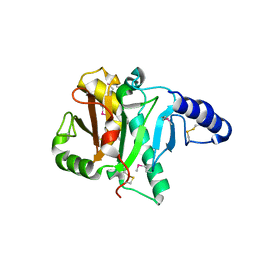
3K85
DownloadVisualize
BU of 3k85 by Molmil
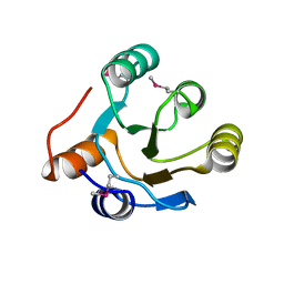
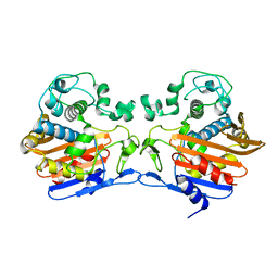
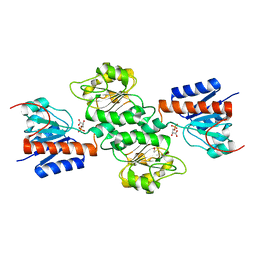
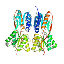
Crystal structure of a D-glycero-D-manno-heptose 1-phosphate kinase from Bacteriodes thetaiotaomicron
Descriptor: D-glycero-D-manno-heptose 1-phosphate kinase
Authors:Palani, K, Kumaran, D, Burley, S.K, Swaminathan, S, New York SGX Research Center for Structural Genomics (NYSGXRC)
Deposit date:2009-10-13
Release date:2009-12-08
Last modified:2021-02-10
Method:X-RAY DIFFRACTION (2.28 Å)
Cite:Crystal structure of a D-glycero-D-manno-heptose 1-phosphate kinase from Bacteriodes thetaiotaomicron
To be Published
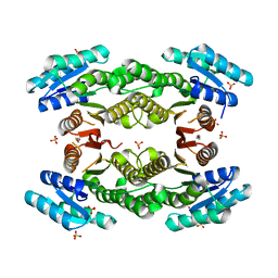
3K9E
DownloadVisualize
BU of 3k9e by Molmil
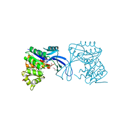
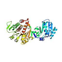
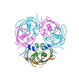
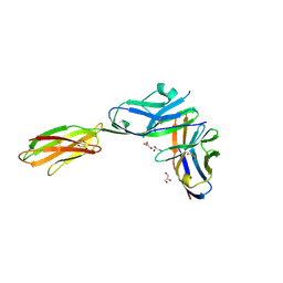
Crystal structure of a putative Ribokinase II (Apo Form) from E.coli
Descriptor: PUTATIVE RIBOKINASE II
Authors:Satyanarayana, L, Burley, S.K, Swaminathan, S, New York SGX Research Center for Structural Genomics (NYSGXRC)
Deposit date:2009-10-15
Release date:2009-11-10
Last modified:2023-11-22
Method:X-RAY DIFFRACTION (2.05 Å)
Cite:Crystal structure of a putative Ribokinase II (Apo Form) from E.coli
To be Published
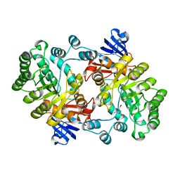
1I4P
DownloadVisualize
BU of 1i4p by Molmil
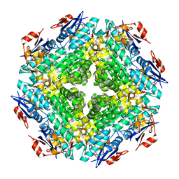
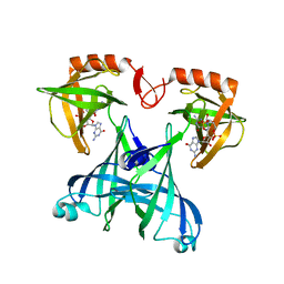
CRYSTAL STRUCTURE OF STAPHYLOCOCCAL ENTEROTOXIN C2 AT 100K CRYSTALLIZED AT PH 5.5
Descriptor: ENTEROTOXIN TYPE C-2, ZINC ION
Authors:Kumaran, D, Swaminathan, S.
Deposit date:2001-02-22
Release date:2001-03-14
Last modified:2023-08-09
Method:X-RAY DIFFRACTION (2 Å)
Cite:Structure of staphylococcal enterotoxin C2 at various pH levels.
Acta Crystallogr.,Sect.D, 57, 2001
2POF
DownloadVisualize
BU of 2pof by Molmil
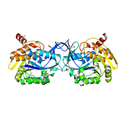
Crystal structure of CDP-diacylglycerol pyrophosphatase
Descriptor: CDP-diacylglycerol pyrophosphatase
Authors:Madegowda, M, Eswaramoorthy, S, Burley, S.K, Swaminathan, S, New York SGX Research Center for Structural Genomics (NYSGXRC)
Deposit date:2007-04-26
Release date:2007-05-15
Last modified:2021-02-03
Method:X-RAY DIFFRACTION (1.4 Å)
Cite:Crystal structure of CDP-diacylglycerol pyrophosphatase.
To be Published
3TFO
DownloadVisualize
BU of 3tfo by Molmil
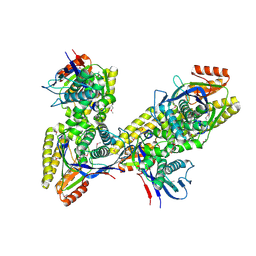
Crystal structure of a putative 3-oxoacyl-(acyl-carrier-protein) reductase from Sinorhizobium meliloti
Descriptor: HEXANE-1,6-DIOL, SULFATE ION, putative 3-oxoacyl-(acyl-carrier-protein) reductase
Authors:Agarwal, R, Chamala, S, Evans, B, Foti, R, Gizzi, A, Hillerich, B, Kar, A, LaFleur, J, Seidel, R, Villigas, G, Zencheck, W, Almo, S.C, Swaminathan, S, New York Structural Genomics Research Consortium (NYSGRC)
Deposit date:2011-08-16
Release date:2011-08-31
Last modified:2024-02-28
Method:X-RAY DIFFRACTION (2.08 Å)
Cite:Crystal structure of a putative 3-oxoacyl-(acyl-carrier-protein) reductase from Sinorhizobium meliloti
To be Published
2PUZ
DownloadVisualize
BU of 2puz by Molmil
Crystal structure of Imidazolonepropionase from Agrobacterium tumefaciens with bound product N-formimino-L-Glutamate
Descriptor: CHLORIDE ION, FE (III) ION, Imidazolonepropionase, ...
Authors:Tyagi, R, Eswaramoorthy, S, Burley, S.K, Swaminathan, S, New York SGX Research Center for Structural Genomics (NYSGXRC)
Deposit date:2007-05-09
Release date:2007-05-22
Last modified:2023-08-30
Method:X-RAY DIFFRACTION (1.83 Å)
Cite:X-ray structure of imidazolonepropionase from Agrobacterium tumefaciens at 1.87 A resolution.
Proteins, 69, 2007
2IOJ
DownloadVisualize
BU of 2ioj by Molmil
Crystal structure of protein AF1212 from Archaeoglobus fulgidus, Pfam DRTGG
Descriptor: Hypothetical protein AF_1212
Authors:Tyagi, R, Burley, S.K, Swaminathan, S, New York SGX Research Center for Structural Genomics (NYSGXRC)
Deposit date:2006-10-10
Release date:2006-10-31
Last modified:2021-02-03
Method:X-RAY DIFFRACTION (2.15 Å)
Cite:The crystal structure of a hypothetical protein (Y1212_ARCFU) from Archeoglobus fulgidus
To be Published
3JUL
DownloadVisualize
BU of 3jul by Molmil
Crystal structure of Listeria innocua D-Tagatose-6-Phosphate Kinase bound with substrate
Descriptor: 6-O-phosphono-beta-D-tagatofuranose, Lin2199 protein, MAGNESIUM ION
Authors:Satyanarayana, L, Burley, S.K, Swaminathan, S, New York SGX Research Center for Structural Genomics (NYSGXRC)
Deposit date:2009-09-15
Release date:2009-10-06
Last modified:2023-11-22
Method:X-RAY DIFFRACTION (2.4 Å)
Cite:Crystal structure of Listeria innocua D-Tagatose-6-Phosphate Kinase bound with substrate
To be Published
3K17
DownloadVisualize
BU of 3k17 by Molmil
Crystal structure of a Lin0012 protein from Listeria innocua
Descriptor: Lin0012 protein, TRIETHYLENE GLYCOL
Authors:Palani, K, Burley, S.K, Swaminathan, S, New York SGX Research Center for Structural Genomics (NYSGXRC)
Deposit date:2009-09-25
Release date:2009-10-06
Last modified:2021-02-10
Method:X-RAY DIFFRACTION (2.1 Å)
Cite:Crystal structure of a Lin0012 protein from Listeria innocua
To be Published
4FHQ
DownloadVisualize
BU of 4fhq by Molmil
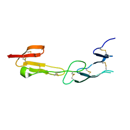
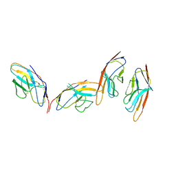
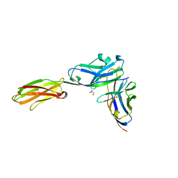
Crystal Structure of HVEM
Descriptor: Tumor necrosis factor receptor superfamily member 14
Authors:Liu, W, Zhan, C, Patskovsky, Y, Bhosle, R.C, Nathenson, S.G, Almo, S.C, Atoms-to-Animals: The Immune Function Network (IFN), New York Structural Genomics Research Consortium (NYSGRC)
Deposit date:2012-06-06
Release date:2012-07-18
Last modified:2023-09-13
Method:X-RAY DIFFRACTION (2.251 Å)
Cite:Increased Heterologous Protein Expression in Drosophila S2 Cells for Massive Production of Immune Ligands/Receptors and Structural Analysis of Human HVEM.
Mol Biotechnol, 57, 2015
3TG9
DownloadVisualize
BU of 3tg9 by Molmil
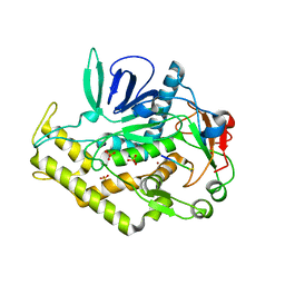
The crystal structure of penicillin binding protein from Bacillus halodurans
Descriptor: Penicillin-binding protein
Authors:Zhang, Z, Satyanarayana, L, Chamala, S, Evans, B, Foti, R, Gizzi, A, Hillerich, B, Kar, A, LaFleur, J, Seidel, R, Villigas, G, Zencheck, W, Almo, S.C, Swaminathan, S, New York Structural Genomics Research Consortium (NYSGRC)
Deposit date:2011-08-17
Release date:2011-08-31
Last modified:2023-09-13
Method:X-RAY DIFFRACTION (2.2 Å)
Cite:The crystal structure of penicillin binding protein from Bacillus halodurans
To be Published
3K5W
DownloadVisualize
BU of 3k5w by Molmil
Crystal structure of a Carbohydrate kinase (YjeF family)from Helicobacter pylori
Descriptor: Carbohydrate kinase, PHOSPHATE ION
Authors:Satyanarayana, L, Burley, S.K, Swaminathan, S, New York SGX Research Center for Structural Genomics (NYSGXRC)
Deposit date:2009-10-08
Release date:2009-12-08
Last modified:2021-02-10
Method:X-RAY DIFFRACTION (2.6 Å)
Cite:Crystal structure of a Carbohydrate kinase (YjeF family)from Helicobacter pylori
To be Published
2QDE
DownloadVisualize
BU of 2qde by Molmil
Crystal structure of mandelate racemase/muconate lactonizing family protein from Azoarcus sp. EbN1
Descriptor: BARIUM ION, Mandelate racemase/muconate lactonizing enzyme family protein
Authors:Agarwal, R, Burley, S.K, Swaminathan, S, New York SGX Research Center for Structural Genomics (NYSGXRC)
Deposit date:2007-06-20
Release date:2007-07-03
Last modified:2024-02-21
Method:X-RAY DIFFRACTION (1.93 Å)
Cite:Crystal structure of mandelate racemase/muconate lactonizing enzyme family protein from Azoarcus sp. EbN1.
To be Published
3D5L
DownloadVisualize
BU of 3d5l by Molmil
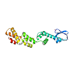
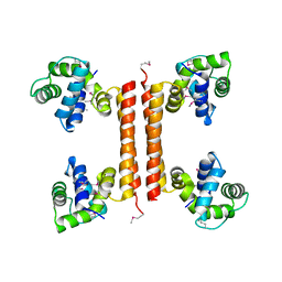
Crystal structure of regulatory protein RecX
Descriptor: Regulatory protein RecX, SULFATE ION
Authors:Agarwal, R, Burley, S.K, Swaminathan, S, New York SGX Research Center for Structural Genomics (NYSGXRC)
Deposit date:2008-05-16
Release date:2008-06-10
Last modified:2024-02-21
Method:X-RAY DIFFRACTION (2.35 Å)
Cite:Crystal structure of DNA repair regulatory protein RecX.
To be Published
4NJM
DownloadVisualize
BU of 4njm by Molmil
Crystal Structure of phosphoglycerate bound 3-phosphoglycerate dehydrogenase in Entamoeba histolytica
Descriptor: 3-PHOSPHOGLYCERIC ACID, D-3-phosphoglycerate dehydrogenase, putative
Authors:Singh, R.K, Gourinath, S.
Deposit date:2013-11-11
Release date:2014-10-08
Last modified:2023-11-08
Method:X-RAY DIFFRACTION (1.79 Å)
Cite:Crystal structures and kinetics of Type III 3-phosphoglycerate dehydrogenase reveal catalysis by lysine.
Febs J., 281, 2014
5BUW
DownloadVisualize
BU of 5buw by Molmil
Crystal Structure of beta-hydroxyacyl-acyl carrier protein dehydratase (FabZ) from Yersinia Pestis
Descriptor: 1,2-ETHANEDIOL, 3-hydroxyacyl-[acyl-carrier-protein] dehydratase FabZ, THIOCYANATE ION
Authors:Mcgillick, B, Kumaran, D, Swaminathan, S.
Deposit date:2015-06-04
Release date:2016-06-15
Last modified:2023-09-27
Method:X-RAY DIFFRACTION (1.8 Å)
Cite:Crystal Structure of beta-hydroxyacyl-acyl carrier protein dehydratase (FabZ) from Yersinia Pestis
To be published
1I4R
DownloadVisualize
BU of 1i4r by Molmil
CRYSTAL STRUCTURE OF STAPHYLOCOCCAL ENTEROTOXIN C2 AT 100K CRYSTALLIZED AT PH 6.5
Descriptor: ENTEROTOXIN TYPE C-2, ZINC ION
Authors:Kumaran, D, Swaminathan, S.
Deposit date:2001-02-22
Release date:2001-03-14
Last modified:2023-08-09
Method:X-RAY DIFFRACTION (2.1 Å)
Cite:Structure of staphylococcal enterotoxin C2 at various pH levels.
Acta Crystallogr.,Sect.D, 57, 2001
1I85
DownloadVisualize
BU of 1i85 by Molmil
CRYSTAL STRUCTURE OF THE CTLA-4/B7-2 COMPLEX
Descriptor: CYTOTOXIC T-LYMPHOCYTE-ASSOCIATED PROTEIN 4, T LYMPHOCYTE ACTIVATION ANTIGEN CD86
Authors:Schwartz, J.-C.D, Zhang, X, Fedorov, A.A, Nathenson, S.G, Almo, S.C.
Deposit date:2001-03-12
Release date:2001-04-04
Last modified:2011-07-13
Method:X-RAY DIFFRACTION (3.2 Å)
Cite:Structural basis for co-stimulation by the human CTLA-4/B7-2 complex.
Nature, 410, 2001
3C4R
DownloadVisualize
BU of 3c4r by Molmil
Crystal structure of an uncharacterized protein encoded by cryptic prophage
Descriptor: Uncharacterized protein
Authors:Sugadev, R, Burley, S.K, Swaminathan, S, Ozyurt, S, Luz, J, New York SGX Research Center for Structural Genomics (NYSGXRC)
Deposit date:2008-01-30
Release date:2008-02-19
Last modified:2021-02-03
Method:X-RAY DIFFRACTION (2.3 Å)
Cite:Crystal structure of an uncharacterized protein encoded by cryptic prophage.
To be Published
3URH
DownloadVisualize
BU of 3urh by Molmil
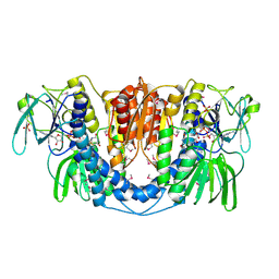
Crystal structure of a dihydrolipoamide dehydrogenase from Sinorhizobium meliloti 1021
Descriptor: 1,2-ETHANEDIOL, Dihydrolipoyl dehydrogenase, FLAVIN-ADENINE DINUCLEOTIDE, ...
Authors:Kumaran, D, Chamala, S, Evans, B, Foti, R, Gizzi, A, Hillerich, B, Kar, A, LaFleur, J, Seidel, R, Villigas, G, Zencheck, W, Almo, S.C, Swaminathan, S, New York Structural Genomics Research Consortium (NYSGRC)
Deposit date:2011-11-22
Release date:2011-12-14
Method:X-RAY DIFFRACTION (1.9 Å)
Cite:Crystal structure of a dihydrolipoamide dehydrogenase from Sinorhizobium meliloti 1021
TO BE PUBLISHED
3BP5
DownloadVisualize
BU of 3bp5 by Molmil
Crystal structure of the mouse PD-1 and PD-L2 complex
Descriptor: GLYCEROL, Programmed cell death 1 ligand 2, Programmed cell death protein 1
Authors:Yan, Q, Lazar-Molnar, E, Cao, E, Ramagopal, U.A, Toro, R, Nathenson, S.G, Almo, S.C.
Deposit date:2007-12-18
Release date:2008-07-15
Last modified:2023-08-30
Method:X-RAY DIFFRACTION (1.8 Å)
Cite:Crystal structure of the complex between programmed death-1 (PD-1) and its ligand PD-L2.
Proc.Natl.Acad.Sci.Usa, 105, 2008
3C8A
DownloadVisualize
BU of 3c8a by Molmil
Crystal structure of the catalytic domain of botulinum neurotoxin serotype A with inhibitory peptide RRGL
Descriptor: Botulinum neurotoxin A light chain, Inhibitor peptide RRGL, SULFATE ION, ...
Authors:Kumaran, D, Swaminathan, S.
Deposit date:2008-02-11
Release date:2008-04-22
Last modified:2011-07-13
Method:X-RAY DIFFRACTION (1.52 Å)
Cite:Structure- and Substrate-based Inhibitor Design for Clostridium botulinum Neurotoxin Serotype A
J.Biol.Chem., 283, 2008
3K9C
DownloadVisualize
BU of 3k9c by Molmil
Crystal structure of LacI Transcriptional regulator from Rhodococcus species.
Descriptor: GLYCEROL, Transcriptional regulator, LacI family protein
Authors:Damodharan, L, Burley, S.K, Swaminathan, S, New York SGX Research Center for Structural Genomics (NYSGXRC)
Deposit date:2009-10-15
Release date:2009-11-03
Last modified:2021-02-10
Method:X-RAY DIFFRACTION (2.14 Å)
Cite:Crystal structure of LacI Transcriptional regulator from Rhodococcus species.
To be Published
3BP6
DownloadVisualize
BU of 3bp6 by Molmil
Crystal structure of the mouse PD-1 Mutant and PD-L2 complex
Descriptor: GLYCEROL, Programmed cell death 1 ligand 2, Programmed cell death protein 1
Authors:Yan, Q, Lazar-Molnar, E, Cao, E, Ramagopal, U.A, Toro, R, Nathenson, S.G, Almo, S.C.
Deposit date:2007-12-18
Release date:2009-02-24
Last modified:2023-12-27
Method:X-RAY DIFFRACTION (1.6 Å)
Cite:Crystal structure of the mouse PD-1 A99L and PD-L2 complex
To be published
2RDE
DownloadVisualize
BU of 2rde by Molmil
Crystal structure of VCA0042 complexed with c-di-GMP
Descriptor: 9,9'-[(2R,3R,3aS,5S,7aR,9R,10R,10aS,12S,14aR)-3,5,10,12-tetrahydroxy-5,12-dioxidooctahydro-2H,7H-difuro[3,2-d:3',2'-j][1,3,7,9,2,8]tetraoxadiphosphacyclododecine-2,9-diyl]bis(2-amino-1,9-dihydro-6H-purin-6-one), Uncharacterized protein VCA0042
Authors:Benach, J, Swaminathan, S.S, Tamayo, R, Seetharaman, J, Handelman, S, Forouhar, F, Neely, H, Camilli, A, Hunt, J.F, Northeast Structural Genomics Consortium (NESG)
Deposit date:2007-09-22
Release date:2007-10-30
Last modified:2023-08-30
Method:X-RAY DIFFRACTION (1.92 Å)
Cite:The structural basis of cyclic diguanylate signal transduction by PilZ domains.
Embo J., 26, 2007

222926

数据于2024-07-24公开中

PDB statisticsPDBj update infoContact PDBjnumon