Loading
PDBj
MenuPDBj@FacebookPDBj@TwitterPDBj@YouTubewwPDB FoundationwwPDB
RCSB PDBPDBeBMRBAdv. SearchSearch help
PDB: 757 results

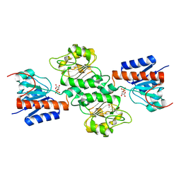
3K85
DownloadVisualize
BU of 3k85 by Molmil
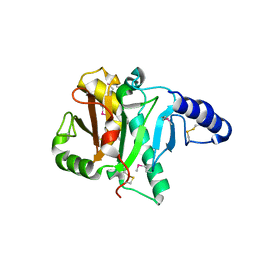
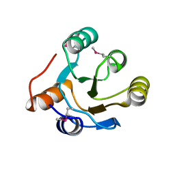
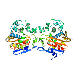
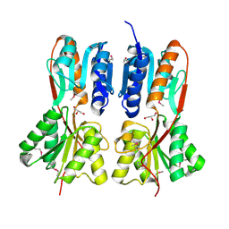
Crystal structure of a D-glycero-D-manno-heptose 1-phosphate kinase from Bacteriodes thetaiotaomicron
Descriptor: D-glycero-D-manno-heptose 1-phosphate kinase
Authors:Palani, K, Kumaran, D, Burley, S.K, Swaminathan, S, New York SGX Research Center for Structural Genomics (NYSGXRC)
Deposit date:2009-10-13
Release date:2009-12-08
Last modified:2021-02-10
Method:X-RAY DIFFRACTION (2.28 Å)
Cite:Crystal structure of a D-glycero-D-manno-heptose 1-phosphate kinase from Bacteriodes thetaiotaomicron
To be Published
3K9E
DownloadVisualize
BU of 3k9e by Molmil
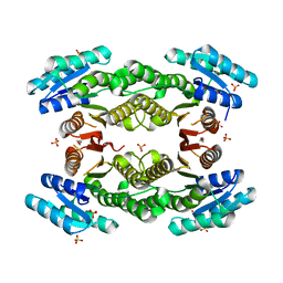
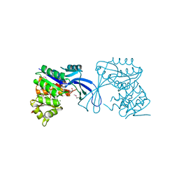
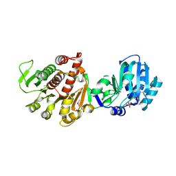
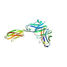
Crystal structure of a putative Ribokinase II (Apo Form) from E.coli
Descriptor: PUTATIVE RIBOKINASE II
Authors:Satyanarayana, L, Burley, S.K, Swaminathan, S, New York SGX Research Center for Structural Genomics (NYSGXRC)
Deposit date:2009-10-15
Release date:2009-11-10
Last modified:2023-11-22
Method:X-RAY DIFFRACTION (2.05 Å)
Cite:Crystal structure of a putative Ribokinase II (Apo Form) from E.coli
To be Published
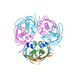
1I4P
DownloadVisualize
BU of 1i4p by Molmil
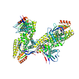
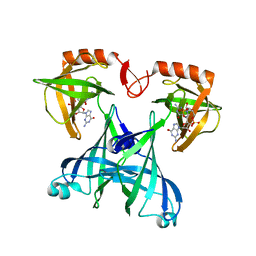
CRYSTAL STRUCTURE OF STAPHYLOCOCCAL ENTEROTOXIN C2 AT 100K CRYSTALLIZED AT PH 5.5
Descriptor: ENTEROTOXIN TYPE C-2, ZINC ION
Authors:Kumaran, D, Swaminathan, S.
Deposit date:2001-02-22
Release date:2001-03-14
Last modified:2023-08-09
Method:X-RAY DIFFRACTION (2 Å)
Cite:Structure of staphylococcal enterotoxin C2 at various pH levels.
Acta Crystallogr.,Sect.D, 57, 2001
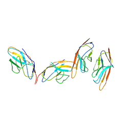
2POF
DownloadVisualize
BU of 2pof by Molmil
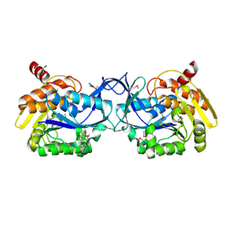
Crystal structure of CDP-diacylglycerol pyrophosphatase
Descriptor: CDP-diacylglycerol pyrophosphatase
Authors:Madegowda, M, Eswaramoorthy, S, Burley, S.K, Swaminathan, S, New York SGX Research Center for Structural Genomics (NYSGXRC)
Deposit date:2007-04-26
Release date:2007-05-15
Last modified:2021-02-03
Method:X-RAY DIFFRACTION (1.4 Å)
Cite:Crystal structure of CDP-diacylglycerol pyrophosphatase.
To be Published
3TFO
DownloadVisualize
BU of 3tfo by Molmil
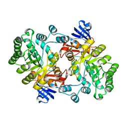
Crystal structure of a putative 3-oxoacyl-(acyl-carrier-protein) reductase from Sinorhizobium meliloti
Descriptor: HEXANE-1,6-DIOL, SULFATE ION, putative 3-oxoacyl-(acyl-carrier-protein) reductase
Authors:Agarwal, R, Chamala, S, Evans, B, Foti, R, Gizzi, A, Hillerich, B, Kar, A, LaFleur, J, Seidel, R, Villigas, G, Zencheck, W, Almo, S.C, Swaminathan, S, New York Structural Genomics Research Consortium (NYSGRC)
Deposit date:2011-08-16
Release date:2011-08-31
Last modified:2024-02-28
Method:X-RAY DIFFRACTION (2.08 Å)
Cite:Crystal structure of a putative 3-oxoacyl-(acyl-carrier-protein) reductase from Sinorhizobium meliloti
To be Published
2PUZ
DownloadVisualize
BU of 2puz by Molmil
Crystal structure of Imidazolonepropionase from Agrobacterium tumefaciens with bound product N-formimino-L-Glutamate
Descriptor: CHLORIDE ION, FE (III) ION, Imidazolonepropionase, ...
Authors:Tyagi, R, Eswaramoorthy, S, Burley, S.K, Swaminathan, S, New York SGX Research Center for Structural Genomics (NYSGXRC)
Deposit date:2007-05-09
Release date:2007-05-22
Last modified:2023-08-30
Method:X-RAY DIFFRACTION (1.83 Å)
Cite:X-ray structure of imidazolonepropionase from Agrobacterium tumefaciens at 1.87 A resolution.
Proteins, 69, 2007
2IOJ
DownloadVisualize
BU of 2ioj by Molmil
Crystal structure of protein AF1212 from Archaeoglobus fulgidus, Pfam DRTGG
Descriptor: Hypothetical protein AF_1212
Authors:Tyagi, R, Burley, S.K, Swaminathan, S, New York SGX Research Center for Structural Genomics (NYSGXRC)
Deposit date:2006-10-10
Release date:2006-10-31
Last modified:2021-02-03
Method:X-RAY DIFFRACTION (2.15 Å)
Cite:The crystal structure of a hypothetical protein (Y1212_ARCFU) from Archeoglobus fulgidus
To be Published
3JUL
DownloadVisualize
BU of 3jul by Molmil
Crystal structure of Listeria innocua D-Tagatose-6-Phosphate Kinase bound with substrate
Descriptor: 6-O-phosphono-beta-D-tagatofuranose, Lin2199 protein, MAGNESIUM ION
Authors:Satyanarayana, L, Burley, S.K, Swaminathan, S, New York SGX Research Center for Structural Genomics (NYSGXRC)
Deposit date:2009-09-15
Release date:2009-10-06
Last modified:2023-11-22
Method:X-RAY DIFFRACTION (2.4 Å)
Cite:Crystal structure of Listeria innocua D-Tagatose-6-Phosphate Kinase bound with substrate
To be Published
3K17
DownloadVisualize
BU of 3k17 by Molmil
Crystal structure of a Lin0012 protein from Listeria innocua
Descriptor: Lin0012 protein, TRIETHYLENE GLYCOL
Authors:Palani, K, Burley, S.K, Swaminathan, S, New York SGX Research Center for Structural Genomics (NYSGXRC)
Deposit date:2009-09-25
Release date:2009-10-06
Last modified:2021-02-10
Method:X-RAY DIFFRACTION (2.1 Å)
Cite:Crystal structure of a Lin0012 protein from Listeria innocua
To be Published
4FHQ
DownloadVisualize
BU of 4fhq by Molmil
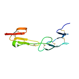
Crystal Structure of HVEM
Descriptor: Tumor necrosis factor receptor superfamily member 14
Authors:Liu, W, Zhan, C, Patskovsky, Y, Bhosle, R.C, Nathenson, S.G, Almo, S.C, Atoms-to-Animals: The Immune Function Network (IFN), New York Structural Genomics Research Consortium (NYSGRC)
Deposit date:2012-06-06
Release date:2012-07-18
Last modified:2023-09-13
Method:X-RAY DIFFRACTION (2.251 Å)
Cite:Increased Heterologous Protein Expression in Drosophila S2 Cells for Massive Production of Immune Ligands/Receptors and Structural Analysis of Human HVEM.
Mol Biotechnol, 57, 2015
3TG9
DownloadVisualize
BU of 3tg9 by Molmil
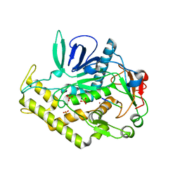
The crystal structure of penicillin binding protein from Bacillus halodurans
Descriptor: Penicillin-binding protein
Authors:Zhang, Z, Satyanarayana, L, Chamala, S, Evans, B, Foti, R, Gizzi, A, Hillerich, B, Kar, A, LaFleur, J, Seidel, R, Villigas, G, Zencheck, W, Almo, S.C, Swaminathan, S, New York Structural Genomics Research Consortium (NYSGRC)
Deposit date:2011-08-17
Release date:2011-08-31
Last modified:2023-09-13
Method:X-RAY DIFFRACTION (2.2 Å)
Cite:The crystal structure of penicillin binding protein from Bacillus halodurans
To be Published
3K5W
DownloadVisualize
BU of 3k5w by Molmil
Crystal structure of a Carbohydrate kinase (YjeF family)from Helicobacter pylori
Descriptor: Carbohydrate kinase, PHOSPHATE ION
Authors:Satyanarayana, L, Burley, S.K, Swaminathan, S, New York SGX Research Center for Structural Genomics (NYSGXRC)
Deposit date:2009-10-08
Release date:2009-12-08
Last modified:2021-02-10
Method:X-RAY DIFFRACTION (2.6 Å)
Cite:Crystal structure of a Carbohydrate kinase (YjeF family)from Helicobacter pylori
To be Published
2QDE
DownloadVisualize
BU of 2qde by Molmil
Crystal structure of mandelate racemase/muconate lactonizing family protein from Azoarcus sp. EbN1
Descriptor: BARIUM ION, Mandelate racemase/muconate lactonizing enzyme family protein
Authors:Agarwal, R, Burley, S.K, Swaminathan, S, New York SGX Research Center for Structural Genomics (NYSGXRC)
Deposit date:2007-06-20
Release date:2007-07-03
Last modified:2024-02-21
Method:X-RAY DIFFRACTION (1.93 Å)
Cite:Crystal structure of mandelate racemase/muconate lactonizing enzyme family protein from Azoarcus sp. EbN1.
To be Published
3D5L
DownloadVisualize
BU of 3d5l by Molmil
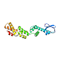
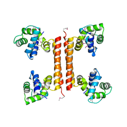
Crystal structure of regulatory protein RecX
Descriptor: Regulatory protein RecX, SULFATE ION
Authors:Agarwal, R, Burley, S.K, Swaminathan, S, New York SGX Research Center for Structural Genomics (NYSGXRC)
Deposit date:2008-05-16
Release date:2008-06-10
Last modified:2024-02-21
Method:X-RAY DIFFRACTION (2.35 Å)
Cite:Crystal structure of DNA repair regulatory protein RecX.
To be Published
4NJM
DownloadVisualize
BU of 4njm by Molmil
Crystal Structure of phosphoglycerate bound 3-phosphoglycerate dehydrogenase in Entamoeba histolytica
Descriptor: 3-PHOSPHOGLYCERIC ACID, D-3-phosphoglycerate dehydrogenase, putative
Authors:Singh, R.K, Gourinath, S.
Deposit date:2013-11-11
Release date:2014-10-08
Last modified:2023-11-08
Method:X-RAY DIFFRACTION (1.79 Å)
Cite:Crystal structures and kinetics of Type III 3-phosphoglycerate dehydrogenase reveal catalysis by lysine.
Febs J., 281, 2014
1I4R
DownloadVisualize
BU of 1i4r by Molmil
CRYSTAL STRUCTURE OF STAPHYLOCOCCAL ENTEROTOXIN C2 AT 100K CRYSTALLIZED AT PH 6.5
Descriptor: ENTEROTOXIN TYPE C-2, ZINC ION
Authors:Kumaran, D, Swaminathan, S.
Deposit date:2001-02-22
Release date:2001-03-14
Last modified:2023-08-09
Method:X-RAY DIFFRACTION (2.1 Å)
Cite:Structure of staphylococcal enterotoxin C2 at various pH levels.
Acta Crystallogr.,Sect.D, 57, 2001
5BUW
DownloadVisualize
BU of 5buw by Molmil
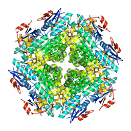
Crystal Structure of beta-hydroxyacyl-acyl carrier protein dehydratase (FabZ) from Yersinia Pestis
Descriptor: 1,2-ETHANEDIOL, 3-hydroxyacyl-[acyl-carrier-protein] dehydratase FabZ, THIOCYANATE ION
Authors:Mcgillick, B, Kumaran, D, Swaminathan, S.
Deposit date:2015-06-04
Release date:2016-06-15
Last modified:2023-09-27
Method:X-RAY DIFFRACTION (1.8 Å)
Cite:Crystal Structure of beta-hydroxyacyl-acyl carrier protein dehydratase (FabZ) from Yersinia Pestis
To be published
1I85
DownloadVisualize
BU of 1i85 by Molmil
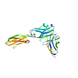
CRYSTAL STRUCTURE OF THE CTLA-4/B7-2 COMPLEX
Descriptor: CYTOTOXIC T-LYMPHOCYTE-ASSOCIATED PROTEIN 4, T LYMPHOCYTE ACTIVATION ANTIGEN CD86
Authors:Schwartz, J.-C.D, Zhang, X, Fedorov, A.A, Nathenson, S.G, Almo, S.C.
Deposit date:2001-03-12
Release date:2001-04-04
Last modified:2011-07-13
Method:X-RAY DIFFRACTION (3.2 Å)
Cite:Structural basis for co-stimulation by the human CTLA-4/B7-2 complex.
Nature, 410, 2001
3C4R
DownloadVisualize
BU of 3c4r by Molmil
Crystal structure of an uncharacterized protein encoded by cryptic prophage
Descriptor: Uncharacterized protein
Authors:Sugadev, R, Burley, S.K, Swaminathan, S, Ozyurt, S, Luz, J, New York SGX Research Center for Structural Genomics (NYSGXRC)
Deposit date:2008-01-30
Release date:2008-02-19
Last modified:2021-02-03
Method:X-RAY DIFFRACTION (2.3 Å)
Cite:Crystal structure of an uncharacterized protein encoded by cryptic prophage.
To be Published
3URH
DownloadVisualize
BU of 3urh by Molmil
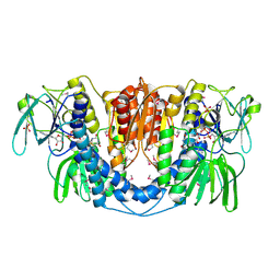
Crystal structure of a dihydrolipoamide dehydrogenase from Sinorhizobium meliloti 1021
Descriptor: 1,2-ETHANEDIOL, Dihydrolipoyl dehydrogenase, FLAVIN-ADENINE DINUCLEOTIDE, ...
Authors:Kumaran, D, Chamala, S, Evans, B, Foti, R, Gizzi, A, Hillerich, B, Kar, A, LaFleur, J, Seidel, R, Villigas, G, Zencheck, W, Almo, S.C, Swaminathan, S, New York Structural Genomics Research Consortium (NYSGRC)
Deposit date:2011-11-22
Release date:2011-12-14
Method:X-RAY DIFFRACTION (1.9 Å)
Cite:Crystal structure of a dihydrolipoamide dehydrogenase from Sinorhizobium meliloti 1021
TO BE PUBLISHED
3BP5
DownloadVisualize
BU of 3bp5 by Molmil
Crystal structure of the mouse PD-1 and PD-L2 complex
Descriptor: GLYCEROL, Programmed cell death 1 ligand 2, Programmed cell death protein 1
Authors:Yan, Q, Lazar-Molnar, E, Cao, E, Ramagopal, U.A, Toro, R, Nathenson, S.G, Almo, S.C.
Deposit date:2007-12-18
Release date:2008-07-15
Last modified:2023-08-30
Method:X-RAY DIFFRACTION (1.8 Å)
Cite:Crystal structure of the complex between programmed death-1 (PD-1) and its ligand PD-L2.
Proc.Natl.Acad.Sci.Usa, 105, 2008
3C8A
DownloadVisualize
BU of 3c8a by Molmil
Crystal structure of the catalytic domain of botulinum neurotoxin serotype A with inhibitory peptide RRGL
Descriptor: Botulinum neurotoxin A light chain, Inhibitor peptide RRGL, SULFATE ION, ...
Authors:Kumaran, D, Swaminathan, S.
Deposit date:2008-02-11
Release date:2008-04-22
Last modified:2011-07-13
Method:X-RAY DIFFRACTION (1.52 Å)
Cite:Structure- and Substrate-based Inhibitor Design for Clostridium botulinum Neurotoxin Serotype A
J.Biol.Chem., 283, 2008
3K9C
DownloadVisualize
BU of 3k9c by Molmil
Crystal structure of LacI Transcriptional regulator from Rhodococcus species.
Descriptor: GLYCEROL, Transcriptional regulator, LacI family protein
Authors:Damodharan, L, Burley, S.K, Swaminathan, S, New York SGX Research Center for Structural Genomics (NYSGXRC)
Deposit date:2009-10-15
Release date:2009-11-03
Last modified:2021-02-10
Method:X-RAY DIFFRACTION (2.14 Å)
Cite:Crystal structure of LacI Transcriptional regulator from Rhodococcus species.
To be Published
3BP6
DownloadVisualize
BU of 3bp6 by Molmil
Crystal structure of the mouse PD-1 Mutant and PD-L2 complex
Descriptor: GLYCEROL, Programmed cell death 1 ligand 2, Programmed cell death protein 1
Authors:Yan, Q, Lazar-Molnar, E, Cao, E, Ramagopal, U.A, Toro, R, Nathenson, S.G, Almo, S.C.
Deposit date:2007-12-18
Release date:2009-02-24
Last modified:2023-12-27
Method:X-RAY DIFFRACTION (1.6 Å)
Cite:Crystal structure of the mouse PD-1 A99L and PD-L2 complex
To be published
2RDE
DownloadVisualize
BU of 2rde by Molmil
Crystal structure of VCA0042 complexed with c-di-GMP
Descriptor: 9,9'-[(2R,3R,3aS,5S,7aR,9R,10R,10aS,12S,14aR)-3,5,10,12-tetrahydroxy-5,12-dioxidooctahydro-2H,7H-difuro[3,2-d:3',2'-j][1,3,7,9,2,8]tetraoxadiphosphacyclododecine-2,9-diyl]bis(2-amino-1,9-dihydro-6H-purin-6-one), Uncharacterized protein VCA0042
Authors:Benach, J, Swaminathan, S.S, Tamayo, R, Seetharaman, J, Handelman, S, Forouhar, F, Neely, H, Camilli, A, Hunt, J.F, Northeast Structural Genomics Consortium (NESG)
Deposit date:2007-09-22
Release date:2007-10-30
Last modified:2023-08-30
Method:X-RAY DIFFRACTION (1.92 Å)
Cite:The structural basis of cyclic diguanylate signal transduction by PilZ domains.
Embo J., 26, 2007

222926

건을2024-07-24부터공개중

PDB statisticsPDBj update infoContact PDBjnumon