Loading
PDBj
MenuPDBj@FacebookPDBj@TwitterPDBj@YouTubewwPDB FoundationwwPDB
RCSB PDBPDBeBMRBAdv. SearchSearch help
PDB: 293 results

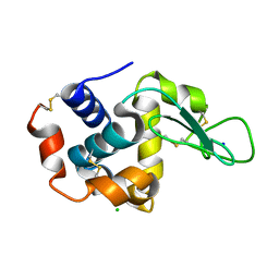
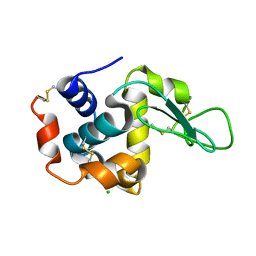
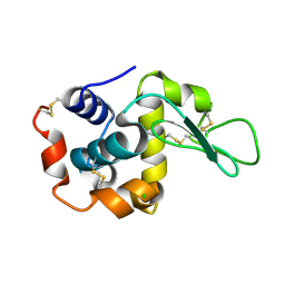
6JXQ
DownloadVisualize
BU of 6jxq by Molmil
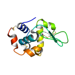
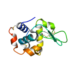
Room temperature structure of lysozyme delivered in polyacrylamide by serial millisecond crystallography
Descriptor: CHLORIDE ION, Lysozyme C
Authors:Nam, K.H.
Deposit date:2019-04-24
Release date:2019-05-15
Last modified:2023-11-22
Method:X-RAY DIFFRACTION (1.76 Å)
Cite:Sample delivery using viscous media, a syringe and a syringe pump for serial crystallography.
J.Synchrotron Radiat., 26, 2019
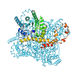
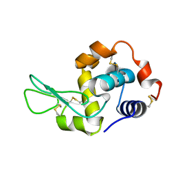
6KD2
DownloadVisualize
BU of 6kd2 by Molmil
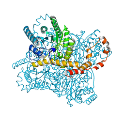
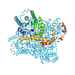
Room temperature structure of glucose isomerase delivered in gelatin by serial millisecond crystallography
Descriptor: MAGNESIUM ION, Xylose isomerase
Authors:Nam, K.H.
Deposit date:2019-06-30
Release date:2019-07-17
Last modified:2023-11-22
Method:X-RAY DIFFRACTION (1.7 Å)
Cite:Stable sample delivery in viscous media via a capillary for serial crystallography.
J.Appl.Crystallogr., 53, 2020
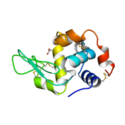
6KCA
DownloadVisualize
BU of 6kca by Molmil
Room temperature structure of glucose isomerase delivered in shortening A by serial millisecond crystallography
Descriptor: MAGNESIUM ION, Xylose isomerase
Authors:Nam, K.H.
Deposit date:2019-06-27
Release date:2020-05-20
Last modified:2023-11-22
Method:X-RAY DIFFRACTION (1.9 Å)
Cite:Shortening injection matrix for serial crystallography.
Sci Rep, 10, 2020
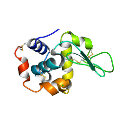
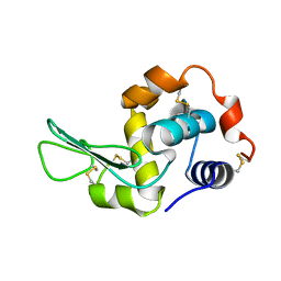
6JXP
DownloadVisualize
BU of 6jxp by Molmil
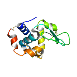
Room temperature structure of lysozyme delivered in LCP by serial millisecond crystallography
Descriptor: CHLORIDE ION, Lysozyme C
Authors:Nam, K.H.
Deposit date:2019-04-24
Release date:2019-05-15
Last modified:2023-11-22
Method:X-RAY DIFFRACTION (1.56 Å)
Cite:Sample delivery using viscous media, a syringe and a syringe pump for serial crystallography.
J.Synchrotron Radiat., 26, 2019
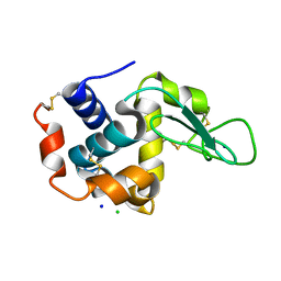
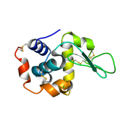
6KD1
DownloadVisualize
BU of 6kd1 by Molmil
Room temperature structure of lysozyme delivered in agarose by serial millisecond crystallography
Descriptor: CHLORIDE ION, Lysozyme C, SODIUM ION
Authors:Nam, K.H.
Deposit date:2019-06-30
Release date:2019-07-17
Last modified:2023-11-22
Method:X-RAY DIFFRACTION (1.85 Å)
Cite:Stable sample delivery in viscous media via a capillary for serial crystallography.
J.Appl.Crystallogr., 53, 2020
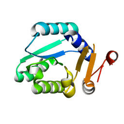
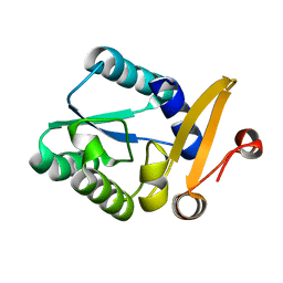
6K1X
DownloadVisualize
BU of 6k1x by Molmil
Crystal structure of Rhodothermus marinus substrate-binding protein at pH 6.0
Descriptor: ABC-type uncharacterized transport system periplasmic component-like protein
Authors:Nam, K.H.
Deposit date:2019-05-13
Release date:2019-08-21
Last modified:2023-11-22
Method:X-RAY DIFFRACTION (1.802 Å)
Cite:Crystal structure of Rhodothermus marinus substrate-binding protein at pH 6.0
To Be Published
6KCD
DownloadVisualize
BU of 6kcd by Molmil
Room temperature structure of lysozyme delivered in shortening B by serial millisecond crystallography
Descriptor: CHLORIDE ION, Lysozyme C, SODIUM ION
Authors:Nam, K.H.
Deposit date:2019-06-27
Release date:2020-05-20
Last modified:2023-11-22
Method:X-RAY DIFFRACTION (1.5 Å)
Cite:Shortening injection matrix for serial crystallography.
Sci Rep, 10, 2020
6KCB
DownloadVisualize
BU of 6kcb by Molmil
Room temperature structure of lysozyme delivered in shortening A by serial millisecond crystallography
Descriptor: CHLORIDE ION, Lysozyme C, SODIUM ION
Authors:Nam, K.H.
Deposit date:2019-06-27
Release date:2020-05-20
Last modified:2023-11-22
Method:X-RAY DIFFRACTION (1.8 Å)
Cite:Shortening injection matrix for serial crystallography.
Sci Rep, 10, 2020
6K1W
DownloadVisualize
BU of 6k1w by Molmil
Crystal structure of Rhodothermus marinus substrate-binding protein at pH 5.5
Descriptor: ABC-type uncharacterized transport system periplasmic component-like protein
Authors:Nam, K.H.
Deposit date:2019-05-13
Release date:2019-08-21
Last modified:2023-11-22
Method:X-RAY DIFFRACTION (1.5 Å)
Cite:Crystal structure of Rhodothermus marinus substrate-binding protein at pH 5.5
To Be Published
6KCC
DownloadVisualize
BU of 6kcc by Molmil
Room temperature structure of glucose isomerase delivered in shortening B by serial millisecond crystallography
Descriptor: MAGNESIUM ION, Xylose isomerase
Authors:Nam, K.H.
Deposit date:2019-06-27
Release date:2020-05-20
Last modified:2023-11-22
Method:X-RAY DIFFRACTION (2 Å)
Cite:Shortening injection matrix for serial crystallography.
Sci Rep, 10, 2020
6K1Y
DownloadVisualize
BU of 6k1y by Molmil
Crystal structure of Rhodothermus marinus substrate-binding protein at pH 7.5
Descriptor: ABC-type uncharacterized transport system periplasmic component-like protein
Authors:Nam, K.H.
Deposit date:2019-05-13
Release date:2019-08-21
Last modified:2023-11-22
Method:X-RAY DIFFRACTION (1.9 Å)
Cite:Crystal structure of Rhodothermus marinus substrate-binding protein at pH 7.5
To Be Published
7WKR
DownloadVisualize
BU of 7wkr by Molmil
Room temperature structure of lysozyme solved by serial synchrotron crystallography
Descriptor: CHLORIDE ION, Lysozyme C
Authors:Nam, K.H.
Deposit date:2022-01-11
Release date:2022-02-02
Last modified:2023-11-29
Method:X-RAY DIFFRACTION (1.6 Å)
Cite:Combination of an inject-and-transfer system for serial femtosecond crystallography.
J.Appl.Crystallogr., 55, 2022
7WUC
DownloadVisualize
BU of 7wuc by Molmil
Room-temperature structure of lysozyme by serial femtosecond crystallography (BITS)
Descriptor: Lysozyme C
Authors:Nam, K.H.
Deposit date:2022-02-08
Release date:2022-03-02
Last modified:2023-11-29
Method:X-RAY DIFFRACTION (2.1 Å)
Cite:Combination of an inject-and-transfer system for serial femtosecond crystallography.
J.Appl.Crystallogr., 55, 2022
7WBE
DownloadVisualize
BU of 7wbe by Molmil
Crystal structure of lysozyme (multilcrystal diffraction, CrystFEL/MOSFLM)
Descriptor: CHLORIDE ION, Lysozyme C
Authors:Nam, K.H.
Deposit date:2021-12-16
Release date:2022-01-19
Last modified:2023-11-29
Method:X-RAY DIFFRACTION (1.9 Å)
Cite:Processing of Multicrystal Diffraction Patterns in Macromolecular Crystallography Using Serial Crystallography Programs.
Crystals, 12, 2022
7WBD
DownloadVisualize
BU of 7wbd by Molmil
Crystal structure of lysozyme (multilcrystal diffraction, CrystFEL/XGANDALF)
Descriptor: CHLORIDE ION, Lysozyme C
Authors:Nam, K.H.
Deposit date:2021-12-16
Release date:2022-01-19
Last modified:2023-11-29
Method:X-RAY DIFFRACTION (1.9 Å)
Cite:Processing of Multicrystal Diffraction Patterns in Macromolecular Crystallography Using Serial Crystallography Programs.
Crystals, 12, 2022
7WBF
DownloadVisualize
BU of 7wbf by Molmil
Crystal structure of lysozyme
Descriptor: CHLORIDE ION, Lysozyme C
Authors:Nam, K.H.
Deposit date:2021-12-16
Release date:2022-01-19
Last modified:2023-11-29
Method:X-RAY DIFFRACTION (1.6 Å)
Cite:Processing of Multicrystal Diffraction Patterns in Macromolecular Crystallography Using Serial Crystallography Programs.
Crystals, 12, 2022
7XF7
DownloadVisualize
BU of 7xf7 by Molmil
Crystal Structure of Human Lysozyme Complexed with N-Acetyl-alpha-D-Glucosamine
Descriptor: 2-acetamido-2-deoxy-alpha-D-glucopyranose, Lysozyme C
Authors:Nam, K.H.
Deposit date:2022-04-01
Release date:2022-04-13
Last modified:2023-11-29
Method:X-RAY DIFFRACTION (1.55 Å)
Cite:Crystal Structure of Human Lysozyme Complexed with N-Acetyl-alpha-d-glucosamine.
Appl Sci (Basel), 12, 2022
7XF6
DownloadVisualize
BU of 7xf6 by Molmil
Crystal Structure of Human Lysozyme
Descriptor: ACETATE ION, Lysozyme C
Authors:Nam, K.H.
Deposit date:2022-04-01
Release date:2022-04-13
Last modified:2023-11-29
Method:X-RAY DIFFRACTION (1.3 Å)
Cite:Crystal Structure of Human Lysozyme Complexed with N-Acetyl-alpha-d-glucosamine.
Appl Sci (Basel), 12, 2022
7XF8
DownloadVisualize
BU of 7xf8 by Molmil
Crystal Structure of Human Lysozyme Complexed with N-Acetyl-alpha-D-Glucosamine
Descriptor: 2-acetamido-2-deoxy-alpha-D-glucopyranose, Lysozyme C
Authors:Nam, K.H.
Deposit date:2022-04-01
Release date:2022-04-13
Last modified:2023-11-29
Method:X-RAY DIFFRACTION (1.6 Å)
Cite:Crystal Structure of Human Lysozyme Complexed with N-Acetyl-alpha-d-glucosamine.
Appl Sci (Basel), 12, 2022
6LNG
DownloadVisualize
BU of 6lng by Molmil
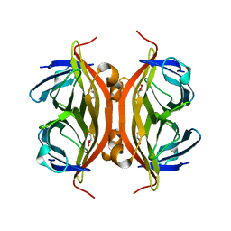
Rapid crystallization of streptavidin using charged peptides
Descriptor: GLYCEROL, Streptavidin
Authors:Minamihata, K, Tsukamoto, K, Adachi, M, Shimizu, R, Mishina, M, Kuroki, R, Nagamune, T.
Deposit date:2019-12-30
Release date:2020-03-18
Last modified:2023-11-22
Method:X-RAY DIFFRACTION (1.8000015 Å)
Cite:Genetically fused charged peptides induce rapid crystallization of proteins.
Chem.Commun.(Camb.), 56, 2020
5L6Q
DownloadVisualize
BU of 5l6q by Molmil
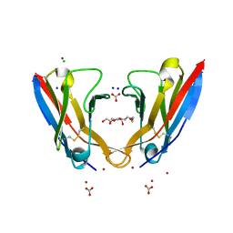
Refolded AL protein from cardiac amyloidosis
Descriptor: CARBONATE ION, CHLORIDE ION, DI(HYDROXYETHYL)ETHER, ...
Authors:Annamalai, K, Liberta, F, Vielberg, M.-T, Lilie, H, Guehrs, K.-H, Schierhorn, A, Koehler, R, Schmidt, A, Haupt, C, Hegenbart, O, Schoenland, S, Groll, M, Faendrich, M.
Deposit date:2016-05-31
Release date:2017-05-31
Last modified:2024-01-10
Method:X-RAY DIFFRACTION (1.4 Å)
Cite:Common Fibril Structures Imply Systemically Conserved Protein Misfolding Pathways In Vivo.
Angew. Chem. Int. Ed. Engl., 56, 2017
6GV1
DownloadVisualize
BU of 6gv1 by Molmil
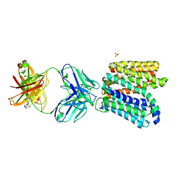
Crystal structure of E.coli Multidrug/H+ antiporter MdfA in outward open conformation with bound Fab fragment
Descriptor: Fab fragment YN1074 heavy chain, Fab fragment YN1074 light chain, Major Facilitator Superfamily multidrug/H+ antiporter MdfA from E.coli, ...
Authors:Nagarathinam, K, Parthier, C, Stubbs, M.T, Tanabe, M.
Deposit date:2018-06-20
Release date:2018-10-03
Last modified:2024-01-17
Method:X-RAY DIFFRACTION (3.4 Å)
Cite:Outward open conformation of a Major Facilitator Superfamily multidrug/H+antiporter provides insights into switching mechanism.
Nat Commun, 9, 2018
1G91
DownloadVisualize
BU of 1g91 by Molmil
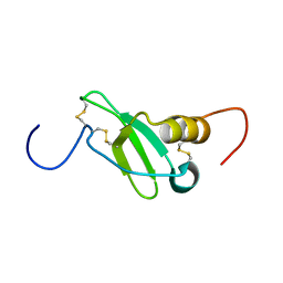
SOLUTION STRUCTURE OF MYELOID PROGENITOR INHIBITORY FACTOR-1 (MPIF-1)
Descriptor: MYELOID PROGENITOR INHIBITORY FACTOR-1
Authors:Rajarathnam, K, Li, Y, Rohrer, T, Gentz, R.
Deposit date:2000-11-21
Release date:2001-03-07
Last modified:2022-12-21
Method:SOLUTION NMR
Cite:Solution structure and dynamics of myeloid progenitor inhibitory factor-1 (MPIF-1), a novel monomeric CC chemokine.
J.Biol.Chem., 276, 2001
7KKF
DownloadVisualize
BU of 7kkf by Molmil
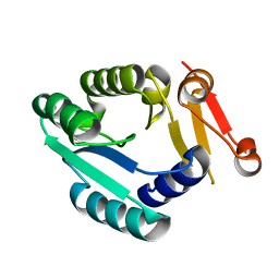
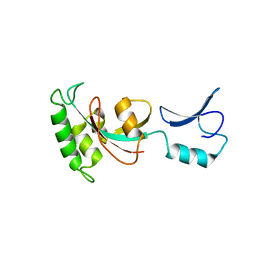
Crystal Structure of S. cerevisiae Ess1
Descriptor: Peptidyl-prolyl cis-trans isomerase ESS1
Authors:Namitz, K.E.W, Alicea-Velazquez, N.L, Cosgrove, M.S, Hanes, S.D.
Deposit date:2020-10-27
Release date:2021-03-31
Last modified:2023-10-18
Method:X-RAY DIFFRACTION (2.4 Å)
Cite:Structure analysis suggests Ess1 isomerizes the carboxy-terminal domain of RNA polymerase II via a bivalent anchoring mechanism.
Commun Biol, 4, 2021
6IAK
DownloadVisualize
BU of 6iak by Molmil
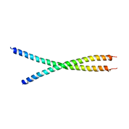
The crystal structure of the chicken CREB3 bZIP
Descriptor: Uncharacterized protein
Authors:Sabaratnam, K, Renner, M.
Deposit date:2018-11-26
Release date:2019-12-11
Last modified:2020-06-24
Method:X-RAY DIFFRACTION (3.95 Å)
Cite:Insights from the crystal structure of the chicken CREB3 bZIP suggest that members of the CREB3 subfamily transcription factors may be activated in response to oxidative stress.
Protein Sci., 28, 2019

222624

PDB entries from 2024-07-17

PDB statisticsPDBj update infoContact PDBjnumon