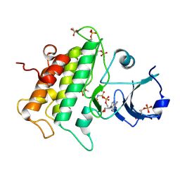
5C8S
 
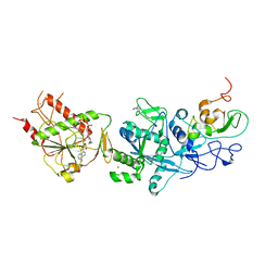 | | Crystal structure of the SARS coronavirus nsp14-nsp10 complex with functional ligands SAH and GpppA | | 分子名称: | GUANOSINE-P3-ADENOSINE-5',5'-TRIPHOSPHATE, Guanine-N7 methyltransferase, MAGNESIUM ION, ... | | 著者 | Ma, Y.Y, Wu, L.J, Zhang, R.G, Rao, Z.H. | | 登録日 | 2015-06-26 | | 公開日 | 2015-07-15 | | 最終更新日 | 2015-08-12 | | 実験手法 | X-RAY DIFFRACTION (3.326 Å) | | 主引用文献 | Structural basis and functional analysis of the SARS coronavirus nsp14-nsp10 complex
Proc.Natl.Acad.Sci.USA, 112, 2015
|
|
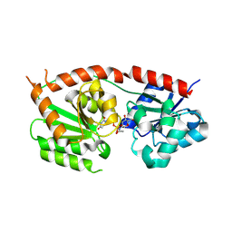
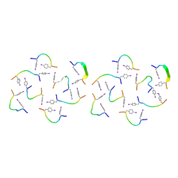
5C8T
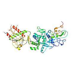 | | Crystal structure of the SARS coronavirus nsp14-nsp10 complex with functional ligand SAM | | 分子名称: | Guanine-N7 methyltransferase, MAGNESIUM ION, Non-structural protein 10, ... | | 著者 | Ma, Y.Y, Wu, L.J, Zhang, R.G, Rao, Z.H. | | 登録日 | 2015-06-26 | | 公開日 | 2015-07-15 | | 最終更新日 | 2015-08-12 | | 実験手法 | X-RAY DIFFRACTION (3.2 Å) | | 主引用文献 | Structural basis and functional analysis of the SARS coronavirus nsp14-nsp10 complex
Proc.Natl.Acad.Sci.USA, 112, 2015
|
|
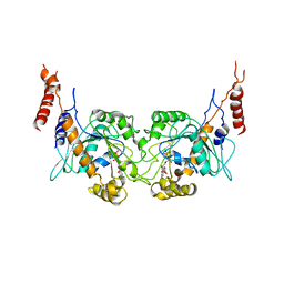
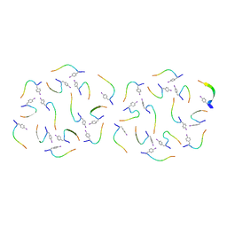
5C8U
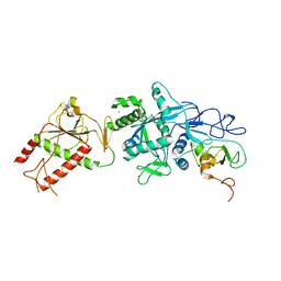 | | Crystal structure of the SARS coronavirus nsp14-nsp10 complex | | 分子名称: | Guanine-N7 methyltransferase, MAGNESIUM ION, Non-structural protein 10, ... | | 著者 | Ma, Y.Y, Wu, L.J, Zhang, R.G, Rao, Z.H. | | 登録日 | 2015-06-26 | | 公開日 | 2015-07-15 | | 最終更新日 | 2024-03-20 | | 実験手法 | X-RAY DIFFRACTION (3.401 Å) | | 主引用文献 | Structural basis and functional analysis of the SARS coronavirus nsp14-nsp10 complex
Proc.Natl.Acad.Sci.USA, 112, 2015
|
|
8RYZ
 | |
8RZ3
 | |
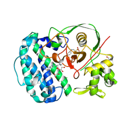
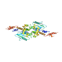
3T35
 | | Arabidopsis thaliana dynamin-related protein 1A in postfission state | | 分子名称: | Dynamin-related protein 1A, LINKER, GUANOSINE-5'-DIPHOSPHATE | | 著者 | Yan, L.M, Ma, Y.Y, Sun, Y.N, Lou, Z.Y. | | 登録日 | 2011-07-24 | | 公開日 | 2012-06-06 | | 最終更新日 | 2023-11-01 | | 実験手法 | X-RAY DIFFRACTION (3.592 Å) | | 主引用文献 | Structural basis for mechanochemical role of Arabidopsis thaliana dynamin-related protein in membrane fission
J Mol Cell Biol, 3, 2011
|
|
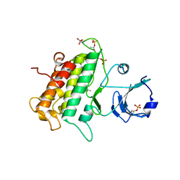
3T34
 | | Arabidopsis thaliana dynamin-related protein 1A (AtDRP1A) in prefission state | | 分子名称: | Dynamin-related protein 1A, LINKER, GUANOSINE-5'-DIPHOSPHATE, ... | | 著者 | Yan, L.M, Ma, Y.Y, Sun, Y.N, Lou, Z.Y. | | 登録日 | 2011-07-24 | | 公開日 | 2012-06-06 | | 最終更新日 | 2023-11-01 | | 実験手法 | X-RAY DIFFRACTION (2.405 Å) | | 主引用文献 | Structural basis for mechanochemical role of Arabidopsis thaliana dynamin-related protein in membrane fission
J Mol Cell Biol, 3, 2011
|
|
3ULZ
 | |
3UIM
 | |
8IKS
 | |
8IKB
 | |
8IK7
 | |
8IZZ
 | |
8IY7
 | |
8IKP
 | |
7DWV
 | | Cryo-EM structure of amyloid fibril formed by familial prion disease-related mutation E196K | | 分子名称: | Major prion protein | | 著者 | Wang, L.Q, Zhao, K, Yuan, H.Y, Li, X.N, Dang, H.B, Ma, Y.Y, Wang, Q, Wang, C, Sun, Y.P, Chen, J, Li, D, Zhang, D.L, Yin, P, Liu, C, Liang, Y. | | 登録日 | 2021-01-18 | | 公開日 | 2021-10-13 | | 実験手法 | ELECTRON MICROSCOPY (3.07 Å) | | 主引用文献 | Genetic prion disease-related mutation E196K displays a novel amyloid fibril structure revealed by cryo-EM.
Sci Adv, 7, 2021
|
|
7DA4
 | | Cryo-EM structure of amyloid fibril formed by human RIPK3 | | 分子名称: | Receptor-interacting serine/threonine-protein kinase 3 | | 著者 | Zhao, K, Ma, Y.Y, Sun, Y.P, Li, D, Liu, C. | | 登録日 | 2020-10-14 | | 公開日 | 2021-04-28 | | 最終更新日 | 2024-03-27 | | 実験手法 | ELECTRON MICROSCOPY (4.24 Å) | | 主引用文献 | The structure of a minimum amyloid fibril core formed by necroptosis-mediating RHIM of human RIPK3.
Proc.Natl.Acad.Sci.USA, 118, 2021
|
|
7VZF
 | | Cryo-EM structure of amyloid fibril formed by full-length human SOD1 | | 分子名称: | Superoxide dismutase [Cu-Zn] | | 著者 | Wang, L.Q, Ma, Y.Y, Yuan, H.Y, Zhao, K, Zhang, M.Y, Wang, Q, Huang, X, Xu, W.C, Chen, J, Li, D, Zhang, D.L, Zou, L.Y, Yin, P, Liu, C, Liang, Y. | | 登録日 | 2021-11-16 | | 公開日 | 2022-06-29 | | 最終更新日 | 2024-06-26 | | 実験手法 | ELECTRON MICROSCOPY (2.95 Å) | | 主引用文献 | Cryo-EM structure of an amyloid fibril formed by full-length human SOD1 reveals its conformational conversion.
Nat Commun, 13, 2022
|
|


