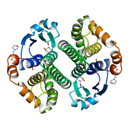
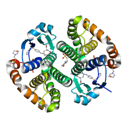
4QM1
 
 | | Crystal Structure of the Inosine 5'-monophosphate Dehydrogenase with an Internal Deletion of the CBS Domain from Bacillus anthracis str. Ames complexed with inhibitor D67 | | 分子名称: | 2-(3-methyl-4-oxo-3,4-dihydrophthalazin-1-yl)-N-(6,7,8,9-tetrahydrodibenzo[b,d]furan-2-yl)acetamide, INOSINIC ACID, Inosine-5'-monophosphate dehydrogenase | | 著者 | Kim, Y, Makowska-Grzyska, M, Gu, M, Mandapati, K, Gollapalli, D, Gorla, S.K, Zhang, M, Hedstrom, L, Anderson, W.F, Joachimiak, A, CSGID, Center for Structural Genomics of Infectious Diseases (CSGID) | | 登録日 | 2014-06-14 | | 公開日 | 2014-07-23 | | 最終更新日 | 2023-09-20 | | 実験手法 | X-RAY DIFFRACTION (2.7964 Å) | | 主引用文献 | Crystal Structure of the Inosine 5'-monophosphate Dehydrogenase with an Internal Deletion of the CBS Domain from Bacillus anthracis str. Ames complexed with inhibitor D67
To be Published, 2014
|
|
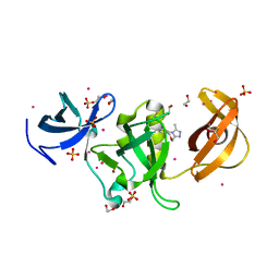
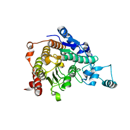
3B6E
 | | Crystal structure of human DECH-box RNA Helicase MDA5 (Melanoma differentiation-associated protein 5), DECH-domain | | 分子名称: | Interferon-induced helicase C domain-containing protein 1, SODIUM ION | | 著者 | Karlberg, T, Welin, M, Arrowsmith, C.H, Berglund, H, Busam, R.D, Collins, R, Dahlgren, L.G, Edwards, A.M, Flodin, S, Flores, A, Graslund, S, Hammarstrom, M, Johansson, I, Kallas, A, Kotenyova, T, Lehtio, L, Moche, M, Nilsson, M.E, Nordlund, P, Nyman, T, Persson, C, Sagemark, J, Svensson, L, Thorsell, A.G, Tresaugues, L, Van Den Berg, S, Weigelt, J, Holmberg-Schiavone, L, Structural Genomics Consortium (SGC) | | 登録日 | 2007-10-29 | | 公開日 | 2007-11-13 | | 最終更新日 | 2024-02-21 | | 実験手法 | X-RAY DIFFRACTION (1.6 Å) | | 主引用文献 | Human DECH-box RNA Helicase MDA5 (Melanoma differentiation-associated protein 5), DECH-domain.
To be Published
|
|
5I2Q
 | | Structure of EF-hand containing protein | | 分子名称: | CALCIUM ION, EF-hand domain-containing protein D2 | | 著者 | Park, K.R, Kwon, M.S, An, J.Y, Lee, J.G, Youn, H.S, Lee, Y, Kang, J.Y, Kim, T.G, Lim, J.J, Park, J.S, Lee, S.H, Song, W.K, Cheong, H, Jun, C, Eom, S.H. | | 登録日 | 2016-02-09 | | 公開日 | 2016-12-28 | | 最終更新日 | 2023-11-08 | | 実験手法 | X-RAY DIFFRACTION (1.935 Å) | | 主引用文献 | Structural implications of Ca(2+)-dependent actin-bundling function of human EFhd2/Swiprosin-1.
Sci Rep, 6, 2016
|
|
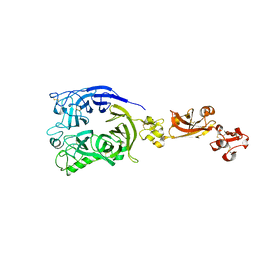
6AI3
 | |
4Q0W
 | | he catalytic core of Rad2 in complex with DNA substrate (complex II) | | 分子名称: | CALCIUM ION, DNA (5'-D(*TP*TP*AP*GP*GP*TP*GP*GP*AP*CP*GP*GP*AP*TP*CP*AP*TP*T)-3'), DNA (5'-D(*TP*TP*TP*GP*AP*TP*CP*CP*GP*TP*CP*CP*AP*CP*CP*TP*TP*T)-3'), ... | | 著者 | Mietus, M, Nowak, E, Jaciuk, M, Kustosz, P, Nowotny, M. | | 登録日 | 2014-04-02 | | 公開日 | 2014-08-27 | | 最終更新日 | 2023-09-20 | | 実験手法 | X-RAY DIFFRACTION (2.1 Å) | | 主引用文献 | Crystal structure of the catalytic core of Rad2: insights into the mechanism of substrate binding.
Nucleic Acids Res., 42, 2014
|
|
5I2O
 | | Structure of EF-hand containing protein | | 分子名称: | CALCIUM ION, EF-hand domain-containing protein D2 | | 著者 | Park, K.R, Kwon, M.S, An, J.Y, Lee, J.G, Youn, H.S, Lee, Y, Kang, J.Y, Kim, T.G, Lim, J.J, Park, J.S, Lee, S.H, Song, W.K, Cheong, H, Jun, C, Eom, S.H. | | 登録日 | 2016-02-09 | | 公開日 | 2016-12-28 | | 最終更新日 | 2023-11-08 | | 実験手法 | X-RAY DIFFRACTION (1.952 Å) | | 主引用文献 | Structural implications of Ca(2+)-dependent actin-bundling function of human EFhd2/Swiprosin-1.
Sci Rep, 6, 2016
|
|
3C0N
 | | Crystal structure of the proaerolysin mutant Y221G at 2.2 A | | 分子名称: | Aerolysin | | 著者 | Pernot, L, Schiltz, M, Thurnheer, S, Burr, S.E, van der Goot, G. | | 登録日 | 2008-01-21 | | 公開日 | 2008-02-12 | | 最終更新日 | 2023-11-01 | | 実験手法 | X-RAY DIFFRACTION (2.2 Å) | | 主引用文献 | Molecular assembly of the aerolysin pore reveals a swirling membrane-insertion mechanism.
Nat.Chem.Biol., 9, 2013
|
|
6ALE
 | | A V-to-F substitution in SK2 channels causes Ca2+ hypersensitivity and improves locomotion in a C. elegans ALS model | | 分子名称: | (3E)-6,7-dichloro-3-(hydroxyimino)-1,3-dihydro-2H-indol-2-one, CALCIUM ION, Calmodulin-2, ... | | 著者 | Nam, Y.W, Zhang, M. | | 登録日 | 2017-08-07 | | 公開日 | 2018-08-08 | | 最終更新日 | 2024-03-13 | | 実験手法 | X-RAY DIFFRACTION (2.5 Å) | | 主引用文献 | A V-to-F substitution in SK2 channels causes Ca2+hypersensitivity and improves locomotion in a C. elegans ALS model.
Sci Rep, 8, 2018
|
|
5UVR
 | |
5UXT
 | | Coiled-coil Trimer with Glu:Trp:Lys Triad | | 分子名称: | GLYCEROL, coiled-coil trimer with Glu:Trp:Lys triad | | 著者 | Smith, M.S, Billings, W.M, Whitby, F.G, Miller, M.B, Price, J.L. | | 登録日 | 2017-02-23 | | 公開日 | 2017-08-09 | | 実験手法 | X-RAY DIFFRACTION (2.197 Å) | | 主引用文献 | Enhancing a long-range salt bridge with intermediate aromatic and nonpolar amino acids.
Org. Biomol. Chem., 15, 2017
|
|
6AH4
 | |
6AI0
 | | Structure of the 328-692 fragment of FlhA (orthorhombic form) | | 分子名称: | (4R)-2-METHYLPENTANE-2,4-DIOL, CALCIUM ION, Flagellar biosynthesis protein FlhA | | 著者 | Ogawa, Y, Kinoshita, M, Minamino, T, Imada, K. | | 登録日 | 2018-08-21 | | 公開日 | 2019-03-20 | | 最終更新日 | 2023-11-22 | | 実験手法 | X-RAY DIFFRACTION (2.4 Å) | | 主引用文献 | Structural Insights into the Substrate Specificity Switch Mechanism of the Type III Protein Export Apparatus.
Structure, 27, 2019
|
|
3C0O
 | | Crystal structure of the proaerolysin mutant Y221G complexed with mannose-6-phosphate | | 分子名称: | 6-O-phosphono-alpha-D-mannopyranose, ACETATE ION, Aerolysin | | 著者 | Pernot, L, Schiltz, M, Thurnheer, S, Burr, S.E, van der Goot, G. | | 登録日 | 2008-01-21 | | 公開日 | 2008-02-12 | | 最終更新日 | 2023-11-01 | | 実験手法 | X-RAY DIFFRACTION (2.5 Å) | | 主引用文献 | Molecular assembly of the aerolysin pore reveals a swirling membrane-insertion mechanism.
Nat.Chem.Biol., 9, 2013
|
|
3C52
 | | Class II fructose-1,6-bisphosphate aldolase from helicobacter pylori in complex with phosphoglycolohydroxamic acid, a competitive inhibitor | | 分子名称: | CALCIUM ION, Fructose-bisphosphate aldolase, PHOSPHOGLYCOLOHYDROXAMIC ACID, ... | | 著者 | Coincon, M, Sygusch, J. | | 登録日 | 2008-01-30 | | 公開日 | 2008-08-26 | | 最終更新日 | 2024-04-03 | | 実験手法 | X-RAY DIFFRACTION (2.3 Å) | | 主引用文献 | Synthesis and Biochemical Evaluation of Selective Inhibitors of Class II Fructose Bisphosphate Aldolases: Towards New Synthetic Antibiotics.
Chemistry, 14, 2008
|
|
3C0M
 | | Crystal structure of the proaerolysin mutant Y221G | | 分子名称: | Aerolysin | | 著者 | Pernot, L, Schiltz, M, Thurnheer, S, Burr, S.E, van der Goot, G. | | 登録日 | 2008-01-21 | | 公開日 | 2008-02-12 | | 最終更新日 | 2023-11-01 | | 実験手法 | X-RAY DIFFRACTION (2.88 Å) | | 主引用文献 | Molecular assembly of the aerolysin pore reveals a swirling membrane-insertion mechanism.
Nat.Chem.Biol., 9, 2013
|
|
18GS
 | | GLUTATHIONE S-TRANSFERASE P1-1 COMPLEXED WITH 1-(S-GLUTATHIONYL)-2,4-DINITROBENZENE | | 分子名称: | 2-(N-MORPHOLINO)-ETHANESULFONIC ACID, GLUTATHIONE S-(2,4 DINITROBENZENE), GLUTATHIONE S-TRANSFERASE | | 著者 | Oakley, A.J, Lo Bello, M, Ricci, G, Federici, G, Parker, M.W. | | 登録日 | 1997-12-07 | | 公開日 | 1999-01-13 | | 最終更新日 | 2024-05-22 | | 実験手法 | X-RAY DIFFRACTION (1.9 Å) | | 主引用文献 | The ligandin (non-substrate) binding site of human Pi class glutathione transferase is located in the electrophile binding site (H-site).
J.Mol.Biol., 291, 1999
|
|
6AU2
 | | Crystal structure of SETDB1 Tudor domain with aryl triazole fragments | | 分子名称: | 1,2-ETHANEDIOL, 1-methyl-4H,6H-[1,2,4]triazolo[4,3-a][4,1]benzoxazepine, BETA-MERCAPTOETHANOL, ... | | 著者 | MADER, P, Mendoza-Sanchez, R, IQBAL, A, DONG, A, DOBROVETSKY, E, CORLESS, V.B, LIEW, S.K, TEMPEL, W, SMIL, D, DELA SENA, C.C, KENNEDY, S, DIAZ, D, HOLOWNIA, A, VEDADI, M, BROWN, P.J, SANTHAKUMAR, V, Bountra, C, Edwards, A.M, YUDIN, A.K, Arrowsmith, C.H, Structural Genomics Consortium (SGC) | | 登録日 | 2017-08-30 | | 公開日 | 2017-10-11 | | 最終更新日 | 2019-10-16 | | 実験手法 | X-RAY DIFFRACTION (1.63 Å) | | 主引用文献 | Identification and characterization of the first fragment hits for SETDB1 Tudor domain.
Bioorg.Med.Chem., 27, 2019
|
|
12GS
 | |
6ATJ
 | | RECOMBINANT HORSERADISH PEROXIDASE C COMPLEX WITH FERULIC ACID | | 分子名称: | 3-(4-HYDROXY-3-METHOXYPHENYL)-2-PROPENOIC ACID, CALCIUM ION, PEROXIDASE C1A, ... | | 著者 | Henriksen, A, Smith, A.T, Gajhede, M. | | 登録日 | 1999-04-23 | | 公開日 | 2000-01-14 | | 最終更新日 | 2023-09-20 | | 実験手法 | X-RAY DIFFRACTION (2 Å) | | 主引用文献 | The structures of the horseradish peroxidase C-ferulic acid complex and the ternary complex with cyanide suggest how peroxidases oxidize small phenolic substrates.
J.Biol.Chem., 274, 1999
|
|
13GS
 | | GLUTATHIONE S-TRANSFERASE COMPLEXED WITH SULFASALAZINE | | 分子名称: | 2-(N-MORPHOLINO)-ETHANESULFONIC ACID, 2-HYDROXY-(5-([4-(2-PYRIDINYLAMINO)SULFONYL]PHENYL)AZO)BENZOIC ACID, GLUTATHIONE, ... | | 著者 | Oakley, A.J, Lo Bello, M, Parker, M.W. | | 登録日 | 1997-11-20 | | 公開日 | 1999-01-13 | | 最終更新日 | 2024-05-22 | | 実験手法 | X-RAY DIFFRACTION (1.9 Å) | | 主引用文献 | The Ligandin (Non-Substrate) Binding Site of Human Pi Class Glutathione Transferase is Located in the Electrophile Binding Site (H-Site).
J.Mol.Biol., 291, 1999
|
|
3C0Y
 | | Crystal structure of catalytic domain of human histone deacetylase HDAC7 | | 分子名称: | Histone deacetylase 7a, POTASSIUM ION, ZINC ION | | 著者 | Min, J.R, Schuetz, A, Allali-Hassani, A, Loppnau, P, Kwiatkowski, N.P, Mazitschek, R, Weigelt, J, Sundstrom, M, Arrowsmith, C.H, Edwards, A.M, Vedadi, M, Bochkarev, A, Plotnikov, A.N, Structural Genomics Consortium (SGC) | | 登録日 | 2008-01-21 | | 公開日 | 2008-02-19 | | 最終更新日 | 2023-08-30 | | 実験手法 | X-RAY DIFFRACTION (2.1 Å) | | 主引用文献 | Human HDAC7 harbors a class IIa histone deacetylase-specific zinc binding motif and cryptic deacetylase activity.
J.Biol.Chem., 283, 2008
|
|
7Y4P
 | | Human Plexin A1, extracellular domains 1-4 | | 分子名称: | 2-acetamido-2-deoxy-beta-D-glucopyranose, 2-acetamido-2-deoxy-beta-D-glucopyranose-(1-4)-2-acetamido-2-deoxy-beta-D-glucopyranose, Plexin-A1 | | 著者 | Tanaka, T, Neyazaki, M, Nogi, T. | | 登録日 | 2022-06-15 | | 公開日 | 2022-10-19 | | 最終更新日 | 2023-11-29 | | 実験手法 | X-RAY DIFFRACTION (3.5 Å) | | 主引用文献 | Hybrid in vitro/in silico analysis of low-affinity protein-protein interactions that regulate signal transduction by Sema6D.
Protein Sci., 31, 2022
|
|
3ELD
 | | Wesselsbron methyltransferase in complex with Sinefungin | | 分子名称: | Methyltransferase, SINEFUNGIN, SULFATE ION | | 著者 | Bollati, M, Milani, M, Mastrangelo, E, Ricagno, S, Bolognesi, M. | | 登録日 | 2008-09-22 | | 公開日 | 2008-11-18 | | 最終更新日 | 2023-11-01 | | 実験手法 | X-RAY DIFFRACTION (1.9 Å) | | 主引用文献 | Recognition of RNA Cap in the Wesselsbron Virus NS5 Methyltransferase Domain: Implications for RNA-Capping Mechanisms in Flavivirus
J.Mol.Biol., 385, 2009
|
|
4QOX
 | | Crystal Structure of CDPK4 from Plasmodium Falciparum, PF3D7_0717500 | | 分子名称: | 3-(3-bromobenzyl)-1-tert-butyl-1H-pyrazolo[3,4-d]pyrimidin-4-amine, Calcium-dependent protein kinase 4, MAGNESIUM ION | | 著者 | Wernimont, A.K, Walker, J.R, Hutchinson, A, Seitova, A, He, H, Loppnau, P, Neculai, M, Amani, M, Lin, Y.H, Ravichandran, M, Arrowsmith, C.H, Edwards, A.M, Bountra, C, Hui, R, Lovato, D.V, Structural Genomics Consortium (SGC) | | 登録日 | 2014-06-20 | | 公開日 | 2014-09-03 | | 最終更新日 | 2024-02-28 | | 実験手法 | X-RAY DIFFRACTION (2.748 Å) | | 主引用文献 | Crystal Structure of CDPK4 from Plasmodium Falciparum, PF3D7_0717500
TO BE PUBLISHED
|
|
3ESZ
 | | K2AK3A Flavodoxin from Anabaena | | 分子名称: | FLAVIN MONONUCLEOTIDE, FLAVODOXIN, GLYCEROL, ... | | 著者 | Herguedas, B, Martinez-Julvez, M, Hermoso, J, Goni, G, Medina, M. | | 登録日 | 2008-10-06 | | 公開日 | 2009-02-10 | | 最終更新日 | 2023-11-01 | | 実験手法 | X-RAY DIFFRACTION (1.94 Å) | | 主引用文献 | Flavodoxin: A compromise between efficiency and versatility in the electron transfer from Photosystem I to Ferredoxin-NADP(+) reductase
Biochim.Biophys.Acta, 1787, 2009
|
|


