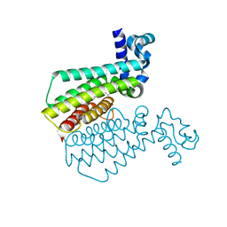
7OXX
 
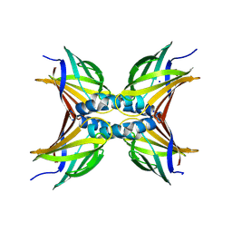 | | CrabP2 mutant R30AK31A | | 分子名称: | Cellular retinoic acid-binding protein 2, SODIUM ION | | 著者 | Tomlinson, C.W.E, Basle, A, Pohl, E. | | 登録日 | 2021-06-23 | | 公開日 | 2022-07-13 | | 最終更新日 | 2024-01-31 | | 実験手法 | X-RAY DIFFRACTION (1.33 Å) | | 主引用文献 | Structural requirements for the specific binding of CRABP2 to cyclin D3
To Be Published
|
|
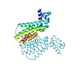
7AA1
 | | Structural comparison of cellular retinoic acid binding proteins I and II in the presence and absence of natural and synthetic ligands | | 分子名称: | 4-[2-(5,5,8,8-tetramethyl-6,7-dihydroquinoxalin-2-yl)ethynyl]benzoic acid, Cellular retinoic acid-binding protein 2 | | 著者 | Tomlinson, C.W.E, Cornish, K.A.S, Pohl, E. | | 登録日 | 2020-09-02 | | 公開日 | 2021-02-17 | | 最終更新日 | 2024-01-31 | | 実験手法 | X-RAY DIFFRACTION (1.71 Å) | | 主引用文献 | Structure-functional relationship of cellular retinoic acid-binding proteins I and II interacting with natural and synthetic ligands.
Acta Crystallogr D Struct Biol, 77, 2021
|
|
7A9Z
 | | Structural comparison of cellular retinoic acid binding protein I and II in the presence and absence of natural and synthetic ligands | | 分子名称: | 4-[2-(5,5,8,8-tetramethyl-6,7-dihydroquinoxalin-2-yl)ethynyl]benzoic acid, Cellular retinoic acid-binding protein 1 | | 著者 | Tomlinson, C.W.E, Cornish, K.A.S, Pohl, E. | | 登録日 | 2020-09-02 | | 公開日 | 2021-02-17 | | 最終更新日 | 2024-01-31 | | 実験手法 | X-RAY DIFFRACTION (2.41 Å) | | 主引用文献 | Structure-functional relationship of cellular retinoic acid-binding proteins I and II interacting with natural and synthetic ligands.
Acta Crystallogr D Struct Biol, 77, 2021
|
|
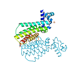
7AA0
 | | Structural comparison of cellular retinoic acid binding protein I and II in the presence and absence of natural and synthetic ligands | | 分子名称: | (~{E})-3-[4-(4,4-dimethyl-1-propan-2-yl-2,3-dihydroquinolin-6-yl)phenyl]prop-2-enoic acid, Cellular retinoic acid-binding protein 2 | | 著者 | Tomlinson, C.W.E, Cornish, K.A.S, Pohl, E. | | 登録日 | 2020-09-02 | | 公開日 | 2021-02-17 | | 最終更新日 | 2024-01-31 | | 実験手法 | X-RAY DIFFRACTION (1.82 Å) | | 主引用文献 | Structure-functional relationship of cellular retinoic acid-binding proteins I and II interacting with natural and synthetic ligands.
Acta Crystallogr D Struct Biol, 77, 2021
|
|
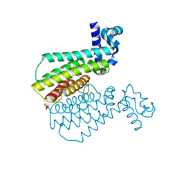
7A9Y
 | | Structural comparison of cellular retinoic acid binding protein I and II in the presence and absence of natural and synthetic ligands | | 分子名称: | Cellular retinoic acid-binding protein 1, GLYCEROL, MYRISTIC ACID, ... | | 著者 | Tomlinson, C.W.E, Cornish, K.A.S, Pohl, E. | | 登録日 | 2020-09-02 | | 公開日 | 2021-02-17 | | 最終更新日 | 2024-01-31 | | 実験手法 | X-RAY DIFFRACTION (1.64 Å) | | 主引用文献 | Structure-functional relationship of cellular retinoic acid-binding proteins I and II interacting with natural and synthetic ligands.
Acta Crystallogr D Struct Biol, 77, 2021
|
|
7NGJ
 | |
7NGD
 | |
7NGO
 | |
7NGR
 | |
7NGS
 | |
7NGX
 | |
7NGK
 | |
7NGT
 | |
7NGU
 | |
7NGN
 | |
7NGG
 | |
7NGM
 | |
7NGI
 | |
7NGY
 | |
7NGW
 | |
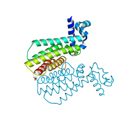
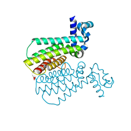
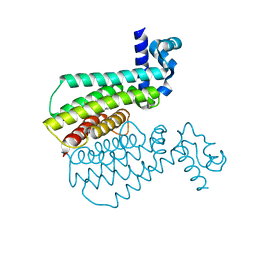
7OCI
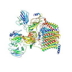 | | Cryo-EM structure of yeast Ost6p containing oligosaccharyltransferase complex | | 分子名称: | 1,2-dioleoyl-sn-glycero-3-phosphoethanolamine, 2-acetamido-2-deoxy-beta-D-glucopyranose, 2-acetamido-2-deoxy-beta-D-glucopyranose-(1-4)-2-acetamido-2-deoxy-beta-D-glucopyranose, ... | | 著者 | Wild, R, Neuhaus, J.D, Eyring, J, Irobalieva, R.N, Kowal, J, Lin, C.W, Locher, K.P, Aebi, M. | | 登録日 | 2021-04-27 | | 公開日 | 2022-02-16 | | 実験手法 | ELECTRON MICROSCOPY (3.46 Å) | | 主引用文献 | Functional analysis of Ost3p and Ost6p containing yeast oligosaccharyltransferases.
Glycobiology, 31, 2021
|
|
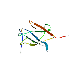
3KCW
 | | Crystal structure of Ganoderma fungal immunomodulatory protein, GMI | | 分子名称: | immunomodulatory protein | | 著者 | Hsu, M.F, Wang, A.H.J, Yang, C.S, Huang, C.T, Hseu, R.S, Lin, C.W, Wu, M.Y, Huang, C.S, Fu, H.Y. | | 登録日 | 2009-10-22 | | 公開日 | 2010-11-03 | | 最終更新日 | 2023-11-01 | | 実験手法 | X-RAY DIFFRACTION (2 Å) | | 主引用文献 | Single cysteine replacement at Leu6 increase the potent and thermostability in Ganoderma fungal immunomodulatory proteins
To be Published
|
|
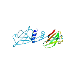
1PJW
 | | Solution Structure of the Domain III of the Japan Encephalitis Virus Envelope Protein | | 分子名称: | envelope protein | | 著者 | Wu, K.P, Wu, C.W, Tsao, Y.P, Lou, Y.C, Lin, C.W. | | 登録日 | 2003-06-04 | | 公開日 | 2003-11-25 | | 最終更新日 | 2022-02-23 | | 実験手法 | SOLUTION NMR | | 主引用文献 | Structural Basis of a Flavivirus Recognized by Its Neutralizing Antibody: SOLUTION STRUCTURE OF THE DOMAIN III OF THE JAPANESE ENCEPHALITIS VIRUS ENVELOPE PROTEIN.
J.Biol.Chem., 278, 2003
|
|
2VPV
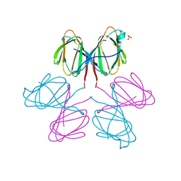 | | Dimerization Domain of Mif2p | | 分子名称: | PROTEIN MIF2, SULFATE ION | | 著者 | Cohen, R.L, Espelin, C.W, Sorger, P.K, Harrison, S.C, Simons, K.T. | | 登録日 | 2008-03-05 | | 公開日 | 2008-08-26 | | 最終更新日 | 2019-05-08 | | 実験手法 | X-RAY DIFFRACTION (2.7 Å) | | 主引用文献 | Structural and Functional Dissection of Mif2P, a Conserved DNA-Binding Kinetochore Protein.
Mol.Biol.Cell, 19, 2008
|
|
8V7R
 | | PanDDA analysis -- Crystal Structure of Zika virus NS3 Helicase in complex with Z56772132 | | 分子名称: | (5R)-5-[2-(4-methoxyphenyl)ethyl]-5-methylimidazolidine-2,4-dione, 1,2-ETHANEDIOL, DIMETHYL SULFOXIDE, ... | | 著者 | Godoy, A.S, Noske, G.D, Fairhead, M, Lithgo, R.M, Koekemoer, L, Aschenbrenner, J.C, Balcomb, B.H, Marples, P.G, Ni, X, Tomlinson, C.W.E, Wild, C, Mesquita, N.C.M.R, Oliva, G, Fearon, D, Walsh, M.A, von Delft, F. | | 登録日 | 2023-12-04 | | 公開日 | 2023-12-20 | | 実験手法 | X-RAY DIFFRACTION (1.41 Å) | | 主引用文献 | PanDDA analysis -- Crystal Structure of Zika virus NS3 Helicase in complex with Z56772132
To Be Published
|
|


