Loading
PDBj
MenuPDBj@FacebookPDBj@TwitterPDBj@YouTubewwPDB FoundationwwPDB
RCSB PDBPDBeBMRBAdv. SearchSearch help
PDB: 205 results

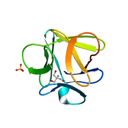
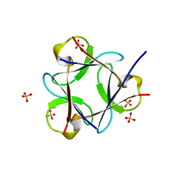
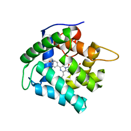
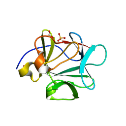
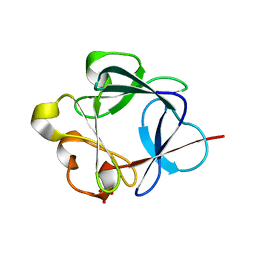
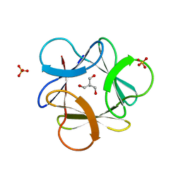
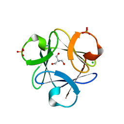
3O4A
DownloadVisualize
BU of 3o4a by Molmil
Crystal structure of Symfoil-2: de novo designed beta-trefoil architecture with symmetric primary structure
Descriptor: 2-AMINO-2-HYDROXYMETHYL-PROPANE-1,3-DIOL, SULFATE ION, de novo designed beta-trefoil architecture with symmetric primary structure
Authors:Lee, J, Blaber, M.
Deposit date:2010-07-26
Release date:2010-12-22
Last modified:2024-02-21
Method:X-RAY DIFFRACTION (1.45 Å)
Cite:Experimental support for the evolution of symmetric protein architecture from a simple peptide motif.
Proc.Natl.Acad.Sci.USA, 108, 2011
3O4D
DownloadVisualize
BU of 3o4d by Molmil
Crystal structure of Symfoil-4P: de novo designed beta-trefoil architecture with symmetric primary structure
Descriptor: 2-AMINO-2-HYDROXYMETHYL-PROPANE-1,3-DIOL, CHLORIDE ION, SULFATE ION, ...
Authors:Lee, J, Blaber, M.
Deposit date:2010-07-26
Release date:2010-12-22
Last modified:2024-02-21
Method:X-RAY DIFFRACTION (1.65 Å)
Cite:Experimental support for the evolution of symmetric protein architecture from a simple peptide motif.
Proc.Natl.Acad.Sci.USA, 108, 2011
3O49
DownloadVisualize
BU of 3o49 by Molmil
Crystal structure of Symfoil-1: de novo designed beta-trefoil architecture with symmetric primary structure
Descriptor: 2-AMINO-2-HYDROXYMETHYL-PROPANE-1,3-DIOL, SULFATE ION, de novo designed beta-trefoil architecture with symmetric primary structure
Authors:Lee, J, Blaber, M.
Deposit date:2010-07-26
Release date:2010-12-22
Last modified:2024-02-21
Method:X-RAY DIFFRACTION (1.45 Å)
Cite:Experimental support for the evolution of symmetric protein architecture from a simple peptide motif.
Proc.Natl.Acad.Sci.USA, 108, 2011
3O3Q
DownloadVisualize
BU of 3o3q by Molmil
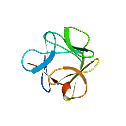
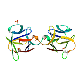
Crystal structure of "L44F/M67I/L73V/A103G/deletion 104-106/F108Y/V109L/L111I/C117V/R119G/deletion 120-122" mutant form of Human acidic fibroblast growth factor
Descriptor: GLYCEROL, Heparin-binding growth factor 1
Authors:Lee, J, Blaber, M.
Deposit date:2010-07-25
Release date:2011-02-23
Last modified:2023-09-06
Method:X-RAY DIFFRACTION (1.6 Å)
Cite:A polypeptide "building block"top-down symmetric deconstruction".
J.Mol.Biol., 407, 2011
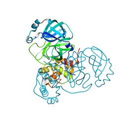
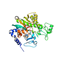
7KHP
DownloadVisualize
BU of 7khp by Molmil
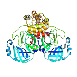
Acyl-enzyme intermediate structure of SARS-CoV-2 Mpro in complex with its C-terminal autoprocessing sequence.
Descriptor: 3C-like proteinase, DIMETHYL SULFOXIDE
Authors:Lee, J, Worrall, L.J, Paetzel, M, Strynadka, N.C.J.
Deposit date:2020-10-21
Release date:2020-10-28
Last modified:2023-10-18
Method:X-RAY DIFFRACTION (1.95 Å)
Cite:Crystallographic structure of wild-type SARS-CoV-2 main protease acyl-enzyme intermediate with physiological C-terminal autoprocessing site.
Nat Commun, 11, 2020
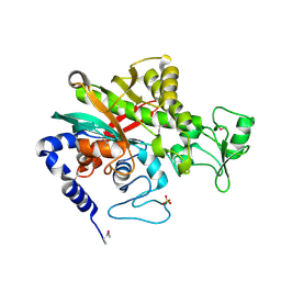
7JOY
DownloadVisualize
BU of 7joy by Molmil
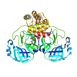
Product structure of SARS-CoV-2 Mpro C145A mutant in complex with its C-terminal autoprocessing sequence.
Descriptor: 3C-like proteinase
Authors:Lee, J, Worrall, L.J, Paetzel, M, Strynadka, N.C.J.
Deposit date:2020-08-07
Release date:2020-10-07
Last modified:2023-10-18
Method:X-RAY DIFFRACTION (2 Å)
Cite:Crystallographic structure of wild-type SARS-CoV-2 main protease acyl-enzyme intermediate with physiological C-terminal autoprocessing site.
Nat Commun, 11, 2020
7JP1
DownloadVisualize
BU of 7jp1 by Molmil
Structure of wild-type substrate free SARS-CoV-2 Mpro.
Descriptor: 3C-like proteinase
Authors:Lee, J, Worrall, L.J, Paetzel, M, Strynadka, N.C.J.
Deposit date:2020-08-07
Release date:2020-10-07
Last modified:2023-10-18
Method:X-RAY DIFFRACTION (1.8 Å)
Cite:Crystallographic structure of wild-type SARS-CoV-2 main protease acyl-enzyme intermediate with physiological C-terminal autoprocessing site.
Nat Commun, 11, 2020
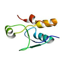
5UMV
DownloadVisualize
BU of 5umv by Molmil
Crystal structure of the BRCT domain of S. cerevisiae Rev1
Descriptor: DNA repair protein REV1
Authors:Lee, J, Xu, C, Thompson, J.R, Botuyan, M.V, Mer, G.
Deposit date:2017-01-29
Release date:2018-01-31
Last modified:2023-10-04
Method:X-RAY DIFFRACTION (1.75 Å)
Cite:Crystal structure of the BRCT domain of S. cerevisiae Rev1
To Be Published
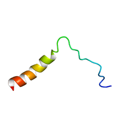
5T42
DownloadVisualize
BU of 5t42 by Molmil
Structure of the Ebola virus envelope protein MPER/TM domain and its interaction with the fusion loop explains their fusion activity
Descriptor: Envelope glycoprotein
Authors:Lee, J, Nyenhuis, D.A, Nelson, E.A, Cafiso, D.S, White, J.M, Tamm, L.K.
Deposit date:2016-08-28
Release date:2017-08-30
Last modified:2024-05-15
Method:SOLUTION NMR
Cite:Structure of the Ebola virus envelope protein MPER/TM domain and its interaction with the fusion loop explains their fusion activity.
Proc. Natl. Acad. Sci. U.S.A., 114, 2017
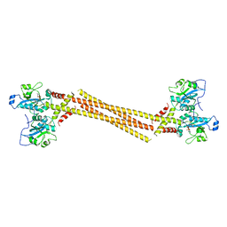
4MVD
DownloadVisualize
BU of 4mvd by Molmil
Crystal Structure of a Mammalian Cytidylyltransferase
Descriptor: Choline-phosphate cytidylyltransferase A, [2-CYTIDYLATE-O'-PHOSPHONYLOXYL]-ETHYL-TRIMETHYL-AMMONIUM
Authors:Lee, J, Cornell, R.B.
Deposit date:2013-09-23
Release date:2013-12-11
Last modified:2024-02-28
Method:X-RAY DIFFRACTION (8 Å)
Cite:Structural Basis for Autoinhibition of CTP:Phosphocholine Cytidylyltransferase (CCT), the Regulatory Enzyme in Phosphatidylcholine Synthesis, by Its Membrane-binding Amphipathic Helix.
J.Biol.Chem., 289, 2014
3OL0
DownloadVisualize
BU of 3ol0 by Molmil
Crystal structure of Monofoil-4P homo-trimer: de novo designed monomer trefoil-fold sub-domain which forms homo-trimer assembly
Descriptor: SULFATE ION, de novo designed monomer trefoil-fold sub-domain which forms homo-trimer assembly
Authors:Lee, J, Blaber, M.
Deposit date:2010-08-25
Release date:2010-12-22
Last modified:2024-02-21
Method:X-RAY DIFFRACTION (1.483 Å)
Cite:Experimental support for the evolution of symmetric protein architecture from a simple peptide motif.
Proc.Natl.Acad.Sci.USA, 108, 2011
3PD7
DownloadVisualize
BU of 3pd7 by Molmil
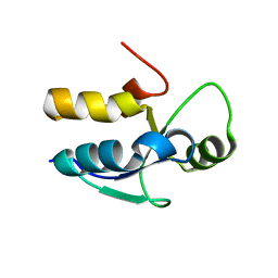
Crystal Structure of the Sixth BRCT Domain of Human TopBP1
Descriptor: DNA topoisomerase 2-binding protein 1
Authors:Lee, J, Xu, C, Cui, G, Thompson, J.R, Mer, G.
Deposit date:2010-10-22
Release date:2010-12-08
Last modified:2024-02-21
Method:X-RAY DIFFRACTION (1.26 Å)
Cite:Crystal Structure of the Sixth BRCT Domain of Human TopBP1
To be Published
3OGF
DownloadVisualize
BU of 3ogf by Molmil
Crystal structure of Difoil-4P homo-trimer: de novo designed dimeric trefoil-fold sub-domain which forms homo-trimer assembly
Descriptor: SULFATE ION, de novo designed dimeric trefoil-fold sub-domain which forms homo-trimer assembly
Authors:Lee, J, Blaber, M.
Deposit date:2010-08-16
Release date:2010-12-22
Last modified:2024-02-21
Method:X-RAY DIFFRACTION (2.864 Å)
Cite:Experimental support for the evolution of symmetric protein architecture from a simple peptide motif.
Proc.Natl.Acad.Sci.USA, 108, 2011
7Y7O
DownloadVisualize
BU of 7y7o by Molmil
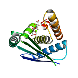
Crystal structure of metallo-endoribonuclease YbeY from Staphylococcus aureus
Descriptor: CITRIC ACID, Endoribonuclease YbeY, ZINC ION
Authors:Lee, J, Ha, N.-C.
Deposit date:2022-06-22
Release date:2023-03-08
Last modified:2024-05-29
Method:X-RAY DIFFRACTION (1.9 Å)
Cite:Crystal Structure of the Metallo-Endoribonuclease YbeY from Staphylococcus aureus.
J Microbiol Biotechnol., 33, 2023
7WBM
DownloadVisualize
BU of 7wbm by Molmil
Crystal structure of Legionella pneumophila effector protein Lpg0081
Descriptor: Lpg0081, [(2R,3S,4R,5R)-5-(6-AMINOPURIN-9-YL)-3,4-DIHYDROXY-OXOLAN-2-YL]METHYL [HYDROXY-[[(2R,3S,4R,5S)-3,4,5-TRIHYDROXYOXOLAN-2-YL]METHOXY]PHOSPHORYL] HYDROGEN PHOSPHATE
Authors:Lee, J, Kim, H, Oh, B.H.
Deposit date:2021-12-17
Release date:2022-06-15
Last modified:2023-11-29
Method:X-RAY DIFFRACTION (2.7 Å)
Cite:Reversible modification of mitochondrial ADP/ATP translocases by paired Legionella effector proteins.
Proc.Natl.Acad.Sci.USA, 119, 2022
7WBK
DownloadVisualize
BU of 7wbk by Molmil
Crystal structure of Legionella pneumophila effector protein Lpg0081
Descriptor: Lpg0081, SULFATE ION
Authors:Lee, J, Kim, H, Oh, B.H.
Deposit date:2021-12-16
Release date:2022-06-15
Method:X-RAY DIFFRACTION (2.74 Å)
Cite:Reversible modification of mitochondrial ADP/ATP translocases by paired Legionella effector proteins.
Proc.Natl.Acad.Sci.USA, 119, 2022
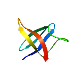
2LXJ
DownloadVisualize
BU of 2lxj by Molmil
Backbone 1H, 13C, and 15N Chemical Shift Assignments for cold shock protein, LmCsp with dT7
Descriptor: Cold shock-like protein CspLA
Authors:Lee, J, Jeong, K, Kim, Y.
Deposit date:2012-08-27
Release date:2013-08-07
Last modified:2024-05-15
Method:SOLUTION NMR
Cite:Structural and Dynamic Features of Cold-Shock Proteins of Listeriamonocytogenes, a Psychrophilic Bacterium
Biochemistry, 52, 2013
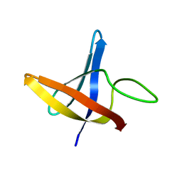
2LXK
DownloadVisualize
BU of 2lxk by Molmil
Backbone 1H, 13C, and 15N Chemical Shift Assignments for cold shock protein, LmCsp
Descriptor: Cold shock-like protein CspLA
Authors:Lee, J, Jeong, K, Kim, Y.
Deposit date:2012-08-27
Release date:2013-08-07
Last modified:2024-05-15
Method:SOLUTION NMR
Cite:Structural and Dynamic Features of Cold-Shock Proteins of Listeriamonocytogenes, a Psychrophilic Bacterium
Biochemistry, 52, 2013
8HU2
DownloadVisualize
BU of 8hu2 by Molmil
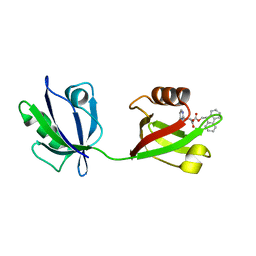
Rattus Syntenin-1 PDZ domain with inhibitor
Descriptor: (2~{S})-2-(9~{H}-fluoren-9-ylmethoxycarbonylamino)-3-(4-oxidanylidene-5~{H}-pyrimidin-2-yl)propanoic acid, Syntenin-1
Authors:Heo, Y, Lee, J, Yun, J.H, Lee, W.
Deposit date:2022-12-22
Release date:2024-01-17
Method:X-RAY DIFFRACTION (1.6 Å)
Cite:Crystal structure of STNPDZ with inhibitor at 1.60 Angstroms resolution.
To Be Published
1LX8
DownloadVisualize
BU of 1lx8 by Molmil
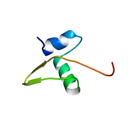
Regulation of directionality in bacteriophage lambda site-specific recombination: structure of the Xis protein
Descriptor: Excisionase
Authors:Sam, M.D, Papagiannis, C, Connolly, K.M, Corselli, L, Iwahara, J, Lee, J, Phillips, M, Wojciak, J.M, Johnson, R.C, Clubb, R.T.
Deposit date:2002-06-04
Release date:2003-06-10
Last modified:2024-05-22
Method:SOLUTION NMR
Cite:Regulation of directionality in bacteriophage lambda site-specific recombination: structure of the Xis protein
J.Mol.Biol., 324, 2002
2PNL
DownloadVisualize
BU of 2pnl by Molmil
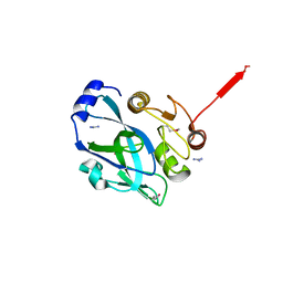
Crystal structure of VP4 protease from infectious pancreatic necrosis virus (IPNV) in space group P1
Descriptor: GUANIDINE, Protease VP4
Authors:Paetzel, M, Lee, J, Feldman, A.R, Delmas, B.
Deposit date:2007-04-24
Release date:2007-06-05
Last modified:2021-10-20
Method:X-RAY DIFFRACTION (2.21 Å)
Cite:Crystal structure of the VP4 protease from infectious pancreatic necrosis virus reveals the acyl-enzyme complex for an intermolecular self-cleavage reaction.
J.Biol.Chem., 282, 2007
2PNM
DownloadVisualize
BU of 2pnm by Molmil
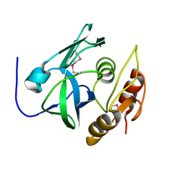
Crystal Structure of VP4 protease from infectious pancreatic necrosis virus (IPNV) in space group P6122
Descriptor: Protease VP4
Authors:Paetzel, M, Lee, J, Feldman, A.R, Delmas, B.
Deposit date:2007-04-24
Release date:2007-06-05
Last modified:2021-10-20
Method:X-RAY DIFFRACTION (2.3 Å)
Cite:Crystal structure of the VP4 protease from infectious pancreatic necrosis virus reveals the acyl-enzyme complex for an intermolecular self-cleavage reaction.
J.Biol.Chem., 282, 2007
4N1F
DownloadVisualize
BU of 4n1f by Molmil
Crystal Structure of F88Y obelin mutant from Obelia longissima at 2.09 Angstrom resolution
Descriptor: C2-HYDROPEROXY-COELENTERAZINE, Obelin
Authors:Natashin, P.V, Markova, S.V, Lee, J, Vysotski, E.S, Liu, Z.J.
Deposit date:2013-10-04
Release date:2014-02-19
Last modified:2023-11-08
Method:X-RAY DIFFRACTION (2.087 Å)
Cite:Crystal structures of the F88Y obelin mutant before and after bioluminescence provide molecular insight into spectral tuning among hydromedusan photoproteins
Febs J., 281, 2014
3BA5
DownloadVisualize
BU of 3ba5 by Molmil
Crystal structure of D28A mutant of Human acidic fibroblast growth factor
Descriptor: FORMIC ACID, Heparin-binding growth factor 1, SULFATE ION
Authors:Blaber, M, Lee, J.
Deposit date:2007-11-07
Release date:2008-04-15
Last modified:2023-08-30
Method:X-RAY DIFFRACTION (1.75 Å)
Cite:A logical OR redundancy within the Asx-Pro-Asx-Gly type I beta-turn motif.
J.Mol.Biol., 377, 2008
3BAD
DownloadVisualize
BU of 3bad by Molmil
Crystal structure of D70A/H93G mutant of Human acidic fibroblast growth factor
Descriptor: Heparin-binding growth factor 1, SULFATE ION
Authors:Blaber, M, Lee, J.
Deposit date:2007-11-07
Release date:2008-04-15
Last modified:2023-08-30
Method:X-RAY DIFFRACTION (2 Å)
Cite:A logical OR redundancy within the Asx-Pro-Asx-Gly type I beta-turn motif.
J.Mol.Biol., 377, 2008

221371

数据于2024-06-19公开中

PDB statisticsPDBj update infoContact PDBjnumon