Loading
PDBj
MenuPDBj@FacebookPDBj@TwitterPDBj@YouTubewwPDB FoundationwwPDB
RCSB PDBPDBeBMRBAdv. SearchSearch help
PDB: 215 results

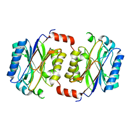
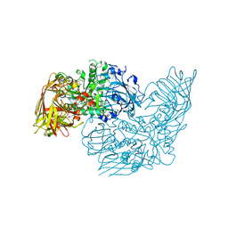
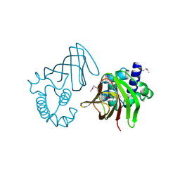
1YT8
DownloadVisualize
BU of 1yt8 by Molmil
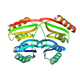
Crystal Structure of Thiosulfate sulfurtransferase from Pseudomonas aeruginosa
Descriptor: GLYCEROL, SULFITE ION, thiosulfate sulfurtransferase
Authors:Kumaran, D, Swaminathan, S, Burley, S.K, New York SGX Research Center for Structural Genomics (NYSGXRC)
Deposit date:2005-02-10
Release date:2005-03-22
Last modified:2021-02-03
Method:X-RAY DIFFRACTION (1.9 Å)
Cite:Crystal Structure of Thiosulfate sulfurtransferase from Pseudomonas aeruginosa
To be Published
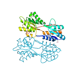
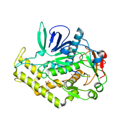
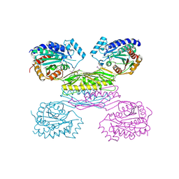
1Y9Q
DownloadVisualize
BU of 1y9q by Molmil
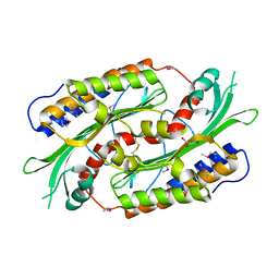
Crystal Structure of HTH_3 family Transcriptional Regulator from Vibrio cholerae
Descriptor: D-METHIONINE, ZINC ION, transcriptional regulator, ...
Authors:Kumaran, D, Swaminathan, S, Burley, S.K, New York SGX Research Center for Structural Genomics (NYSGXRC)
Deposit date:2004-12-16
Release date:2005-01-04
Last modified:2021-02-03
Method:X-RAY DIFFRACTION (1.9 Å)
Cite:Crystal Structure of HTH_3 family Transcriptional Regulator from Vibrio cholerae
To be Published
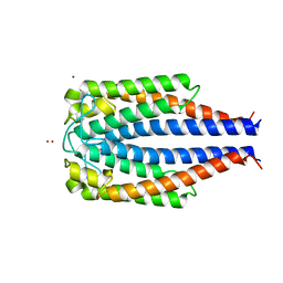
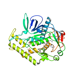
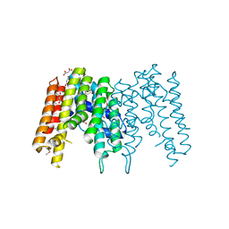
1F1M
DownloadVisualize
BU of 1f1m by Molmil
CRYSTAL STRUCTURE OF OUTER SURFACE PROTEIN C (OSPC)
Descriptor: OUTER SURFACE PROTEIN C, ZINC ION
Authors:Kumaran, D, Eswaramoorthy, S, Dunn, J.J, Swaminathan, S.
Deposit date:2000-05-19
Release date:2001-04-04
Last modified:2024-02-07
Method:X-RAY DIFFRACTION (1.8 Å)
Cite:Crystal structure of outer surface protein C (OspC) from the Lyme disease spirochete, Borrelia burgdorferi.
EMBO J., 20, 2001
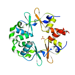
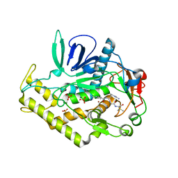
1YAV
DownloadVisualize
BU of 1yav by Molmil
Crystal structure of CBS domain-containing protein ykuL from Bacillus subtilis
Descriptor: SULFATE ION, hypothetical protein BSU14130
Authors:Kumaran, D, Swaminathan, S, Burley, S.K, New York SGX Research Center for Structural Genomics (NYSGXRC)
Deposit date:2004-12-17
Release date:2004-12-28
Last modified:2024-02-14
Method:X-RAY DIFFRACTION (2.1 Å)
Cite:Crystal Structure of a hypothetical protein (ykul) containing CBS domains from Bacillus subtilis.
To be Published
1F89
DownloadVisualize
BU of 1f89 by Molmil
Crystal structure of Saccharomyces cerevisiae Nit3, a member of branch 10 of the nitrilase superfamily
Descriptor: 32.5 KDA PROTEIN YLR351C
Authors:Kumaran, D, Eswaramoorthy, S, Studier, F.W, Swaminathan, S, Burley, S.K, New York SGX Research Center for Structural Genomics (NYSGXRC)
Deposit date:2000-06-29
Release date:2001-10-04
Last modified:2024-02-07
Method:X-RAY DIFFRACTION (2.4 Å)
Cite:Crystal structure of a putative CN hydrolase from yeast
Proteins, 52, 2003
3BBL
DownloadVisualize
BU of 3bbl by Molmil
Crystal structure of a regulatory protein of LacI family from Chloroflexus aggregans
Descriptor: 1,2-ETHANEDIOL, Regulatory protein of LacI family
Authors:Kumaran, D, Burley, S.K, Swaminathan, S, New York SGX Research Center for Structural Genomics (NYSGXRC)
Deposit date:2007-11-09
Release date:2007-11-27
Last modified:2021-02-03
Method:X-RAY DIFFRACTION (2.35 Å)
Cite:Crystal structure of a regulatory protein of LacI family from the Chloroflexus aggregans.
To be Published
3DEC
DownloadVisualize
BU of 3dec by Molmil
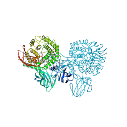
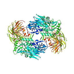
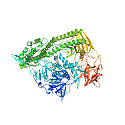
Crystal structure of a glycosyl hydrolases family 2 protein from Bacteroides thetaiotaomicron
Descriptor: Beta-galactosidase, POTASSIUM ION
Authors:Kumaran, D, Bonanno, J, Romero, R, Burley, S.K, Swaminathan, S, New York SGX Research Center for Structural Genomics (NYSGXRC)
Deposit date:2008-06-09
Release date:2008-06-17
Last modified:2023-11-15
Method:X-RAY DIFFRACTION (2.8 Å)
Cite:Crystal Structure of a Glycosyl Hydrolases Family 2 protein from Bacteroides thetaiotaomicron.
To be Published
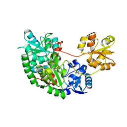
3DDA
DownloadVisualize
BU of 3dda by Molmil
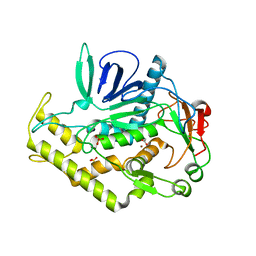
Crystal structure of the catalytic domain of Botulinum neurotoxin serotype a with a snap-25 peptide
Descriptor: Botulinum neurotoxin A light chain, SULFATE ION, Synaptosomal-associated protein 25, ...
Authors:Kumaran, D, Swaminathan, S.
Deposit date:2008-06-05
Release date:2008-09-16
Last modified:2023-08-30
Method:X-RAY DIFFRACTION (1.5 Å)
Cite:Substrate binding mode and its implication on drug design for botulinum neurotoxin A
Plos Pathog., 4, 2008
3C8B
DownloadVisualize
BU of 3c8b by Molmil
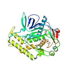
Crystal structure of the catalytic domain of botulinum neurotoxin serotype A with inhibitory peptide RRGI
Descriptor: Botulinum neurotoxin A light chain, Inhibitor peptide RRGI, SULFATE ION, ...
Authors:Kumaran, D, Swaminathan, S.
Deposit date:2008-02-11
Release date:2008-04-22
Last modified:2023-08-30
Method:X-RAY DIFFRACTION (1.47 Å)
Cite:Structure- and Substrate-based Inhibitor Design for Clostridium botulinum Neurotoxin Serotype A
J.Biol.Chem., 283, 2008
3C89
DownloadVisualize
BU of 3c89 by Molmil
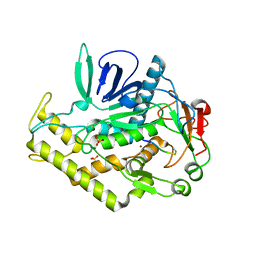
Crystal structure of the catalytic domain of botulinum neurotoxin serotype A with inhibitory peptide RRGM
Descriptor: Botulinum neurotoxin A light chain, Inhibitor peptide RRGM, SULFATE ION, ...
Authors:Kumaran, D, Swaminathan, S.
Deposit date:2008-02-11
Release date:2008-04-22
Last modified:2023-08-30
Method:X-RAY DIFFRACTION (1.58 Å)
Cite:Structure- and Substrate-based Inhibitor Design for Clostridium botulinum Neurotoxin Serotype A
J.Biol.Chem., 283, 2008
3CIH
DownloadVisualize
BU of 3cih by Molmil
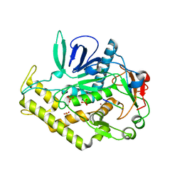
Crystal structure of a putative alpha-rhamnosidase from Bacteroides thetaiotaomicron
Descriptor: 1,2-ETHANEDIOL, 2-AMINO-2-HYDROXYMETHYL-PROPANE-1,3-DIOL, Putative alpha-rhamnosidase
Authors:Kumaran, D, Burley, S.K, Swaminathan, S, New York SGX Research Center for Structural Genomics (NYSGXRC)
Deposit date:2008-03-11
Release date:2008-04-01
Last modified:2021-02-03
Method:X-RAY DIFFRACTION (2.33 Å)
Cite:Crystal structure of a putative alpha-rhamnosidase from Bacteroides thetaiotaomicron.
To be Published
3C8A
DownloadVisualize
BU of 3c8a by Molmil
Crystal structure of the catalytic domain of botulinum neurotoxin serotype A with inhibitory peptide RRGL
Descriptor: Botulinum neurotoxin A light chain, Inhibitor peptide RRGL, SULFATE ION, ...
Authors:Kumaran, D, Swaminathan, S.
Deposit date:2008-02-11
Release date:2008-04-22
Last modified:2011-07-13
Method:X-RAY DIFFRACTION (1.52 Å)
Cite:Structure- and Substrate-based Inhibitor Design for Clostridium botulinum Neurotoxin Serotype A
J.Biol.Chem., 283, 2008
3EUW
DownloadVisualize
BU of 3euw by Molmil
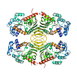
Crystal Structure of a Myo-inositol dehydrogenase from Corynebacterium glutamicum ATCC 13032
Descriptor: Myo-inositol dehydrogenase, SODIUM ION
Authors:Kumaran, D, Mahmood, A, Burley, S.K, Swaminathan, S, New York SGX Research Center for Structural Genomics (NYSGXRC)
Deposit date:2008-10-11
Release date:2008-10-21
Last modified:2023-12-27
Method:X-RAY DIFFRACTION (2.3 Å)
Cite:Crystal Structure of a Myo-inositol dehydrogenase from Corynebacterium glutamicum ATCC 13032
To be Published
1RRM
DownloadVisualize
BU of 1rrm by Molmil
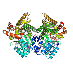
Crystal Structure of Lactaldehyde reductase
Descriptor: ADENOSINE-5-DIPHOSPHORIBOSE, FE (II) ION, Lactaldehyde reductase, ...
Authors:Kumaran, D, Swaminathan, S, Burley, S.K, New York SGX Research Center for Structural Genomics (NYSGXRC)
Deposit date:2003-12-08
Release date:2004-08-17
Last modified:2021-02-03
Method:X-RAY DIFFRACTION (1.6 Å)
Cite:Crystal structure of Lactaldehyde reductase
To be Published
3BWI
DownloadVisualize
BU of 3bwi by Molmil
Crystal structure of the catalytic domain of botulinum neurotoxin serotype A with an acetate ion bound at the active site
Descriptor: ACETATE ION, Botulinum neurotoxin A light chain, SULFATE ION, ...
Authors:Kumaran, D, Rawat, R, Swaminathan, S.
Deposit date:2008-01-09
Release date:2008-04-22
Last modified:2023-08-30
Method:X-RAY DIFFRACTION (1.7 Å)
Cite:Structure- and Substrate-based Inhibitor Design for Clostridium botulinum Neurotoxin Serotype A
J.Biol.Chem., 283, 2008
3C88
DownloadVisualize
BU of 3c88 by Molmil
Crystal structure of the catalytic domain of botulinum neurotoxin serotype A with inhibitory peptide RRGC
Descriptor: Botulinum neurotoxin A light chain, Inhibitor peptide RRGC, SODIUM ION, ...
Authors:Kumaran, D, Swaminathan, S.
Deposit date:2008-02-11
Release date:2008-04-22
Last modified:2023-08-30
Method:X-RAY DIFFRACTION (1.6 Å)
Cite:Structure- and Substrate-based Inhibitor Design for Clostridium botulinum Neurotoxin Serotype A
J.Biol.Chem., 283, 2008
3BGA
DownloadVisualize
BU of 3bga by Molmil
Crystal structure of beta-galactosidase from Bacteroides thetaiotaomicron VPI-5482
Descriptor: Beta-galactosidase, CHLORIDE ION, MAGNESIUM ION, ...
Authors:Kumaran, D, Burley, S.K, Swaminathan, S, New York SGX Research Center for Structural Genomics (NYSGXRC)
Deposit date:2007-11-26
Release date:2007-12-11
Last modified:2021-02-03
Method:X-RAY DIFFRACTION (2.1 Å)
Cite:Crystal structure analysis of beta-galactosidase from Bacteroides thetaiotaomicron VPI-5482.
To be Published
3DDB
DownloadVisualize
BU of 3ddb by Molmil
Crystal structure of the catalytic domain of Botulinum neurotoxin serotype a with a substrate analog peptide
Descriptor: Botulinum neurotoxin A light chain, SULFATE ION, Synaptosomal-associated protein 25, ...
Authors:Kumaran, D, Swaminathan, S.
Deposit date:2008-06-05
Release date:2008-09-16
Last modified:2023-08-30
Method:X-RAY DIFFRACTION (1.6 Å)
Cite:Substrate binding mode and its implication on drug design for botulinum neurotoxin A
Plos Pathog., 4, 2008
3FFZ
DownloadVisualize
BU of 3ffz by Molmil
Domain organization in Clostridium butulinum neurotoxin type E is unique: Its implication in faster translocation
Descriptor: ACETATE ION, Botulinum neurotoxin type E, SODIUM ION, ...
Authors:Kumaran, D, Eswaramoorthy, S, Swaminathan, S.
Deposit date:2008-12-04
Release date:2008-12-16
Last modified:2023-09-06
Method:X-RAY DIFFRACTION (2.65 Å)
Cite:Domain organization in Clostridium botulinum neurotoxin type E is unique: its implication in faster translocation.
J.Mol.Biol., 386, 2009
1TXN
DownloadVisualize
BU of 1txn by Molmil
Crystal structure of coproporphyrinogen III oxidase
Descriptor: Coproporphyrinogen III oxidase, GLYCEROL
Authors:Kumaran, D, Swaminathan, S, Burley, S.K, New York SGX Research Center for Structural Genomics (NYSGXRC)
Deposit date:2004-07-05
Release date:2004-11-30
Last modified:2021-02-03
Method:X-RAY DIFFRACTION (1.7 Å)
Cite:Crystal structure of coproporphyrinogen III oxidase
To be Published
1U8S
DownloadVisualize
BU of 1u8s by Molmil
Crystal structure of putative glycine cleavage system transcriptional repressor
Descriptor: glycine cleavage system transcriptional repressor, putative
Authors:Kumaran, D, Swaminathan, S.
Deposit date:2004-08-06
Release date:2004-08-17
Last modified:2024-02-14
Method:X-RAY DIFFRACTION (2.45 Å)
Cite:Crystal structure of putative glycine cleavage system transcriptional repressor
To be Published
1TXZ
DownloadVisualize
BU of 1txz by Molmil
Crystal structure of yeast ymx7, an ADP-ribose-1''-monophosphatase, complexed with ADP-ribose
Descriptor: 1,2-ETHANEDIOL, ADENOSINE-5-DIPHOSPHORIBOSE, Hypothetical 32.1 kDa protein in ADH3-RCA1 intergenic region, ...
Authors:Kumaran, D, Swaminathan, S, Burley, S.K, New York SGX Research Center for Structural Genomics (NYSGXRC)
Deposit date:2004-07-06
Release date:2004-11-30
Last modified:2023-08-23
Method:X-RAY DIFFRACTION (2.05 Å)
Cite:Structure and mechanism of ADP-ribose-1''-monophosphatase (Appr-1''-pase), a ubiquitous cellular processing enzyme.
Protein Sci., 14, 2005
3IO1
DownloadVisualize
BU of 3io1 by Molmil
Crystal Structure of Aminobenzoyl-glutamate utilization protein from Klebsiella pneumoniae
Descriptor: Aminobenzoyl-glutamate utilization protein, SODIUM ION, YTTRIUM (III) ION
Authors:Kumaran, D, Baumann, K, Burley, S.K, Swaminathan, S, New York SGX Research Center for Structural Genomics (NYSGXRC)
Deposit date:2009-08-13
Release date:2009-08-25
Last modified:2021-02-10
Method:X-RAY DIFFRACTION (2.5 Å)
Cite:Crystal Structure of Aminobenzoyl-glutamate utilization protein from Klebsiella pneumoniae
To be Published
3IPI
DownloadVisualize
BU of 3ipi by Molmil
Crystal Structure of a Geranyltranstransferase from the Methanosarcina mazei
Descriptor: Geranyltranstransferase, MALONIC ACID
Authors:Kumaran, D, Mohammed, M.B, Brown, A, Burley, S.K, Swaminathan, S, New York SGX Research Center for Structural Genomics (NYSGXRC)
Deposit date:2009-08-17
Release date:2009-09-08
Last modified:2021-02-10
Method:X-RAY DIFFRACTION (1.9 Å)
Cite:Crystal Structure of a Geranyltranstransferase from the Methanosarcina mazei
To be Published
2G59
DownloadVisualize
BU of 2g59 by Molmil
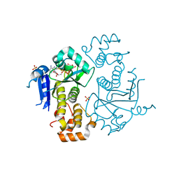
Crystal Structure of the Catalytic Domain of Protein Tyrosine Phosphatase from Homo sapiens
Descriptor: PHOSPHATE ION, Receptor-type tyrosine-protein phosphatase O
Authors:Kumaran, D, Swaminathan, S, Burley, S.K, New York SGX Research Center for Structural Genomics (NYSGXRC)
Deposit date:2006-02-22
Release date:2006-03-14
Last modified:2023-08-30
Method:X-RAY DIFFRACTION (2.19 Å)
Cite:Structural genomics of protein phosphatases.
J.Struct.Funct.Genom., 8, 2007

223166

数据于2024-07-31公开中

PDB statisticsPDBj update infoContact PDBjnumon