8QWN
 
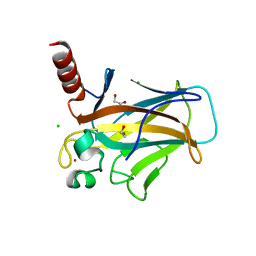 | | Structure of p53 cancer mutant Y220C (hexagonal crystal form) | | 分子名称: | 1,2-ETHANEDIOL, CHLORIDE ION, Cellular tumor antigen p53, ... | | 著者 | Balourdas, D.I, Knapp, S, Joerger, A.C, Structural Genomics Consortium (SGC) | | 登録日 | 2023-10-19 | | 公開日 | 2024-06-19 | | 実験手法 | X-RAY DIFFRACTION (1.44 Å) | | 主引用文献 | Structural basis of p53 inactivation by cavity-creating cancer mutations and its implications for the development of mutant p53 reactivators.
Cell Death Dis, 15, 2024
|
|
8QWK
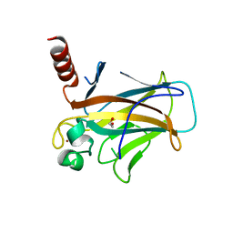 | | Structure of p53 cancer mutant Y126C | | 分子名称: | 1,2-ETHANEDIOL, Cellular tumor antigen p53, ZINC ION | | 著者 | Markl, A.M, Balourdas, D.I, Kraemer, A, Knapp, S, Joerger, A.C, Structural Genomics Consortium (SGC) | | 登録日 | 2023-10-19 | | 公開日 | 2024-06-19 | | 実験手法 | X-RAY DIFFRACTION (1.69 Å) | | 主引用文献 | Structural basis of p53 inactivation by cavity-creating cancer mutations and its implications for the development of mutant p53 reactivators.
Cell Death Dis, 15, 2024
|
|
8QWO
 | | Structure of p53 cancer mutant Y234C | | 分子名称: | Cellular tumor antigen p53, GLYCEROL, ZINC ION | | 著者 | Balourdas, D.I, Knapp, S, Joerger, A.C, Structural Genomics Consortium (SGC) | | 登録日 | 2023-10-19 | | 公開日 | 2024-06-19 | | 実験手法 | X-RAY DIFFRACTION (1.38 Å) | | 主引用文献 | Structural basis of p53 inactivation by cavity-creating cancer mutations and its implications for the development of mutant p53 reactivators.
Cell Death Dis, 15, 2024
|
|
8QWP
 | | Structure of p53 cancer mutant Y236C | | 分子名称: | 1,2-ETHANEDIOL, Cellular tumor antigen p53, L(+)-TARTARIC ACID, ... | | 著者 | Balourdas, D.I, Knapp, S, Joerger, A.C, Structural Genomics Consortium (SGC) | | 登録日 | 2023-10-19 | | 公開日 | 2024-06-19 | | 実験手法 | X-RAY DIFFRACTION (2.1 Å) | | 主引用文献 | Structural basis of p53 inactivation by cavity-creating cancer mutations and its implications for the development of mutant p53 reactivators.
Cell Death Dis, 15, 2024
|
|
8QWL
 | | Structure of p53 cancer mutant Y163C | | 分子名称: | 1,2-ETHANEDIOL, Cellular tumor antigen p53, MALONATE ION, ... | | 著者 | Balourdas, D.I, Markl, A.M, Kraemer, A, Knapp, S, Joerger, A.C, Structural Genomics Consortium (SGC) | | 登録日 | 2023-10-19 | | 公開日 | 2024-06-19 | | 実験手法 | X-RAY DIFFRACTION (1.65 Å) | | 主引用文献 | Structural basis of p53 inactivation by cavity-creating cancer mutations and its implications for the development of mutant p53 reactivators.
Cell Death Dis, 15, 2024
|
|
8QWM
 | | Structure of p53 cancer mutant Y205C | | 分子名称: | 1,2-ETHANEDIOL, Cellular tumor antigen p53, L(+)-TARTARIC ACID, ... | | 著者 | Balourdas, D.I, Knapp, S, Joerger, A.C, Structural Genomics Consortium (SGC) | | 登録日 | 2023-10-19 | | 公開日 | 2024-06-19 | | 実験手法 | X-RAY DIFFRACTION (1.54 Å) | | 主引用文献 | Structural basis of p53 inactivation by cavity-creating cancer mutations and its implications for the development of mutant p53 reactivators.
Cell Death Dis, 15, 2024
|
|
6Y2D
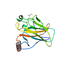
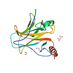
 | | Crystal structure of the second KH domain of FUBP1 | | 分子名称: | Far upstream element-binding protein 1, GLYCEROL, SULFATE ION | | 著者 | Ni, X, Chaikuad, A, Joerger, A.C, Arrowsmith, C.H, Edwards, A.M, Bountra, C, Knapp, S, Structural Genomics Consortium (SGC) | | 登録日 | 2020-02-15 | | 公開日 | 2020-03-25 | | 最終更新日 | 2024-01-24 | | 実験手法 | X-RAY DIFFRACTION (1.9 Å) | | 主引用文献 | Comparative structural analyses and nucleotide-binding characterization of the four KH domains of FUBP1.
Sci Rep, 10, 2020
|
|
6Y2C
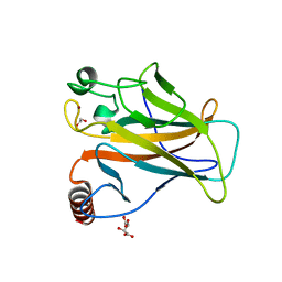
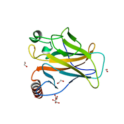
 | | Crystal structure of the third KH domain of FUBP1 | | 分子名称: | 1,2-ETHANEDIOL, Far upstream element-binding protein 1, ZINC ION | | 著者 | Ni, X, Joerger, A.C, Chaikuad, A, Arrowsmith, C.H, Edwards, A.M, Bountra, C, Knapp, S, Structural Genomics Consortium (SGC) | | 登録日 | 2020-02-15 | | 公開日 | 2020-03-25 | | 最終更新日 | 2024-01-24 | | 実験手法 | X-RAY DIFFRACTION (2 Å) | | 主引用文献 | Comparative structural analyses and nucleotide-binding characterization of the four KH domains of FUBP1.
Sci Rep, 10, 2020
|
|
6Y24
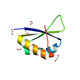 | | Crystal structure of fourth KH domain of FUBP1 | | 分子名称: | 1,2-ETHANEDIOL, Far upstream element-binding protein 1 | | 著者 | Ni, X, Joerger, A.C, Chaikuad, A, Arrowsmith, C.H, Edwards, A.M, Bountra, C, Knapp, S, Structural Genomics Consortium (SGC) | | 登録日 | 2020-02-14 | | 公開日 | 2020-03-25 | | 最終更新日 | 2024-01-24 | | 実験手法 | X-RAY DIFFRACTION (1.86 Å) | | 主引用文献 | Comparative structural analyses and nucleotide-binding characterization of the four KH domains of FUBP1.
Sci Rep, 10, 2020
|
|


