Loading
PDBj
MenuPDBj@FacebookPDBj@TwitterPDBj@YouTubewwPDB FoundationwwPDB
RCSB PDBPDBeBMRBAdv. SearchSearch help
PDB: 84 results

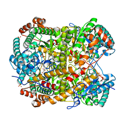
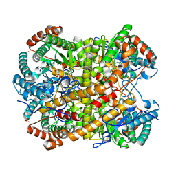
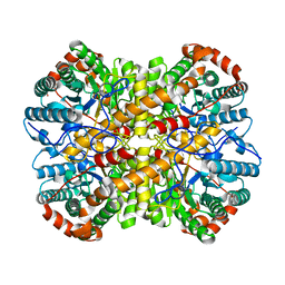
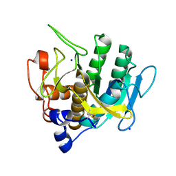
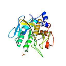
7AM5
DownloadVisualize
BU of 7am5 by Molmil
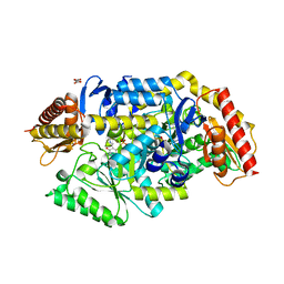
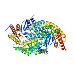
Crystal structure of Peptiligase mutant - L217H/M222P/A225N
Descriptor: SODIUM ION, Subtilisin BPN'
Authors:Rozeboom, H.J, Janssen, D.J.
Deposit date:2020-10-08
Release date:2021-02-17
Last modified:2021-03-24
Method:X-RAY DIFFRACTION (2.3 Å)
Cite:From thiol-subtilisin to omniligase: Design and structure of a broadly applicable peptide ligase.
Comput Struct Biotechnol J, 19, 2021
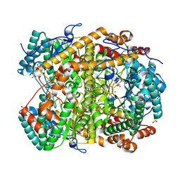
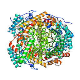
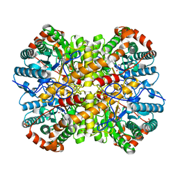
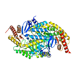
7AM7
DownloadVisualize
BU of 7am7 by Molmil
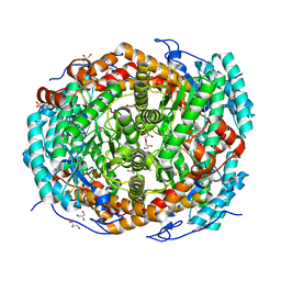
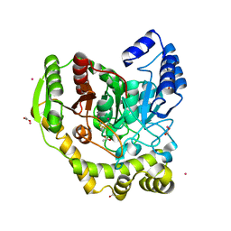
Crystal structure of Peptiligase mutant - M222P/L217H/A225N/F189W/N218D
Descriptor: Eglin C fragment, GLYCEROL, SULFATE ION, ...
Authors:Rozeboom, H.J, Janssen, D.J.
Deposit date:2020-10-08
Release date:2021-02-17
Last modified:2024-05-01
Method:X-RAY DIFFRACTION (2.61 Å)
Cite:From thiol-subtilisin to omniligase: Design and structure of a broadly applicable peptide ligase.
Comput Struct Biotechnol J, 19, 2021
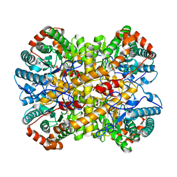
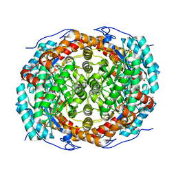
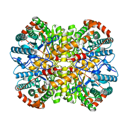
5NH8
DownloadVisualize
BU of 5nh8 by Molmil
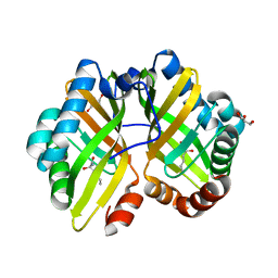
Crystal structure of xylose isomerase from Piromyces E2 in complex with two Ca2+ ions and xylose
Descriptor: CALCIUM ION, D-xylose, SULFATE ION, ...
Authors:Rozeboom, H.J, Janssen, D.B.
Deposit date:2017-03-21
Release date:2017-11-01
Last modified:2024-05-01
Method:X-RAY DIFFRACTION (1.86 Å)
Cite:Metal Dependence of the Xylose Isomerase from Piromyces sp. E2 Explored by Activity Profiling and Protein Crystallography.
Biochemistry, 56, 2017
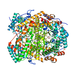
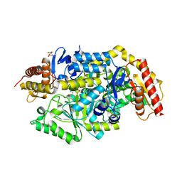
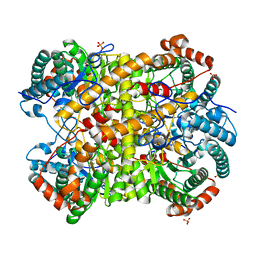
5NHE
DownloadVisualize
BU of 5nhe by Molmil
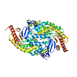
Crystal structure of xylose isomerase from Piromyces E2 in complex with two Cd2+ ions and xylose
Descriptor: CADMIUM ION, D-xylose, SULFATE ION, ...
Authors:Rozeboom, H.J, Janssen, D.B.
Deposit date:2017-03-21
Release date:2017-11-01
Last modified:2024-05-01
Method:X-RAY DIFFRACTION (1.86 Å)
Cite:Metal Dependence of the Xylose Isomerase from Piromyces sp. E2 Explored by Activity Profiling and Protein Crystallography.
Biochemistry, 56, 2017
5NH9
DownloadVisualize
BU of 5nh9 by Molmil
Crystal structure of xylose isomerase from Piromyces E2 in complex with two Mn2+ ions and xylose
Descriptor: D-xylose, MANGANESE (II) ION, SULFATE ION, ...
Authors:Rozeboom, H.J, Janssen, D.B.
Deposit date:2017-03-21
Release date:2017-11-01
Last modified:2024-05-01
Method:X-RAY DIFFRACTION (2.08 Å)
Cite:Metal Dependence of the Xylose Isomerase from Piromyces sp. E2 Explored by Activity Profiling and Protein Crystallography.
Biochemistry, 56, 2017
5NH7
DownloadVisualize
BU of 5nh7 by Molmil
Crystal structure of xylose isomerase from Piromyces E2 in complex with two Mg2+ ions and xylose
Descriptor: D-xylose, MAGNESIUM ION, SULFATE ION, ...
Authors:Rozeboom, H.J, Janssen, D.B.
Deposit date:2017-03-21
Release date:2017-11-01
Last modified:2024-05-01
Method:X-RAY DIFFRACTION (1.9 Å)
Cite:Metal Dependence of the Xylose Isomerase from Piromyces sp. E2 Explored by Activity Profiling and Protein Crystallography.
Biochemistry, 56, 2017
5NHD
DownloadVisualize
BU of 5nhd by Molmil
Crystal structure of xylose isomerase from Piromyces E2 in complex with 2 Ni2+ ions and xylose
Descriptor: D-xylose, NICKEL (II) ION, SULFATE ION, ...
Authors:Rozeboom, H.J, Janssen, D.B.
Deposit date:2017-03-21
Release date:2017-11-01
Last modified:2024-05-01
Method:X-RAY DIFFRACTION (1.8 Å)
Cite:Metal Dependence of the Xylose Isomerase from Piromyces sp. E2 Explored by Activity Profiling and Protein Crystallography.
Biochemistry, 56, 2017
6T8F
DownloadVisualize
BU of 6t8f by Molmil
Crystal structure of mutant xylose isomerase (V270A/A273G) from Piromyces E2 grown in yeast, in complex with xylose
Descriptor: CALCIUM ION, D-xylose, SULFATE ION, ...
Authors:Rozeboom, H.J, Janssen, D.B.
Deposit date:2019-10-24
Release date:2020-01-29
Last modified:2024-01-24
Method:X-RAY DIFFRACTION (2 Å)
Cite:Structure-based directed evolution improves S. cerevisiae growth on xylose by influencing in vivo enzyme performance.
Biotechnol Biofuels, 13, 2020
6T8E
DownloadVisualize
BU of 6t8e by Molmil
Crystal structure of native xylose isomerase from Piromyces E2 grown in yeast, in complex with xylose
Descriptor: CALCIUM ION, D-xylose, SULFATE ION, ...
Authors:Rozeboom, H.J, Janssen, D.B.
Deposit date:2019-10-24
Release date:2020-01-29
Last modified:2024-01-24
Method:X-RAY DIFFRACTION (1.86 Å)
Cite:Structure-based directed evolution improves S. cerevisiae growth on xylose by influencing in vivo enzyme performance.
Biotechnol Biofuels, 13, 2020
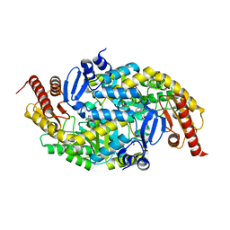
7B4J
DownloadVisualize
BU of 7b4j by Molmil
Thermostable omega transaminase PjTA-R6 variant W58M/F86L/R417L engineered for asymmetric synthesis of enantiopure bulky amines
Descriptor: 4'-DEOXY-4'-AMINOPYRIDOXAL-5'-PHOSPHATE, Aspartate aminotransferase family protein, SUCCINIC ACID
Authors:Capra, N, Rozeboom, H.J, Thunnissen, A.M.W.H, Janssen, D.B.
Deposit date:2020-12-02
Release date:2021-09-01
Last modified:2024-06-19
Method:X-RAY DIFFRACTION (1.9 Å)
Cite:Computational Redesign of an omega-Transaminase from Pseudomonas jessenii for Asymmetric Synthesis of Enantiopure Bulky Amines.
Acs Catalysis, 11, 2021
7B4I
DownloadVisualize
BU of 7b4i by Molmil
Thermostable omega transaminase PjTA-R6 variant W58G engineered for asymmetric synthesis of enantiopure bulky amines
Descriptor: Aspartate aminotransferase family protein, PYRIDOXAL-5'-PHOSPHATE, SUCCINIC ACID
Authors:Capra, N, Rozeboom, H.J, Thunnissen, A.M.W.H, Janssen, D.B.
Deposit date:2020-12-02
Release date:2021-09-01
Last modified:2021-09-22
Method:X-RAY DIFFRACTION (1.7 Å)
Cite:Computational Redesign of an omega-Transaminase from Pseudomonas jessenii for Asymmetric Synthesis of Enantiopure Bulky Amines.
Acs Catalysis, 11, 2021
5NH5
DownloadVisualize
BU of 5nh5 by Molmil
Crystal structure of native xylose isomerase from Piromyces E2
Descriptor: CALCIUM ION, FE (II) ION, GLYCEROL, ...
Authors:Rozeboom, H.J, Janssen, D.B.
Deposit date:2017-03-21
Release date:2017-11-01
Last modified:2024-01-17
Method:X-RAY DIFFRACTION (1.8 Å)
Cite:Metal Dependence of the Xylose Isomerase from Piromyces sp. E2 Explored by Activity Profiling and Protein Crystallography.
Biochemistry, 56, 2017
6TP2
DownloadVisualize
BU of 6tp2 by Molmil
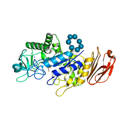
Crystal structure of Bacillus paralicheniformis alpha-amylase in complex with beta-cyclodextrin
Descriptor: Amylase, CALCIUM ION, Cycloheptakis-(1-4)-(alpha-D-glucopyranose), ...
Authors:Rozeboom, H.J, Janssen, D.B.
Deposit date:2019-12-12
Release date:2020-10-14
Last modified:2024-01-24
Method:X-RAY DIFFRACTION (1.94 Å)
Cite:Characterization of the starch surface binding site on Bacillus paralicheniformis alpha-amylase.
Int.J.Biol.Macromol., 165, 2020
5NHC
DownloadVisualize
BU of 5nhc by Molmil
Crystal structure of xylose isomerase from Piromyces E2 in complex with two Co2+ ions and xylulose
Descriptor: 4-HYDROXYPROLINE, COBALT (II) ION, D-XYLULOSE, ...
Authors:Rozeboom, H.J, Janssen, D.B.
Deposit date:2017-03-21
Release date:2017-11-01
Last modified:2024-05-01
Method:X-RAY DIFFRACTION (1.93 Å)
Cite:Metal Dependence of the Xylose Isomerase from Piromyces sp. E2 Explored by Activity Profiling and Protein Crystallography.
Biochemistry, 56, 2017
5NH6
DownloadVisualize
BU of 5nh6 by Molmil
Crystal structure of xylose isomerase from Piromyces E2 Complexed with one Mg2+ ion and xylitol
Descriptor: MAGNESIUM ION, SULFATE ION, Xylitol, ...
Authors:Rozeboom, H.J, Janssen, D.B.
Deposit date:2017-03-21
Release date:2017-11-01
Last modified:2024-05-01
Method:X-RAY DIFFRACTION (1.75 Å)
Cite:Metal Dependence of the Xylose Isomerase from Piromyces sp. E2 Explored by Activity Profiling and Protein Crystallography.
Biochemistry, 56, 2017
5NHM
DownloadVisualize
BU of 5nhm by Molmil
Crystal structure of apo xylose isomerase from Piromyces E2
Descriptor: ACETIC ACID, GLYCEROL, SULFATE ION, ...
Authors:Rozeboom, H.J, Janssen, D.B.
Deposit date:2017-03-21
Release date:2017-11-01
Last modified:2024-05-01
Method:X-RAY DIFFRACTION (1.67 Å)
Cite:Metal Dependence of the Xylose Isomerase from Piromyces sp. E2 Explored by Activity Profiling and Protein Crystallography.
Biochemistry, 56, 2017
5NHB
DownloadVisualize
BU of 5nhb by Molmil
Crystal structure of xylose isomerase from Piromyces E2 in complex with two Fe2+ ions
Descriptor: FE (II) ION, SULFATE ION, Xylose isomerase
Authors:Rozeboom, H.J, Janssen, D.B.
Deposit date:2017-03-21
Release date:2017-11-01
Last modified:2024-05-01
Method:X-RAY DIFFRACTION (2.4 Å)
Cite:Metal Dependence of the Xylose Isomerase from Piromyces sp. E2 Explored by Activity Profiling and Protein Crystallography.
Biochemistry, 56, 2017
5NHA
DownloadVisualize
BU of 5nha by Molmil
Crystal structure of xylose isomerase from Piromyces sp. E2 in complex with two Mn2+ ions and sorbitol
Descriptor: MANGANESE (II) ION, SULFATE ION, Xylose isomerase, ...
Authors:Rozeboom, H.J, Janssen, D.B.
Deposit date:2017-03-21
Release date:2017-11-01
Last modified:2024-05-01
Method:X-RAY DIFFRACTION (1.8 Å)
Cite:Metal Dependence of the Xylose Isomerase from Piromyces sp. E2 Explored by Activity Profiling and Protein Crystallography.
Biochemistry, 56, 2017
6G4E
DownloadVisualize
BU of 6g4e by Molmil
Crystal structure of the omega TRANSAMINASE FROM PSEUDOMONAS Jessenii in complex with PLP and 6-aminohexanoate (6-ACA)
Descriptor: 6-AMINOHEXANOIC ACID, Aspartate aminotransferase family protein, GLYCEROL, ...
Authors:Rozeboom, H.J, Janssen, D.B.
Deposit date:2018-03-27
Release date:2019-04-10
Last modified:2024-05-01
Method:X-RAY DIFFRACTION (2.45 Å)
Cite:Biochemical properties of a Pseudomonas aminotransferase involved in caprolactam metabolism.
Febs J., 286, 2019
5AC3
DownloadVisualize
BU of 5ac3 by Molmil
Crystal structure of PAM12A
Descriptor: ACETIC ACID, CADMIUM ION, PEPTIDE AMIDASE
Authors:Wu, B, Wijma, H.J, Song, L, Rozeboom, H.J, Poloni, C, Tian, Y, Arif, M.I, Nuijens, T, Quadflieg, P.J.L.M, Szymanski, W, Feringa, B.L, Janssen, D.B.
Deposit date:2015-08-11
Release date:2016-07-20
Last modified:2024-01-10
Method:X-RAY DIFFRACTION (1.8 Å)
Cite:Versatile Peptide C-Terminal Functionalization Via a Computationally Peptide Amidase
Acs Catalysis, 2016
4R9K
DownloadVisualize
BU of 4r9k by Molmil
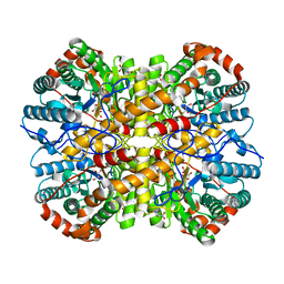
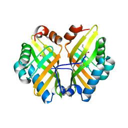
Structure of thermostable eightfold mutant of limonene epoxide hydrolase from Rhodococcus erythropolis
Descriptor: (2R)-2-hydroxyhexanamide, GLYCEROL, Limonene-1,2-epoxide hydrolase
Authors:Floor, R.J, Wijma, H.J, Jekel, P.A, Terwisscha van Scheltinga, A.C, Dijkstra, B.W, Janssen, D.B.
Deposit date:2014-09-05
Release date:2014-09-24
Last modified:2023-09-20
Method:X-RAY DIFFRACTION (1.5 Å)
Cite:X-ray crystallographic validation of structure predictions used in computational design for protein stabilization.
Proteins, 83, 2015
6G4C
DownloadVisualize
BU of 6g4c by Molmil
Crystal structure of the omega transaminase from Pseudomonas jessenii in the apo form, crystallized from ammonium phosphate
Descriptor: Aspartate aminotransferase family protein, PHOSPHATE ION
Authors:Rozeboom, H.J, Janssen, D.B.
Deposit date:2018-03-27
Release date:2019-04-10
Last modified:2024-05-01
Method:X-RAY DIFFRACTION (1.87 Å)
Cite:Biochemical properties of a Pseudomonas aminotransferase involved in caprolactam metabolism.
Febs J., 286, 2019
6G4F
DownloadVisualize
BU of 6g4f by Molmil
Crystal structure of the omega TRANSAMINASE FROM PSEUDOMONAS Jessenii in complex with PMP
Descriptor: 4'-DEOXY-4'-AMINOPYRIDOXAL-5'-PHOSPHATE, Aspartate aminotransferase family protein, GLYCEROL, ...
Authors:Rozeboom, H.J, Janssen, D.B.
Deposit date:2018-03-27
Release date:2019-04-10
Last modified:2024-05-01
Method:X-RAY DIFFRACTION (2.5 Å)
Cite:Biochemical properties of a Pseudomonas aminotransferase involved in caprolactam metabolism.
Febs J., 286, 2019
4R9L
DownloadVisualize
BU of 4r9l by Molmil
Structure of a thermostable elevenfold mutant of limonene epoxide hydrolase from Rhodococcus erythropolis, containing two stabilizing disulfide bonds
Descriptor: (2R)-2-hydroxyhexanamide, Limonene-1,2-epoxide hydrolase
Authors:Floor, R.J, Wijma, H.J, Jekel, P.A, Terwisscha van Scheltinga, A.C, Dijkstra, B.W, Janssen, D.B.
Deposit date:2014-09-05
Release date:2014-09-24
Last modified:2023-09-20
Method:X-RAY DIFFRACTION (1.8 Å)
Cite:X-ray crystallographic validation of structure predictions used in computational design for protein stabilization.
Proteins, 83, 2015
6G4D
DownloadVisualize
BU of 6g4d by Molmil
Crystal structure of the omega TRANSAMINASE FROM PSEUDOMONAS Jessenii in complex with PLP
Descriptor: Aspartate aminotransferase family protein, GLYCEROL, PYRIDOXAL-5'-PHOSPHATE
Authors:Rozeboom, H.J, Janssen, D.B.
Deposit date:2018-03-27
Release date:2019-04-10
Last modified:2024-05-01
Method:X-RAY DIFFRACTION (2.15 Å)
Cite:Biochemical properties of a Pseudomonas aminotransferase involved in caprolactam metabolism.
Febs J., 286, 2019

223166

PDB entries from 2024-07-31

PDB statisticsPDBj update infoContact PDBjnumon