5IXO
 
 | |
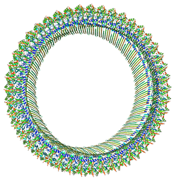
8JYZ
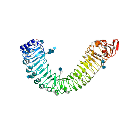
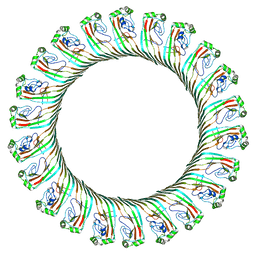 | | Cryo-EM structure of RCD-1 pore from Neurospora crassa | | 分子名称: | Gasdermin-like protein rcd-1-1, Gasdermin-like protein rcd-1-2 | | 著者 | Hou, Y.J, Sun, Q, Li, Y, Ding, J. | | 登録日 | 2023-07-04 | | 公開日 | 2024-05-01 | | 最終更新日 | 2024-05-29 | | 実験手法 | ELECTRON MICROSCOPY (3.63 Å) | | 主引用文献 | Cleavage-independent activation of ancient eukaryotic gasdermins and structural mechanisms.
Science, 384, 2024
|
|
7LHL
 | |
5IY7
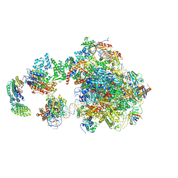 | | Human holo-PIC in the open state | | 分子名称: | DNA-directed RNA polymerase II subunit RPB1, DNA-directed RNA polymerase II subunit RPB10, DNA-directed RNA polymerase II subunit RPB11-a, ... | | 著者 | He, Y, Yan, C, Fang, J, Inouye, C, Tjian, R, Ivanov, I, Nogales, E. | | 登録日 | 2016-03-24 | | 公開日 | 2016-05-18 | | 最終更新日 | 2024-03-06 | | 実験手法 | ELECTRON MICROSCOPY (8.6 Å) | | 主引用文献 | Near-atomic resolution visualization of human transcription promoter opening.
Nature, 533, 2016
|
|
8JYW
 | |
8JYX
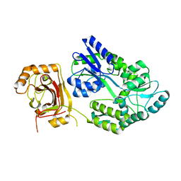 | |
8B81
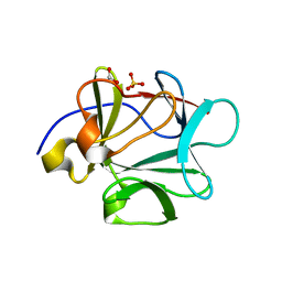
 | | The structure of Gan1D W433A in complex with cellobiose-6-phosphate | | 分子名称: | 6-O-phosphono-beta-D-glucopyranose-(1-4)-beta-D-glucopyranose, IMIDAZOLE, Putative 6-phospho-beta-galactobiosidase | | 著者 | Snyder, J, Lansky, S, Zehavi, A, Shoham, Y, Shoham, G. | | 登録日 | 2022-10-04 | | 公開日 | 2022-10-19 | | 最終更新日 | 2024-01-31 | | 実験手法 | X-RAY DIFFRACTION (1.585 Å) | | 主引用文献 | The structure of Gan1D W433A in complex with cellobiose-6-phosphate
To Be Published
|
|
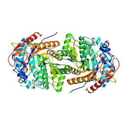
8B80
 | | The structure of Gan1D W433A in complex with galactose-6P | | 分子名称: | 6-O-phosphono-beta-D-galactopyranose, GLYCEROL, IMIDAZOLE, ... | | 著者 | Snyder, J, Lansky, S, Zehavi, A, Shoham, Y, Shoham, G. | | 登録日 | 2022-10-04 | | 公開日 | 2022-10-19 | | 最終更新日 | 2024-01-31 | | 実験手法 | X-RAY DIFFRACTION (1.78 Å) | | 主引用文献 | The structure of Gan1D W433A in complex with galactose-6P
To Be Published
|
|
3BAV
 | |
5J1P
 | |
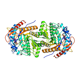
5IZW
 | | Crystal structure of RNA editing specific factor of designer PLS-type PPR-9R protein | | 分子名称: | PLS9-PPR | | 著者 | Yan, J, Zhang, Q, Guan, Z, Zou, T, Yin, P. | | 登録日 | 2016-03-26 | | 公開日 | 2017-03-29 | | 最終更新日 | 2024-03-20 | | 実験手法 | X-RAY DIFFRACTION (1.738 Å) | | 主引用文献 | MORF9 increases the RNA-binding activity of PLS-type pentatricopeptide repeat protein in plastid RNA editing
Nat Plants, 3, 2017
|
|
8K70
 | |
1H12
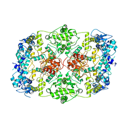
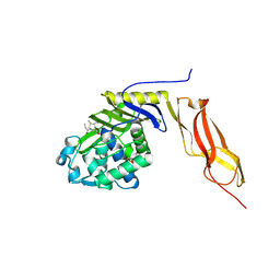
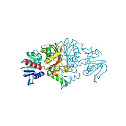
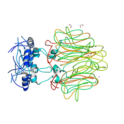
 | | Structure of a cold-adapted family 8 xylanase | | 分子名称: | ENDO-1,4-BETA-XYLANASE, alpha-D-xylopyranose, beta-D-xylopyranose | | 著者 | Van Petegem, F, Collins, T, Meuwis, M.A, Feller, G, Gerday, C, Van Beeumen, J. | | 登録日 | 2002-07-02 | | 公開日 | 2003-03-13 | | 最終更新日 | 2024-05-01 | | 実験手法 | X-RAY DIFFRACTION (1.2 Å) | | 主引用文献 | The Structure of a Cold-Adapted Family 8 Xylanase at 1.3 A Resolution: Structural Adaptations to Cold and Investigation of the Active Site
J.Biol.Chem., 278, 2003
|
|
3BEB
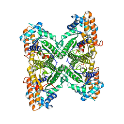
 | | Crystal structure of E. coli penicillin-binding protein 5 in complex with a peptide-mimetic penicillin | | 分子名称: | (2R,4S)-2-[(1R)-1-{[(6S)-6-amino-6-carboxyhexanoyl]amino}-2-oxoethyl]-5,5-dimethyl-1,3-thiazolidine-4-carboxylic acid, GLYCEROL, Penicillin-binding protein 5 | | 著者 | Heilemann, J, Powell, A.J, Davies, C. | | 登録日 | 2007-11-16 | | 公開日 | 2008-08-26 | | 最終更新日 | 2023-08-30 | | 実験手法 | X-RAY DIFFRACTION (2 Å) | | 主引用文献 | Crystal structures of complexes of bacterial DD-peptidases with peptidoglycan-mimetic ligands: the substrate specificity puzzle
J.Mol.Biol., 381, 2008
|
|
3B7Y
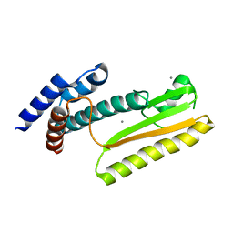
 | | Crystal structure of the C2 Domain of the E3 Ubiquitin-Protein Ligase NEDD4 | | 分子名称: | CALCIUM ION, E3 ubiquitin-protein ligase NEDD4 | | 著者 | Walker, J.R, Ruzanov, M, Butler-Cole, C, Weigelt, J, Arrowsmith, C.H, Edwards, A.M, Bochkarev, A, Dhe-Paganon, S, Structural Genomics Consortium (SGC) | | 登録日 | 2007-10-31 | | 公開日 | 2007-11-27 | | 最終更新日 | 2023-08-30 | | 実験手法 | X-RAY DIFFRACTION (1.8 Å) | | 主引用文献 | C2 Domain of the Human E3 Ubiquitin-Protein Ligase NEDD4.
To be Published
|
|
3B8D
 | |
1HAX
 | | Snapshots of serine protease catalysis: (A) acyl-enzyme intermediate between porcine pancreatic elastase and human beta-casomorphin-7 at pH 5 | | 分子名称: | BETA-CASOMORPHIN-7, CALCIUM ION, ELASTASE 1, ... | | 著者 | Wilmouth, R.C, Edman, K, Neutze, R, Wright, P.A, Clifton, I.J, Schneider, T.R, Schofield, C.J, Hajdu, J. | | 登録日 | 2001-04-10 | | 公開日 | 2001-08-02 | | 最終更新日 | 2023-12-13 | | 実験手法 | X-RAY DIFFRACTION (1.6 Å) | | 主引用文献 | X-Ray Snapshots of Serine Protease Catalysis Reveal a Tetrahedral Intermediate
Nat.Struct.Biol., 8, 2001
|
|
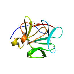
1H5B
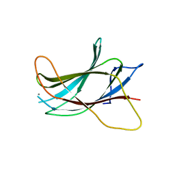
 | | T cell receptor Valpha11 (AV11S5) domain | | 分子名称: | CHLORIDE ION, GLYCEROL, MURINE T CELL RECEPTOR (TCR) VALPHA DOMAIN | | 著者 | Machius, M, Cianga, P, Deisenhofer, J, Sally Ward, E. | | 登録日 | 2001-05-21 | | 公開日 | 2001-06-21 | | 最終更新日 | 2019-03-06 | | 実験手法 | X-RAY DIFFRACTION (1.85 Å) | | 主引用文献 | Crystal Structure of a T Cell Receptor Valpha11 (Av11S5) Domain: New Canonical Forms for the First and Second Complementarity Determining Regions
J.Mol.Biol., 310, 2001
|
|
8BK0
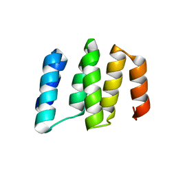
 | | Crystal structure of human Ephrin type-A receptor 2 (EPHA2) Kinase domain in complex with LDN-211904 | | 分子名称: | 1,2-ETHANEDIOL, Ephrin type-A receptor 2, ~{N}-(2-chlorophenyl)-6-piperidin-4-yl-imidazo[1,2-a]pyridine-3-carboxamide | | 著者 | Zhubi, R, Gerninghaus, J, Knapp, S, Kraemer, A, Structural Genomics Consortium (SGC) | | 登録日 | 2022-11-08 | | 公開日 | 2022-11-16 | | 最終更新日 | 2024-01-31 | | 実験手法 | X-RAY DIFFRACTION (1.7 Å) | | 主引用文献 | Crystal structure of human Ephrin type-A receptor 2 (EPHA2) Kinase domain in complex with LDN-211904
To Be Published
|
|
8KAJ
 | |
5J6H
 | |
3BB2
 | |
8JUO
 | |
4DMI
 | | Crystal Structure of a Pentameric Capsid Protein Isolated from Metagenomic Phage Sequences (CASP) | | 分子名称: | 1,2-ETHANEDIOL, Capsid Protein, SODIUM ION | | 著者 | Craig, T.K, Abendroth, J, Lorimer, D, Burgin Jr, A.B, Segall, A, Rohwer, F. | | 登録日 | 2012-02-07 | | 公開日 | 2012-08-29 | | 最終更新日 | 2024-02-28 | | 実験手法 | X-RAY DIFFRACTION (1.5 Å) | | 主引用文献 | Crystal Structure of a Pentameric Capsid Protein Isolated from Metagenomic Phage Sequences
To be Published
|
|
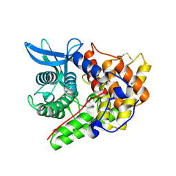
1GR1
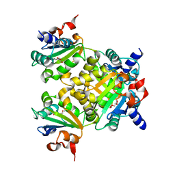
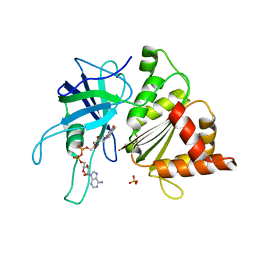 | | Structure of Ferredoxin-NADP+ Reductase with Glu 139 replaced by Lys (E139K) | | 分子名称: | FERREDOXIN--NADP+ REDUCTASE, FLAVIN-ADENINE DINUCLEOTIDE, SULFATE ION | | 著者 | Hermoso, J.A, Mayoral, T, Medina, M, Sanz-Aparicio, J, Gomez-Moreno, C. | | 登録日 | 2001-12-12 | | 公開日 | 2002-10-24 | | 最終更新日 | 2023-12-13 | | 実験手法 | X-RAY DIFFRACTION (2.5 Å) | | 主引用文献 | Probing the Role of Glutamic Acid 139 of Anabena Ferrodoxin-Nadp+ Reductase in the Interaction with Substrates
Eur.J.Biochem., 269, 2002
|
|


