3CF0
 
 | |
5KLM
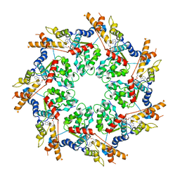
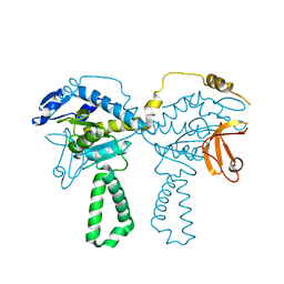
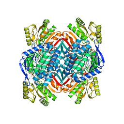
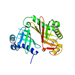
 | | Crystal structure of 2-hydroxymuconate-6-semialdehyde derived intermediate in NAD(+)-bound 2-aminomuconate 6-semialdehyde dehydrogenase N169D | | 分子名称: | 2-aminomuconate 6-semialdehyde dehydrogenase, NICOTINAMIDE-ADENINE-DINUCLEOTIDE, SODIUM ION | | 著者 | Yang, Y, Davis, I, Ha, U, Wang, Y, Shin, I, Liu, A. | | 登録日 | 2016-06-24 | | 公開日 | 2016-11-09 | | 最終更新日 | 2023-09-27 | | 実験手法 | X-RAY DIFFRACTION (2.102 Å) | | 主引用文献 | A Pitcher-and-Catcher Mechanism Drives Endogenous Substrate Isomerization by a Dehydrogenase in Kynurenine Metabolism.
J.Biol.Chem., 291, 2016
|
|
6WPX
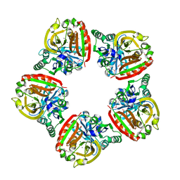
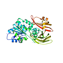 | | Crystal structure of Bacillus licheniformis lipase BlEst2 in propetide form | | 分子名称: | BlEst2, IODIDE ION | | 著者 | Nakamura, A.M, Godoy, A.S, Kadowaki, M.A.S, Polikarpov, I. | | 登録日 | 2020-04-28 | | 公開日 | 2021-06-09 | | 最終更新日 | 2024-08-07 | | 実験手法 | X-RAY DIFFRACTION (2 Å) | | 主引用文献 | Structures of BlEst2 from Bacillus licheniformis in its propeptide and mature forms reveal autoinhibitory effects of the C-terminal domain
Febs J., n/a, 2024
|
|
6X87
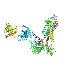
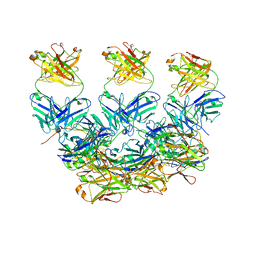 | | CryoEM structure of the Plasmodium berghei circumsporozoite protein in complex with inhibitory mouse antibody 3D11. | | 分子名称: | 3D11 Fab heavy chain, 3D11 Fab kappa chain, Circumsporozoite protein | | 著者 | Kucharska, I, Thai, E, Rubinstein, J, Julien, J.P. | | 登録日 | 2020-06-01 | | 公開日 | 2020-12-02 | | 最終更新日 | 2020-12-16 | | 実験手法 | ELECTRON MICROSCOPY (3.2 Å) | | 主引用文献 | Structural ordering of the Plasmodium berghei circumsporozoite protein repeats by inhibitory antibody 3D11.
Elife, 9, 2020
|
|
4C3I
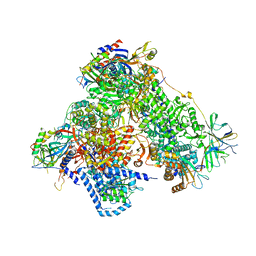 | | Structure of 14-subunit RNA polymerase I at 3.0 A resolution, crystal form C2-100 | | 分子名称: | (4S)-2-METHYL-2,4-PENTANEDIOL, DNA-DIRECTED RNA POLYMERASE I SUBUNIT RPA12, DNA-DIRECTED RNA POLYMERASE I SUBUNIT RPA135, ... | | 著者 | Fernandez-Tornero, C, Moreno-Morcillo, M, Rashid, U.J, Taylor, N.M.I, Ruiz, F.M, Gruene, T, Legrand, P, Steuerwald, U, Muller, C.W. | | 登録日 | 2013-08-24 | | 公開日 | 2013-10-23 | | 最終更新日 | 2024-05-08 | | 実験手法 | X-RAY DIFFRACTION (3 Å) | | 主引用文献 | Crystal Structure of the 14-Subunit RNA Polymerase I
Nature, 502, 2013
|
|
6QNO
 | | Rhodopsin-Gi protein complex | | 分子名称: | 2-acetamido-2-deoxy-beta-D-glucopyranose-(1-4)-2-acetamido-2-deoxy-beta-D-glucopyranose, Fab antibody fragment heavy chain, Fab antibody fragment light chain, ... | | 著者 | Tsai, C.-J, Marino, J, Adaixo, R.J, Pamula, F, Muehle, J, Maeda, S, Flock, T, Taylor, N.M.I, Mohammed, I, Matile, H, Dawson, R.J.P, Deupi, X, Stahlberg, H, Schertler, G.F.X. | | 登録日 | 2019-02-11 | | 公開日 | 2019-07-10 | | 最終更新日 | 2020-07-29 | | 実験手法 | ELECTRON MICROSCOPY (4.38 Å) | | 主引用文献 | Cryo-EM structure of the rhodopsin-G alpha i-beta gamma complex reveals binding of the rhodopsin C-terminal tail to the G beta subunit.
Elife, 8, 2019
|
|
4TOI
 | | Crystal structure of E.coli ribosomal protein S2 in complex with N-terminal domain of S1 | | 分子名称: | 30S ribosomal protein S2,Ribosomal protein S1, ZINC ION | | 著者 | Grishkovskaya, I, Byrgazov, K, Moll, I, Djinovic-Carugo, K. | | 登録日 | 2014-06-05 | | 公開日 | 2014-12-31 | | 最終更新日 | 2023-12-20 | | 実験手法 | X-RAY DIFFRACTION (2.3 Å) | | 主引用文献 | Structural basis for the interaction of protein S1 with the Escherichia coli ribosome.
Nucleic Acids Res., 43, 2015
|
|
5KLN
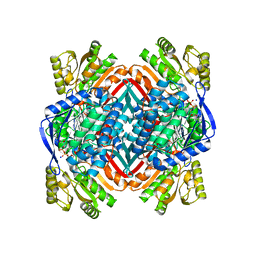
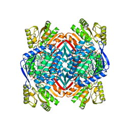 | | Crystal structure of 2-aminomuconate 6-semialdehyde dehydrogenase N169A in complex with NAD+ | | 分子名称: | 2-aminomuconate 6-semialdehyde dehydrogenase, NICOTINAMIDE-ADENINE-DINUCLEOTIDE, SODIUM ION | | 著者 | Yang, Y, Davis, I, Ha, U, Wang, Y, Shin, I, Liu, A. | | 登録日 | 2016-06-24 | | 公開日 | 2016-11-09 | | 最終更新日 | 2023-09-27 | | 実験手法 | X-RAY DIFFRACTION (1.992 Å) | | 主引用文献 | A Pitcher-and-Catcher Mechanism Drives Endogenous Substrate Isomerization by a Dehydrogenase in Kynurenine Metabolism.
J.Biol.Chem., 291, 2016
|
|
5KLK
 | | Crystal structure of 2-aminomuconate 6-semialdehyde dehydrogenase N169D in complex with NAD+ and 2-hydroxymuconate-6-semialdehyde | | 分子名称: | (2E,4E)-2-hydroxy-6-oxohexa-2,4-dienoic acid, 2-aminomuconate 6-semialdehyde dehydrogenase, NICOTINAMIDE-ADENINE-DINUCLEOTIDE, ... | | 著者 | Yang, Y, Davis, I, Ha, U, Wang, Y, Shin, I, Liu, A. | | 登録日 | 2016-06-24 | | 公開日 | 2016-11-09 | | 最終更新日 | 2023-09-27 | | 実験手法 | X-RAY DIFFRACTION (2.006 Å) | | 主引用文献 | A Pitcher-and-Catcher Mechanism Drives Endogenous Substrate Isomerization by a Dehydrogenase in Kynurenine Metabolism.
J.Biol.Chem., 291, 2016
|
|
2VBL
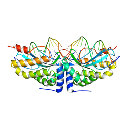 | | Molecular basis of human XPC gene recognition and cleavage by engineered homing endonuclease heterodimers | | 分子名称: | 5'-D(*DA*DA*DA*DA*DG*DG*DC*DA*DG*DAP)-3', 5'-D(*DA*DG*DG*DA*DT*DC*DC*DT*DA*DAP)-3', 5'-D(*DT*DC*DT*DG*DC*DC*DT*DT*DT*DT*DT*DT *DGP*DAP)-3', ... | | 著者 | Redondo, P, Prieto, J, Munoz, I.G, Alibes, A, Stricher, F, Serrano, L, Arnould, S, Perez, C, Cabaniols, J.P, Duchateau, P, Paques, F, Blanco, F.J, Montoya, G. | | 登録日 | 2007-09-14 | | 公開日 | 2008-10-28 | | 最終更新日 | 2023-12-13 | | 実験手法 | X-RAY DIFFRACTION (1.8 Å) | | 主引用文献 | Molecular Basis of Xeroderma Pigmentosum Group C DNA Recognition by Engineered Meganucleases
Nature, 456, 2008
|
|
3DEO
 | |
2VBJ
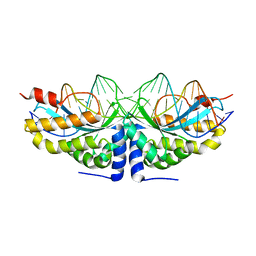 | | Molecular basis of human XPC gene recognition and cleavage by engineered homing endonuclease heterodimers | | 分子名称: | 5'-D(*TP*CP*TP*GP*CP*CP*TP*TP*TP*TP *TP*TP*GP*AP*AP*GP*GP*AP*TP*CP*CP*TP*AP*A)-3', 5'-D(*TP*TP*AP*GP*GP*AP*TP*CP*CP*TP *TP*CP*AP*AP*AP*AP*AP*AP*GP*GP*CP*AP*GP*A)-3', CALCIUM ION, ... | | 著者 | Redondo, P, Prieto, J, Munoz, I.G, Alibes, A, Stricher, F, Serrano, L, Arnould, S, Perez, C, Cabaniols, J.P, Duchateau, P, Paques, F, Blanco, F.J, Montoya, G. | | 登録日 | 2007-09-14 | | 公開日 | 2008-10-28 | | 最終更新日 | 2023-12-13 | | 実験手法 | X-RAY DIFFRACTION (1.95 Å) | | 主引用文献 | Molecular Basis of Xeroderma Pigmentosum Group C DNA Recognition by Engineered Meganucleases
Nature, 456, 2008
|
|
4RZM
 | | Crystal structure of the Lsd19-lasalocid A complex | | 分子名称: | CHLORIDE ION, Epoxide hydrolase LasB, FORMIC ACID, ... | | 著者 | Mathews, I.I, Hotta, K, Chen, X, Kim, C.-Y. | | 登録日 | 2014-12-22 | | 公開日 | 2015-01-21 | | 最終更新日 | 2023-09-20 | | 実験手法 | X-RAY DIFFRACTION (2.33 Å) | | 主引用文献 | Epoxide hydrolase-lasalocid a structure provides mechanistic insight into polyether natural product biosynthesis.
J.Am.Chem.Soc., 137, 2015
|
|
5M86
 | | Crystal Structure of the Thermoplasma acidophilum Protein Ta1207 | | 分子名称: | CALCIUM ION, CHLORIDE ION, GLYCEROL, ... | | 著者 | Pathare, G.R, Nagy, I, Bracher, A. | | 登録日 | 2016-10-28 | | 公開日 | 2017-06-14 | | 最終更新日 | 2017-06-21 | | 実験手法 | X-RAY DIFFRACTION (2.4 Å) | | 主引用文献 | Crystal structure of the Thermoplasma acidophilum protein Ta1207.
Acta Crystallogr F Struct Biol Commun, 73, 2017
|
|
5L8I
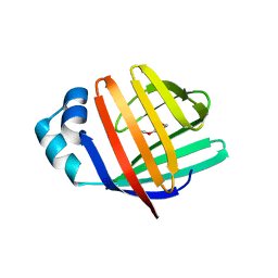 | | crystal structure of human FABP6 apo-protein | | 分子名称: | 3,6,9,12,15,18-HEXAOXAICOSANE-1,20-DIOL, DI(HYDROXYETHYL)ETHER, Gastrotropin, ... | | 著者 | Hendrick, A, Mueller, I, Leonard, P.M, Davenport, R, Mitchell, P. | | 登録日 | 2016-06-08 | | 公開日 | 2016-08-24 | | 最終更新日 | 2024-01-10 | | 実験手法 | X-RAY DIFFRACTION (1.88 Å) | | 主引用文献 | Identification and Investigation of Novel Binding Fragments in the Fatty Acid Binding Protein 6 (FABP6).
J.Med.Chem., 59, 2016
|
|
5MUV
 | | Atomic structure fitted into a localized reconstruction of bacteriophage phi6 packaging hexamer P4 | | 分子名称: | ADENOSINE-5'-DIPHOSPHATE, CALCIUM ION, Packaging enzyme P4 | | 著者 | Sun, Z, El Omari, K, Sun, X, Ilca, S, Kotecha, A, Stuart, D.I, Poranen, M.M, Huiskonen, J.T. | | 登録日 | 2017-01-14 | | 公開日 | 2017-03-22 | | 最終更新日 | 2024-05-15 | | 実験手法 | ELECTRON MICROSCOPY (9.1 Å) | | 主引用文献 | Double-stranded RNA virus outer shell assembly by bona fide domain-swapping.
Nat Commun, 8, 2017
|
|
7A01
 | | The Halastavi arva virus intergenic region IRES promotes translation by the simplest possible initiation mechanism | | 分子名称: | 18S RIBOSOMAL RNA, 28S RIBOSOMAL RNA, 40S RIBOSOMAL PROTEIN ES21, ... | | 著者 | Abaeva, I, Vicens, Q, Bochler, A, Soufari, H, Simonetti, A, Pestova, T.V, Hashem, Y, Hellen, C.U.T. | | 登録日 | 2020-08-05 | | 公開日 | 2020-12-30 | | 実験手法 | ELECTRON MICROSCOPY (3.6 Å) | | 主引用文献 | The Halastavi arva Virus Intergenic Region IRES Promotes Translation by the Simplest Possible Initiation Mechanism.
Cell Rep, 33, 2020
|
|
6HCV
 | | Crystal Structure of Lysyl-tRNA Synthetase from Plasmodium falciparum complexed with a chromone ligand | | 分子名称: | 6-fluoranyl-~{N}-[(1-oxidanylcyclohexyl)methyl]-4-oxidanylidene-chromene-2-carboxamide, Lysine--tRNA ligase | | 著者 | Robinson, D.A, Baragana, B, Norcross, N, Forte, B, Walpole, C, Gilbert, I.H. | | 登録日 | 2018-08-16 | | 公開日 | 2019-04-03 | | 最終更新日 | 2024-01-17 | | 実験手法 | X-RAY DIFFRACTION (2.2 Å) | | 主引用文献 | Lysyl-tRNA synthetase as a drug target in malaria and cryptosporidiosis.
Proc.Natl.Acad.Sci.USA, 116, 2019
|
|
5L8N
 | | crystal structure of human FABP6 protein with fragment 1 | | 分子名称: | 3,6,9,12,15,18-HEXAOXAICOSANE-1,20-DIOL, 5,6-dimethyl-1~{H}-benzimidazol-2-amine, DI(HYDROXYETHYL)ETHER, ... | | 著者 | Hendrick, A, Mueller, I, Leonard, P.M, Davenport, R, Mitchell, P. | | 登録日 | 2016-06-08 | | 公開日 | 2016-08-24 | | 最終更新日 | 2024-01-10 | | 実験手法 | X-RAY DIFFRACTION (2.12 Å) | | 主引用文献 | Identification and Investigation of Novel Binding Fragments in the Fatty Acid Binding Protein 6 (FABP6).
J.Med.Chem., 59, 2016
|
|
3DEF
 | | Crystal structure of Toc33 from Arabidopsis thaliana, dimerization deficient mutant R130A | | 分子名称: | GUANOSINE-5'-DIPHOSPHATE, MAGNESIUM ION, T7I23.11 protein | | 著者 | Koenig, P, Schleiff, E, Sinning, I, Tews, I. | | 登録日 | 2008-06-10 | | 公開日 | 2008-06-24 | | 最終更新日 | 2023-11-01 | | 実験手法 | X-RAY DIFFRACTION (1.96 Å) | | 主引用文献 | On the significance of Toc-GTPase homodimers
J.Biol.Chem., 283, 2008
|
|
6XJ0
 | | Crystal structure of multi-copper oxidase from Pediococcus pentosaceus | | 分子名称: | CHLORIDE ION, COPPER (II) ION, CU-O-CU LINKAGE, ... | | 著者 | Pardo, I, Soares, A.S, Collins, R, Partowmah, S.H, Coler, E.A. | | 登録日 | 2020-06-22 | | 公開日 | 2021-03-10 | | 最終更新日 | 2021-05-12 | | 実験手法 | X-RAY DIFFRACTION (2.34 Å) | | 主引用文献 | Structural analysis and biochemical properties of laccase enzymes from two Pediococcus species.
Microb Biotechnol, 14, 2021
|
|
6GO2
 | |
2VBO
 | | Molecular basis of human XPC gene recognition and cleavage by engineered homing endonuclease heterodimers | | 分子名称: | 5'-D(*TP*CP*TP*GP*CP*CP*TP*TP*TP*TP *TP*TP*GP*AP*AP*GP*GP*AP*TP*CP*CP*TP*AP*A)-3', 5'-D(*TP*TP*AP*GP*GP*AP*TP*CP*CP*TP *TP*CP*AP*AP*AP*AP*AP*AP*GP*GP*CP*AP*GP*A)-3', CALCIUM ION, ... | | 著者 | Redondo, P, Prieto, J, Munoz, I.G, Alibes, A, Stricher, F, Serrano, L, Arnould, S, Perez, C, Cabaniols, J.P, Duchateau, P, Paques, F, Blanco, F.J, Montoya, G. | | 登録日 | 2007-09-14 | | 公開日 | 2008-10-28 | | 最終更新日 | 2023-12-13 | | 実験手法 | X-RAY DIFFRACTION (1.8 Å) | | 主引用文献 | Molecular Basis of Xeroderma Pigmentosum Group C DNA Recognition by Engineered Meganucleases
Nature, 456, 2008
|
|
5I3C
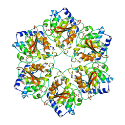
 | | Crystal structure of E.coli purine nucleoside phosphorylase with acycloguanosine | | 分子名称: | 9-HYROXYETHOXYMETHYLGUANINE, Purine nucleoside phosphorylase DeoD-type, SULFATE ION | | 著者 | Timofeev, V.I, Abramchik, Y.A, Esipov, R.S, Kuranova, I.P. | | 登録日 | 2016-02-10 | | 公開日 | 2017-02-22 | | 最終更新日 | 2024-01-10 | | 実験手法 | X-RAY DIFFRACTION (2.32 Å) | | 主引用文献 | Crystal structure of Escherichia coli purine nucleoside phosphorylase complexed with acyclovir.
Acta Crystallogr F Struct Biol Commun, 74, 2018
|
|
6WH2
 | | Structure of the C-terminal BRCT domain of human XRCC1 | | 分子名称: | X-ray repair cross complementing protein 1 variant | | 著者 | Pourfarjam, Y, Ellenberger, T, Tainer, J.A, Tomkinson, A.E, Kim, I.K. | | 登録日 | 2020-04-07 | | 公開日 | 2020-12-02 | | 最終更新日 | 2023-10-18 | | 実験手法 | X-RAY DIFFRACTION (2.414 Å) | | 主引用文献 | An atypical BRCT-BRCT interaction with the XRCC1 scaffold protein compacts human DNA Ligase III alpha within a flexible DNA repair complex.
Nucleic Acids Res., 49, 2021
|
|


