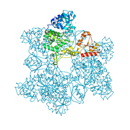
1JD2
 
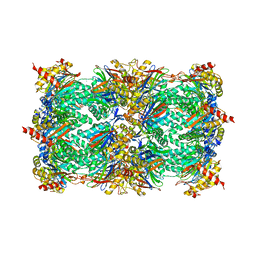 | | Crystal Structure of the yeast 20S Proteasome:TMC-95A complex: A non-covalent Proteasome Inhibitor | | 分子名称: | MAGNESIUM ION, PROTEASOME COMPONENT C1, PROTEASOME COMPONENT C11, ... | | 著者 | Groll, M, Koguchi, Y, Huber, R, Kohno, J. | | 登録日 | 2001-06-12 | | 公開日 | 2002-02-13 | | 最終更新日 | 2017-10-04 | | 実験手法 | X-RAY DIFFRACTION (3 Å) | | 主引用文献 | Crystal structure of the 20 S proteasome:TMC-95A complex: a non-covalent proteasome inhibitor.
J.Mol.Biol., 311, 2001
|
|
1JQG
 | | Crystal Structure of the Carboxypeptidase A from Helicoverpa Armigera | | 分子名称: | ZINC ION, carboxypeptidase A | | 著者 | Estebanez-Perpina, E, Bayes, A, Vendrell, J, Jongsma, M.A, Bown, D.P, Gatehouse, J.A, Huber, R, Bode, W, Aviles, F.X, Reverter, D. | | 登録日 | 2001-08-07 | | 公開日 | 2002-08-07 | | 最終更新日 | 2023-08-16 | | 実験手法 | X-RAY DIFFRACTION (2.5 Å) | | 主引用文献 | Crystal structure of a novel mid-gut procarboxypeptidase from the cotton pest Helicoverpa armigera.
J.Mol.Biol., 313, 2001
|
|
1JED
 | | Crystal Structure of ATP Sulfurylase in complex with ADP | | 分子名称: | 2-AMINO-2-HYDROXYMETHYL-PROPANE-1,3-DIOL, ACETIC ACID, ADENOSINE-5'-DIPHOSPHATE, ... | | 著者 | Ullrich, T.C, Huber, R. | | 登録日 | 2001-06-17 | | 公開日 | 2001-11-14 | | 最終更新日 | 2023-08-16 | | 実験手法 | X-RAY DIFFRACTION (2.95 Å) | | 主引用文献 | The complex structures of ATP sulfurylase with thiosulfate, ADP and chlorate reveal new insights in inhibitory effects and the catalytic cycle.
J.Mol.Biol., 313, 2001
|
|
1JG5
 | | CRYSTAL STRUCTURE OF RAT GTP CYCLOHYDROLASE I FEEDBACK REGULATORY PROTEIN, GFRP | | 分子名称: | GTP CYCLOHYDROLASE I FEEDBACK REGULATORY PROTEIN, POTASSIUM ION | | 著者 | Bader, G, Schiffmann, S, Herrmann, A, Fischer, M, Gutlich, M, Auerbach, G, Ploom, T, Bacher, A, Huber, R, Lemm, T. | | 登録日 | 2001-06-23 | | 公開日 | 2001-10-10 | | 最終更新日 | 2024-03-13 | | 実験手法 | X-RAY DIFFRACTION (2.6 Å) | | 主引用文献 | Crystal structure of rat GTP cyclohydrolase I feedback regulatory protein, GFRP.
J.Mol.Biol., 312, 2001
|
|
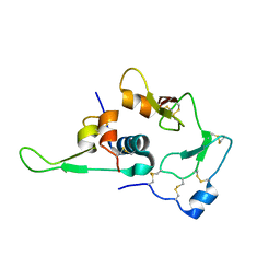
1JHJ
 | | Crystal structure of the APC10/Doc1 subunit of the human anaphase-promoting complex | | 分子名称: | APC10, NICKEL (II) ION | | 著者 | Wendt, K.S, Vodermaier, H.C, Jacob, U, Gieffers, C, Gmachl, M, Peters, J.-M, Huber, R, Sondermann, P. | | 登録日 | 2001-06-28 | | 公開日 | 2001-10-24 | | 最終更新日 | 2024-05-29 | | 実験手法 | X-RAY DIFFRACTION (1.6 Å) | | 主引用文献 | Crystal structure of the APC10/DOC1 subunit of the human anaphase-promoting complex
Nat.Struct.Biol., 8, 2001
|
|
1JEC
 | |
1JEE
 | | Crystal Structure of ATP Sulfurylase in complex with chlorate | | 分子名称: | 2-AMINO-2-HYDROXYMETHYL-PROPANE-1,3-DIOL, ACETIC ACID, ADENOSINE-5'-PHOSPHOSULFATE, ... | | 著者 | Ullrich, T.C, Huber, R. | | 登録日 | 2001-06-17 | | 公開日 | 2001-11-14 | | 最終更新日 | 2023-08-16 | | 実験手法 | X-RAY DIFFRACTION (2.8 Å) | | 主引用文献 | The complex structures of ATP sulfurylase with thiosulfate, ADP and chlorate reveal new insights in inhibitory effects and the catalytic cycle.
J.Mol.Biol., 313, 2001
|
|
1K32
 | |
1KFX
 | | Crystal Structure of Human m-Calpain Form I | | 分子名称: | M-CALPAIN LARGE SUBUNIT, M-CALPAIN SMALL SUBUNIT | | 著者 | Strobl, S, Fernandez-Catalan, C, Braun, M, Huber, R, Masumoto, H, Nakagawa, K, Irie, A, Sorimachi, H, Bourenkow, G, Bartunik, H, Suzuki, K, Bode, W. | | 登録日 | 2001-11-23 | | 公開日 | 2001-12-07 | | 最終更新日 | 2023-08-16 | | 実験手法 | X-RAY DIFFRACTION (3.15 Å) | | 主引用文献 | The crystal structure of calcium-free human m-calpain suggests an electrostatic switch mechanism for activation by calcium.
Proc.Natl.Acad.Sci.USA, 97, 2000
|
|
1KLI
 | | Cofactor-and substrate-assisted activation of factor VIIa | | 分子名称: | BENZAMIDINE, CALCIUM ION, GLYCEROL, ... | | 著者 | Sichler, K, Banner, D.W, D'Arcy, A, Hopfner, K.P, Huber, R, Bode, W, Kresse, G.B, Kopetzki, E, Brandstetter, H. | | 登録日 | 2001-12-12 | | 公開日 | 2002-09-18 | | 最終更新日 | 2011-07-13 | | 実験手法 | X-RAY DIFFRACTION (1.69 Å) | | 主引用文献 | Crystal Structure of Uninhibited Factor VIIa Link its Cofactor and Substrate-assisted Activation to Specific Interactions
J.Mol.Biol., 322, 2002
|
|
1KFU
 | | Crystal Structure of Human m-Calpain Form II | | 分子名称: | M-CALPAIN LARGE SUBUNIT, M-CALPAIN SMALL SUBUNIT | | 著者 | Strobl, S, Fernandez-Catalan, C, Braun, M, Huber, R, Masumoto, H, Nakagawa, K, Irie, A, Sorimachi, H, Bourenkow, G, Bartunik, H, Suzuki, K, Bode, W. | | 登録日 | 2001-11-23 | | 公開日 | 2001-12-07 | | 最終更新日 | 2024-02-07 | | 実験手法 | X-RAY DIFFRACTION (2.5 Å) | | 主引用文献 | The crystal structure of calcium-free human m-calpain suggests an electrostatic switch mechanism for activation by calcium.
Proc.Natl.Acad.Sci.USA, 97, 2000
|
|
1KL7
 | | Crystal Structure of Threonine Synthase from Yeast | | 分子名称: | PYRIDOXAL-5'-PHOSPHATE, Threonine Synthase | | 著者 | Garrido-Franco, M, Ehlert, S, Messerschmidt, A, Marinkovic, S, Huber, R, Laber, B, Bourenkov, G.P, Clausen, T. | | 登録日 | 2001-12-11 | | 公開日 | 2002-04-24 | | 最終更新日 | 2018-01-31 | | 実験手法 | X-RAY DIFFRACTION (2.7 Å) | | 主引用文献 | Structure and function of threonine synthase from yeast.
J.Biol.Chem., 277, 2002
|
|
1KLJ
 | | Crystal structure of uninhibited factor VIIa | | 分子名称: | 1,2-ETHANEDIOL, CALCIUM ION, factor VIIa | | 著者 | Sichler, K, Banner, D, D'Arcy, A, Hopfner, K.P, Huber, R, Bode, W, Kresse, G.B, Kopetzki, E, Brandstetter, H. | | 登録日 | 2001-12-12 | | 公開日 | 2002-10-09 | | 最終更新日 | 2023-08-16 | | 実験手法 | X-RAY DIFFRACTION (2.44 Å) | | 主引用文献 | Crystal structures of uninhibited factor VIIa link its cofactor and substrate-assisted activation to specific interactions.
J.Mol.Biol., 322, 2002
|
|
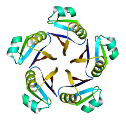
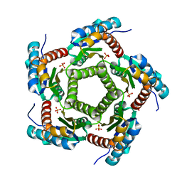
1KZ6
 | | Mutant enzyme W63Y/L119F Lumazine Synthase from S.pombe | | 分子名称: | 6,7-Dimethyl-8-ribityllumazine Synthase, PHOSPHATE ION | | 著者 | Gerhardt, S, Haase, I, Steinbacher, S, Kaiser, J.T, Cushman, M, Bacher, A, Huber, R, Fischer, M. | | 登録日 | 2002-02-06 | | 公開日 | 2002-07-24 | | 最終更新日 | 2024-05-29 | | 実験手法 | X-RAY DIFFRACTION (2.7 Å) | | 主引用文献 | The structural basis of riboflavin binding to Schizosaccharomyces pombe 6,7-dimethyl-8-ribityllumazine synthase.
J.Mol.Biol., 318, 2002
|
|
1WQJ
 | | Structural Basis for the Regulation of Insulin-Like Growth Factors (IGFs) by IGF Binding Proteins (IGFBPs) | | 分子名称: | Insulin-like growth factor IB, Insulin-like growth factor binding protein 4 | | 著者 | Siwanowicz, I, Popowicz, G.M, Wisniewska, M, Huber, R, Kuenkele, K.P, Lang, K, Engh, R.A, Holak, T.A. | | 登録日 | 2004-09-29 | | 公開日 | 2005-03-01 | | 最終更新日 | 2023-10-25 | | 実験手法 | X-RAY DIFFRACTION (1.6 Å) | | 主引用文献 | Structural basis for the regulation of insulin-like growth factors by IGF binding proteins
Structure, 13, 2005
|
|
1KYY
 | | Lumazine Synthase from S.pombe bound to nitropyrimidinedione | | 分子名称: | 5-NITRO-6-RIBITYL-AMINO-2,4(1H,3H)-PYRIMIDINEDIONE, 6,7-Dimethyl-8-ribityllumazine Synthase, PHOSPHATE ION | | 著者 | Gerhardt, S, Haase, I, Steinbacher, S, Kaiser, J.T, Cushman, M, Bacher, A, Huber, R, Fischer, M. | | 登録日 | 2002-02-06 | | 公開日 | 2002-07-24 | | 最終更新日 | 2024-03-13 | | 実験手法 | X-RAY DIFFRACTION (2.4 Å) | | 主引用文献 | The structural basis of riboflavin binding to Schizosaccharomyces pombe 6,7-dimethyl-8-ribityllumazine synthase.
J.Mol.Biol., 318, 2002
|
|
1KZ1
 | | Mutant enzyme W27G Lumazine Synthase from S.pombe | | 分子名称: | 6,7-Dimethyl-8-ribityllumazine Synthase | | 著者 | Gerhardt, S, Haase, I, Steinbacher, S, Kaiser, J.T, Cushman, M, Bacher, A, Huber, R, Fischer, M. | | 登録日 | 2002-02-06 | | 公開日 | 2002-07-24 | | 最終更新日 | 2024-05-29 | | 実験手法 | X-RAY DIFFRACTION (2 Å) | | 主引用文献 | The structural basis of riboflavin binding to Schizosaccharomyces pombe 6,7-dimethyl-8-ribityllumazine synthase.
J.Mol.Biol., 318, 2002
|
|
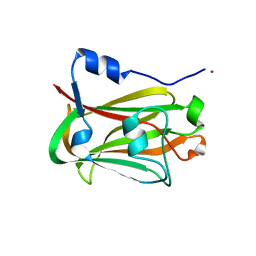
1WYX
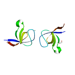 | | The Crystal Structure of the p130Cas SH3 Domain at 1.1 A Resolution | | 分子名称: | 1,2-ETHANEDIOL, CRK-associated substrate, MAGNESIUM ION | | 著者 | Wisniewska, M, Bossenmaier, B, Georges, G, Hesse, F, Dangl, M, Kuenkele, K.P, Ioannidis, I, Huber, R, Engh, R.A. | | 登録日 | 2005-02-17 | | 公開日 | 2005-04-26 | | 最終更新日 | 2024-03-13 | | 実験手法 | X-RAY DIFFRACTION (1.14 Å) | | 主引用文献 | The 1.1 A resolution crystal structure of the p130cas SH3 domain and ramifications for ligand selectivity
J.Mol.Biol., 347, 2005
|
|
1KZ9
 | | Mutant Enzyme L119F Lumazine Synthase from S.pombe | | 分子名称: | 6,7-Dimethyl-8-ribityllumazine Synthase, PHOSPHATE ION | | 著者 | Gerhardt, S, Haase, I, Steinbacher, S, Kaiser, J.T, Cushman, M, Bacher, A, Huber, R, Fischer, M. | | 登録日 | 2002-02-06 | | 公開日 | 2002-07-24 | | 最終更新日 | 2024-05-29 | | 実験手法 | X-RAY DIFFRACTION (3.1 Å) | | 主引用文献 | The structural basis of riboflavin binding to Schizosaccharomyces pombe 6,7-dimethyl-8-ribityllumazine synthase.
J.Mol.Biol., 318, 2002
|
|
1KYX
 | | Lumazine Synthase from S.pombe bound to carboxyethyllumazine | | 分子名称: | 3-[8-((2S,3S,4R)-2,3,4,5-TETRAHYDROXYPENTYL)-2,4,7-TRIOXO-1,3,8-TRIHYDROPTERIDIN-6-YL]PROPANOIC ACID, 6,7-Dimethyl-8-ribityllumazine Synthase, PHOSPHATE ION | | 著者 | Gerhardt, S, Haase, I, Steinbacher, S, Kaiser, J.T, Cushman, M, Bacher, A, Huber, R, Fischer, M. | | 登録日 | 2002-02-06 | | 公開日 | 2002-07-24 | | 最終更新日 | 2024-03-13 | | 実験手法 | X-RAY DIFFRACTION (2.6 Å) | | 主引用文献 | The structural basis of riboflavin binding to Schizosaccharomyces pombe 6,7-dimethyl-8-ribityllumazine synthase.
J.Mol.Biol., 318, 2002
|
|
1KZ4
 | | Mutant enzyme W63Y Lumazine Synthase from S.pombe | | 分子名称: | 6,7-Dimethyl-8-ribityllumazine Synthase, PHOSPHATE ION | | 著者 | Gerhardt, S, Haase, I, Steinbacher, S, Kaiser, J.T, Cushman, M, Bacher, A, Huber, R, Fischer, M. | | 登録日 | 2002-02-06 | | 公開日 | 2002-07-24 | | 最終更新日 | 2024-05-29 | | 実験手法 | X-RAY DIFFRACTION (3.1 Å) | | 主引用文献 | The structural basis of riboflavin binding to Schizosaccharomyces pombe 6,7-dimethyl-8-ribityllumazine synthase.
J.Mol.Biol., 318, 2002
|
|
1KY9
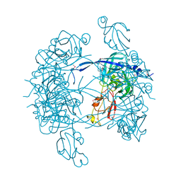 | | Crystal Structure of DegP (HtrA) | | 分子名称: | PROTEASE DO | | 著者 | Krojer, T, Garrido-Franco, M, Huber, R, Ehrmann, M, Clausen, T. | | 登録日 | 2002-02-04 | | 公開日 | 2002-04-03 | | 最終更新日 | 2021-10-27 | | 実験手法 | X-RAY DIFFRACTION (2.8 Å) | | 主引用文献 | Crystal structure of DegP (HtrA) reveals a new protease-chaperone machine.
Nature, 416, 2002
|
|
1KYV
 | | Lumazine Synthase from S.pombe bound to riboflavin | | 分子名称: | 6,7-Dimethyl-8-ribityllumazine Synthase, PHOSPHATE ION, RIBOFLAVIN | | 著者 | Gerhardt, S, Haase, I, Steinbacher, S, Kaiser, J.T, Cushman, M, Bacher, A, Huber, R, Fischer, M. | | 登録日 | 2002-02-06 | | 公開日 | 2002-07-24 | | 最終更新日 | 2024-03-13 | | 実験手法 | X-RAY DIFFRACTION (2.4 Å) | | 主引用文献 | The structural basis of riboflavin binding to Schizosaccharomyces pombe 6,7-dimethyl-8-ribityllumazine synthase.
J.Mol.Biol., 318, 2002
|
|
1KZL
 | | Riboflavin Synthase from S.pombe bound to Carboxyethyllumazine | | 分子名称: | 3-[8-((2S,3S,4R)-2,3,4,5-TETRAHYDROXYPENTYL)-2,4,7-TRIOXO-1,3,8-TRIHYDROPTERIDIN-6-YL]PROPANOIC ACID, MERCURY (II) ION, Riboflavin Synthase | | 著者 | Gerhardt, S, Schott, A.K, Kairies, N, Cushman, M, Illarionov, B, Eisenreich, W, Bacher, A, Huber, R, Steinbacher, S, Fischer, M. | | 登録日 | 2002-02-07 | | 公開日 | 2002-11-06 | | 最終更新日 | 2024-03-13 | | 実験手法 | X-RAY DIFFRACTION (2.1 Å) | | 主引用文献 | Studies on the Reaction Mechanism of Riboflavin Synthase; X-Ray Crystal Structure of a Complex with 6-Carboxyethyl-7-Oxo-8-Ribityllumazine
STRUCTURE, 10, 2002
|
|
1XRN
 | | Crystal structure of active site F1-mutant E213Q soaked with peptide Phe-Ala | | 分子名称: | ALANINE, Proline iminopeptidase | | 著者 | Goettig, P, Brandstetter, H, Groll, M, Goehring, W, Konarev, P.V, Svergun, D.I, Huber, R, Kim, J.-S. | | 登録日 | 2004-10-15 | | 公開日 | 2005-07-12 | | 最終更新日 | 2021-11-10 | | 実験手法 | X-RAY DIFFRACTION (2.8 Å) | | 主引用文献 | X-ray snapshots of peptide processing in mutants of tricorn-interacting factor F1 from Thermoplasma acidophilum
J.Biol.Chem., 280, 2005
|
|


