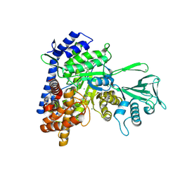
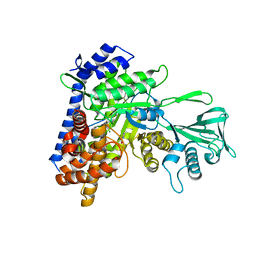
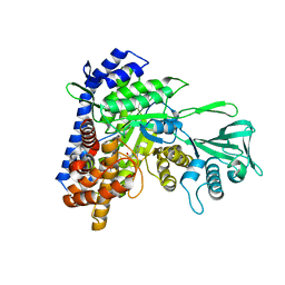
8JYU
 
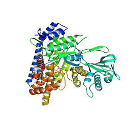
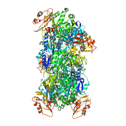 | | Acyl-ACP Synthetase structure bound to Decanoyl-AMP | | Descriptor: | ADENOSINE MONOPHOSPHATE, Acyl-acyl carrier protein synthetase, DECANOIC ACID, ... | | Authors: | Huang, H, Chang, S, Huang, M, Zhang, H, Zhou, C, Zhang, X, Feng, Y. | | Deposit date: | 2023-07-03 | | Release date: | 2024-07-10 | | Method: | ELECTRON MICROSCOPY (2.19 Å) | | Cite: | Acyl-ACP Synthetase structure bound to Decanoyl-AMP
To Be Published
|
|
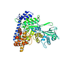
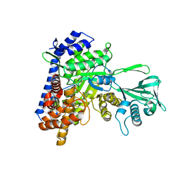
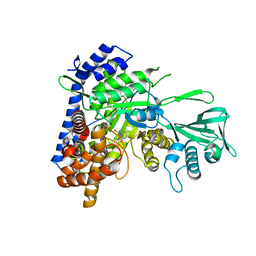
8JYL
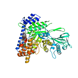
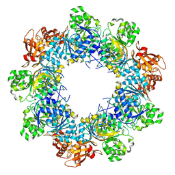 | | Acyl-ACP Synthetase structure bound to C10-AMS | | Descriptor: | Acyl-acyl carrier protein synthetase, MAGNESIUM ION, [(2R,3S,4R,5R)-5-(6-aminopurin-9-yl)-3,4-bis(oxidanyl)oxolan-2-yl]methyl N-decanoylsulfamate | | Authors: | Huang, H, Chang, S, Huang, M, Zhang, H, Zhou, C, Zhang, X, Feng, Y. | | Deposit date: | 2023-07-03 | | Release date: | 2024-07-10 | | Method: | ELECTRON MICROSCOPY (2.33 Å) | | Cite: | Acyl-ACP Synthetase structure bound to C10-AMS
To Be Published
|
|
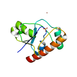
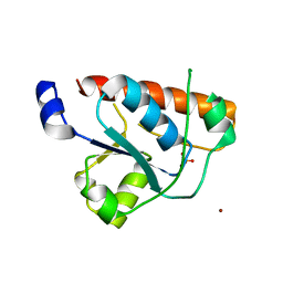
2L4I
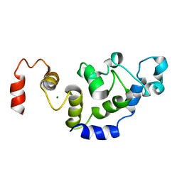 | | The Solution Structure of Magnesium bound CIB1 | | Descriptor: | Calcium and integrin-binding protein 1, MAGNESIUM ION | | Authors: | Huang, H, Vogel, H.J. | | Deposit date: | 2010-10-06 | | Release date: | 2011-03-09 | | Last modified: | 2024-05-01 | | Method: | SOLUTION NMR | | Cite: | Solution Structures of Ca2+-CIB1 and Mg2+-CIB1 and Their Interactions with the Platelet Integrin {alpha}IIb Cytoplasmic Domain.
J.Biol.Chem., 286, 2011
|
|
2L4H
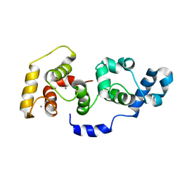 | | The Solution Structure of Calcium Bound CIB1 | | Descriptor: | CALCIUM ION, Calcium and integrin-binding protein 1 | | Authors: | Huang, H, Vogel, H.J. | | Deposit date: | 2010-10-06 | | Release date: | 2011-03-09 | | Last modified: | 2024-05-01 | | Method: | SOLUTION NMR | | Cite: | Solution Structures of Ca2+-CIB1 and Mg2+-CIB1 and Their Interactions with the Platelet Integrin {alpha}IIb Cytoplasmic Domain.
J.Biol.Chem., 286, 2011
|
|
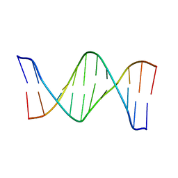
2LG3
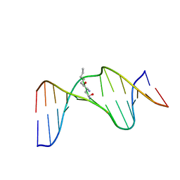 | | Structure of the duplex containing HNE derived (6S,8R,11S) gamma-HO-PdG when placed opposite dT | | Descriptor: | (4S)-nonane-1,4-diol, DNA (5'-D(*GP*CP*TP*AP*GP*CP*GP*AP*GP*TP*CP*C)-3'), DNA (5'-D(*GP*GP*AP*CP*TP*TP*GP*CP*TP*AP*GP*C)-3') | | Authors: | Huang, H, Wang, H, Kozekova, A, Lloyd, R.S, Rizzo, C.J, Stone, M.P. | | Deposit date: | 2011-07-19 | | Release date: | 2012-08-01 | | Last modified: | 2024-05-15 | | Method: | SOLUTION NMR | | Cite: | Ring-chain tautomerization of trans-4-hydroxynonenal derived (6S,8R,11S) gamma-hydroxy-1,N2-propano-deoxyguanosine adduct when placed opposite 2'-deoxythymidine in duplex
To be Published
|
|
2KV4
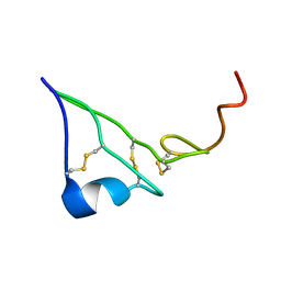 | | EGF | | Descriptor: | Epidermal growth factor | | Authors: | Huang, H.W, Mohan, S.K, Yu, C. | | Deposit date: | 2010-03-08 | | Release date: | 2011-02-23 | | Last modified: | 2023-06-14 | | Method: | SOLUTION NMR | | Cite: | The NMR solution structure of human epidermal growth factor (hEGF) at physiological pH and its interactions with suramin
Biochem.Biophys.Res.Commun., 402, 2010
|
|
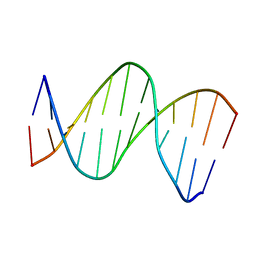
2KV6
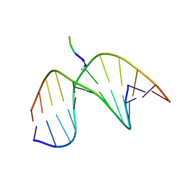 | | Tetrapeptide KWKK conjugated to oligonucleotide duplex by a trimethylene tether | | Descriptor: | 5'-D(*GP*CP*TP*AP*GP*CP*GP*AP*GP*TP*CP*C)-3', 5'-D(*GP*GP*AP*CP*TP*CP*GP*CP*TP*AP*GP*C)-3', KWKK Tetrapeptide | | Authors: | Huang, H, Kozekov, I.D, Kozekova, A, Rizzo, C.J, McCullough, A, LLoyd, R.S, Stone, M.P. | | Deposit date: | 2010-03-08 | | Release date: | 2010-07-21 | | Last modified: | 2011-07-13 | | Method: | SOLUTION NMR | | Cite: | Minor Groove Orientation of the KWKK Peptide Tethered via the N-Terminal Amine to the Acrolein-Derived 1,N(2)-gamma-Hydroxypropanodeoxyguanosine Lesion with a Trimethylene Linkage .
Biochemistry, 49, 2010
|
|
2LFA
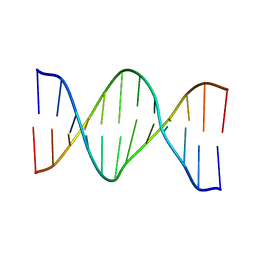 | | Oligonucleotide duplex contaning (5'S)-8,5'-cyclo-2'-deoxyguansine | | Descriptor: | DNA (5'-D(*AP*CP*AP*AP*AP*CP*AP*CP*GP*CP*AP*C)-3'), DNA (5'-D(*GP*TP*GP*CP*(2LF)P*TP*GP*TP*TP*TP*GP*T)-3') | | Authors: | Huang, H, Das, R.S, Basu, A, Stone, M.P. | | Deposit date: | 2011-06-29 | | Release date: | 2012-01-04 | | Last modified: | 2024-05-01 | | Method: | SOLUTION NMR | | Cite: | Structure of (5'S)-8,5'-Cyclo-2'-deoxyguanosine in DNA.
J.Am.Chem.Soc., 133, 2011
|
|
2LFX
 | | Structure of the duplex when (5'S)-8,5'-cyclo-2'-deoxyguanosine is placed opposite dT | | Descriptor: | DNA (5'-D(*AP*CP*AP*AP*AP*CP*AP*TP*GP*CP*AP*C)-3'), DNA (5'-D(*GP*TP*GP*CP*(2LF)P*TP*GP*TP*TP*TP*GP*T)-3') | | Authors: | Huang, H, Das, R.S, Basu, A, Stone, M.P. | | Deposit date: | 2011-07-18 | | Release date: | 2012-06-27 | | Last modified: | 2024-05-15 | | Method: | SOLUTION NMR | | Cite: | Structures of (5'S)-8,5'-Cyclo-2'-deoxyguanosine Mismatched with dA or dT.
Chem.Res.Toxicol., 25, 2012
|
|
2LG0
 | | structure of the duplex containing (5'S)-8,5'-cyclo-2'-deoxyadenosine | | Descriptor: | DNA (5'-D(*AP*CP*AP*AP*AP*CP*AP*TP*GP*CP*AP*C)-3'), DNA (5'-D(*GP*TP*GP*CP*(02I)P*TP*GP*TP*TP*TP*GP*T)-3') | | Authors: | Huang, H, Das, R.S, Basu, A, Stone, M.P. | | Deposit date: | 2011-07-18 | | Release date: | 2012-06-27 | | Last modified: | 2024-05-15 | | Method: | SOLUTION NMR | | Cite: | Structure of (5'S)-8,5'-cyclo-2'-deoxyguanosine in DNA.
J.Am.Chem.Soc., 133, 2011
|
|
5T8G
 | |
2WMY
 | |
5CJN
 | |
5E9X
 | |
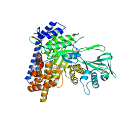
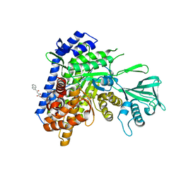
2WJA
 | | Crystal structure of the tyrosine phosphatase Wzb from Escherichia coli K30 in complex with phosphate. | | Descriptor: | NICKEL (II) ION, PHOSPHATE ION, PUTATIVE ACID PHOSPHATASE WZB | | Authors: | Huang, H, Hagelueken, G, Whitfield, C, Naismith, J.H. | | Deposit date: | 2009-05-25 | | Release date: | 2009-07-14 | | Last modified: | 2023-12-13 | | Method: | X-RAY DIFFRACTION (2.5 Å) | | Cite: | Crystal Structures of Wzb of Escherichia Coli and Cpsb of Streptococcus Pneumoniae, Representatives of Two Families of Tyrosine Phosphatases that Regulate Capsule Assembly.
J.Mol.Biol., 392, 2009
|
|
5DRC
 | |
5CAH
 | |
5C7V
 | |
5C9X
 | |
5CBB
 | |
5CAK
 | |
5C9W
 | |
5CEW
 | |
5C9R
 | |
5CBJ
 | |


