3C6Q
 
 | |
8TG1
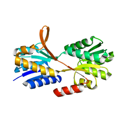
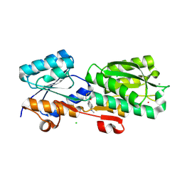
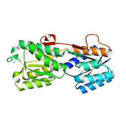
 | | Caldicellulosiruptor saccharolyticus periplasmic urea-binding protein | | 分子名称: | BROMIDE ION, Extracellular ligand-binding receptor, UREA | | 著者 | Allert, M.J, Kumar, S, Wang, Y, Beese, L.S, Hellinga, H.W. | | 登録日 | 2023-07-12 | | 公開日 | 2024-06-19 | | 実験手法 | X-RAY DIFFRACTION (2.097 Å) | | 主引用文献 | Structure-based functional analysis reveals multiple roles and widespread use of urea-binding proteins in nitrogen metabolism
To Be Published
|
|
3I5O
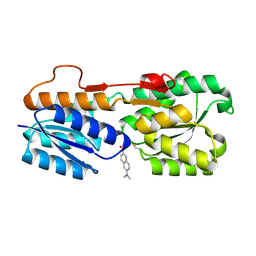
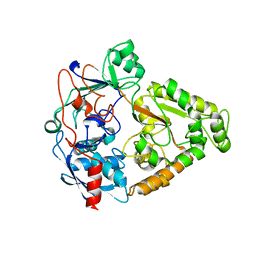
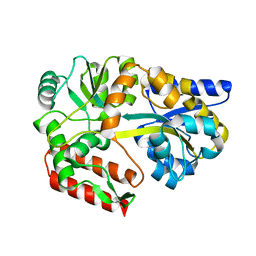
 | | The X-ray crystal structure of a thermophilic cellobiose binding protein bound with cellopentaose | | 分子名称: | Oligopeptide ABC transporter, periplasmic oligopeptide-binding protein, beta-D-glucopyranose-(1-4)-beta-D-glucopyranose-(1-4)-beta-D-glucopyranose-(1-4)-beta-D-glucopyranose-(1-4)-beta-D-glucopyranose | | 著者 | Cuneo, M.J, Hellinga, H.W. | | 登録日 | 2009-07-06 | | 公開日 | 2009-07-21 | | 最終更新日 | 2023-09-06 | | 実験手法 | X-RAY DIFFRACTION (1.5 Å) | | 主引用文献 | Structural Analysis of Semi-specific Oligosaccharide Recognition by a Cellulose-binding Protein of Thermotoga maritima Reveals Adaptations for Functional Diversification of the Oligopeptide Periplasmic Binding Protein Fold.
J.Biol.Chem., 284, 2009
|
|
8FXT
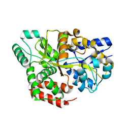
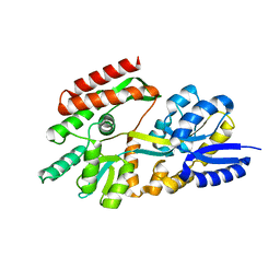
 | | Escherichia coli periplasmic Glucose-Binding Protein glucose complex: Acrylodan conjugate attached at W183C | | 分子名称: | 1-[6-(dimethylamino)naphthalen-2-yl]propan-1-one, CALCIUM ION, D-galactose/methyl-galactoside binding periplasmic protein MglB, ... | | 著者 | Allert, M.J, Kumar, S, Wang, Y, Beese, L.S, Hellinga, H.W. | | 登録日 | 2023-01-25 | | 公開日 | 2023-08-30 | | 最終更新日 | 2024-04-10 | | 実験手法 | X-RAY DIFFRACTION (1.53 Å) | | 主引用文献 | Chromophore carbonyl twisting in fluorescent biosensors encodes direct readout of protein conformations with multicolor switching.
Commun Chem, 6, 2023
|
|
8FXU
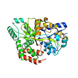
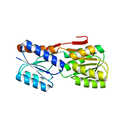
 | | Thermoanaerobacter thermosaccharolyticum periplasmic Glucose-Binding Protein glucose complex: Badan conjugate attached at F17C | | 分子名称: | 2-bromo-1-[6-(dimethylamino)naphthalen-2-yl]ethan-1-one, CALCIUM ION, CHLORIDE ION, ... | | 著者 | Allert, M.J, Kumar, S, Beese, L.S, Hellinga, H.W. | | 登録日 | 2023-01-25 | | 公開日 | 2023-08-30 | | 実験手法 | X-RAY DIFFRACTION (1.59 Å) | | 主引用文献 | Chromophore carbonyl twisting in fluorescent biosensors encodes direct readout of protein conformations with multicolor switching.
Commun Chem, 6, 2023
|
|
2O7I
 | |
2B3B
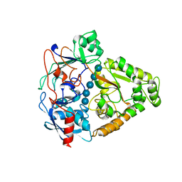
 | | Thermus thermophilus Glucose/Galactose Binding Protein With Bound Glucose | | 分子名称: | alpha-D-glucopyranose, beta-D-glucopyranose, glucose-binding protein | | 著者 | Cuneo, M.J, Changela, A, Warren, J.J, Beese, L.S, Hellinga, H.W. | | 登録日 | 2005-09-20 | | 公開日 | 2006-07-18 | | 最終更新日 | 2024-02-14 | | 実験手法 | X-RAY DIFFRACTION (1.95 Å) | | 主引用文献 | The crystal structure of a thermophilic glucose binding protein reveals adaptations that interconvert mono and di-saccharide binding sites.
J.Mol.Biol., 362, 2006
|
|
2B3F
 | | Thermus thermophilus Glucose/Galactose Binding Protein Bound With Galactose | | 分子名称: | beta-D-galactopyranose, glucose-binding protein | | 著者 | Cuneo, M.J, Changela, A, Warren, J.J, Beese, L.S, Hellinga, H.W. | | 登録日 | 2005-09-20 | | 公開日 | 2006-07-18 | | 最終更新日 | 2024-02-14 | | 実験手法 | X-RAY DIFFRACTION (1.56 Å) | | 主引用文献 | The crystal structure of a thermophilic glucose binding protein reveals adaptations that interconvert mono and di-saccharide binding sites.
J.Mol.Biol., 362, 2006
|
|
3QEB
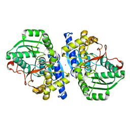
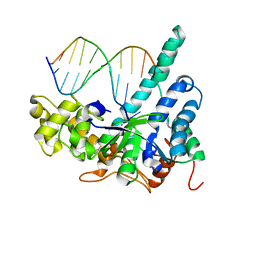 | | Crystal structure of human exonuclease 1 Exo1 (WT) in complex with DNA and Mn2+ (complex III) | | 分子名称: | DNA (5'-D(*CP*GP*CP*TP*AP*GP*TP*CP*GP*AP*CP*AP*T)-3'), DNA (5'-D(P*TP*CP*GP*AP*CP*TP*AP*GP*CP*G)-3'), Exonuclease 1, ... | | 著者 | Orans, J, McSweeney, E.A, Iyer, R.R, Hast, M.A, Hellinga, H.W, Modrich, P, Beese, L.S. | | 登録日 | 2011-01-20 | | 公開日 | 2011-04-20 | | 最終更新日 | 2024-02-21 | | 実験手法 | X-RAY DIFFRACTION (3 Å) | | 主引用文献 | Structures of human exonuclease 1 DNA complexes suggest a unified mechanism for nuclease family.
Cell(Cambridge,Mass.), 145, 2011
|
|
3QE9
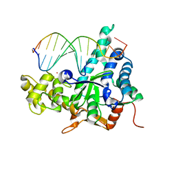 | | Crystal structure of human exonuclease 1 Exo1 (D173A) in complex with DNA (complex I) | | 分子名称: | CALCIUM ION, DNA (5'-D(*CP*GP*CP*TP*AP*GP*TP*CP*GP*AP*CP*AP*T)-3'), DNA (5'-D(P*TP*CP*GP*AP*CP*TP*AP*GP*CP*G)-3'), ... | | 著者 | Orans, J, McSweeney, E.A, Iyer, R.R, Hast, M.A, Hellinga, H.W, Modrich, P, Beese, L.S. | | 登録日 | 2011-01-20 | | 公開日 | 2011-04-20 | | 最終更新日 | 2024-02-21 | | 実験手法 | X-RAY DIFFRACTION (2.51 Å) | | 主引用文献 | Structures of human exonuclease 1 DNA complexes suggest a unified mechanism for nuclease family.
Cell(Cambridge,Mass.), 145, 2011
|
|
3QEA
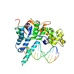 | | Crystal structure of human exonuclease 1 Exo1 (WT) in complex with DNA (complex II) | | 分子名称: | BARIUM ION, DNA (5'-D(P*CP*GP*CP*TP*AP*GP*TP*CP*GP*AP*CP*AP*T)-3'), DNA (5'-D(P*TP*CP*GP*AP*CP*TP*AP*GP*CP*G)-3'), ... | | 著者 | Orans, J, McSweeney, E.A, Iyer, R.R, Hast, M.A, Hellinga, H.W, Modrich, P, Beese, L.S. | | 登録日 | 2011-01-20 | | 公開日 | 2011-04-20 | | 最終更新日 | 2024-02-21 | | 実験手法 | X-RAY DIFFRACTION (3.1 Å) | | 主引用文献 | Structures of human exonuclease 1 DNA complexes suggest a unified mechanism for nuclease family.
Cell(Cambridge,Mass.), 145, 2011
|
|
2GX6
 | |
2HPG
 | |
2HPH
 | |
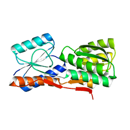
2FN9
 | | Thermotoga maritima Ribose Binding Protein Unliganded Form | | 分子名称: | ribose ABC transporter, periplasmic ribose-binding protein | | 著者 | Cuneo, M.J, Changela, A, Tian, Y, Beese, L.S, Hellinga, H.W. | | 登録日 | 2006-01-10 | | 公開日 | 2007-01-16 | | 最終更新日 | 2023-08-30 | | 実験手法 | X-RAY DIFFRACTION (1.4 Å) | | 主引用文献 | Ligand-induced conformational changes in a thermophilic ribose-binding protein.
Bmc Struct.Biol., 8, 2008
|
|
2IPM
 | |
2IPN
 | |
2GH9
 | | Thermus thermophilus maltotriose binding protein bound with maltotriose | | 分子名称: | alpha-D-glucopyranose-(1-4)-alpha-D-glucopyranose-(1-4)-alpha-D-glucopyranose, maltose/maltodextrin-binding protein | | 著者 | Cuneo, M.J, Changela, A, Beese, L.S, Hellinga, H.W. | | 登録日 | 2006-03-27 | | 公開日 | 2007-02-06 | | 最終更新日 | 2024-02-14 | | 実験手法 | X-RAY DIFFRACTION (1.95 Å) | | 主引用文献 | Structural adaptations that modulate monosaccharide, disaccharide, and trisaccharide specificities in periplasmic maltose-binding proteins.
J.Mol.Biol., 389, 2009
|
|
2GHB
 | | Thermotoga maritima maltotriose binding protein, ligand free form | | 分子名称: | maltose ABC transporter, periplasmic maltose-binding protein | | 著者 | Cuneo, M.J, Changela, A, Hocker, B, Beese, L.S, Hellinga, H.W. | | 登録日 | 2006-03-27 | | 公開日 | 2007-02-06 | | 最終更新日 | 2024-02-14 | | 実験手法 | X-RAY DIFFRACTION (2.1 Å) | | 主引用文献 | T. maritima maltotriose binding protein open form
To be Published
|
|
2IOY
 | |
2IPL
 | |
2GHA
 | | Thermotoga maritima maltotriose binding protein bound with maltotriose | | 分子名称: | alpha-D-glucopyranose-(1-4)-alpha-D-glucopyranose-(1-4)-alpha-D-glucopyranose, maltose ABC transporter, periplasmic maltose-binding protein | | 著者 | Cuneo, M.J, Changela, A, Hocker, B, Beese, L.S, Hellinga, H.W. | | 登録日 | 2006-03-27 | | 公開日 | 2007-02-06 | | 最終更新日 | 2024-02-14 | | 実験手法 | X-RAY DIFFRACTION (1.6 Å) | | 主引用文献 | T. maritima maltotriose binding protein
To be Published
|
|
3SRY
 | |
3SVB
 | |
3SST
 | |


