2CU9
 
 | |
4B2D
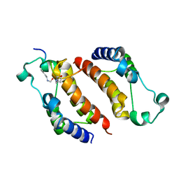
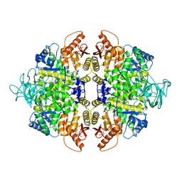 | | human PKM2 with L-serine and FBP bound. | | 分子名称: | 1,6-di-O-phosphono-beta-D-fructofuranose, MAGNESIUM ION, PYRUVATE KINASE ISOZYMES M1/M2, ... | | 著者 | Chaneton, B, Hillmann, P, Zheng, L, Martin, A.C.L, Maddocks, O.D.K, Chokkathukalam, A, Coyle, J.E, Jankevics, A, Holding, F.P, Vousden, K.H, Frezza, C, O'Reilly, M, Gottlieb, E. | | 登録日 | 2012-07-13 | | 公開日 | 2012-10-10 | | 最終更新日 | 2023-12-20 | | 実験手法 | X-RAY DIFFRACTION (2.3 Å) | | 主引用文献 | Serine is a natural ligand and allosteric activator of pyruvate kinase M2.
Nature, 491, 2012
|
|
7C60
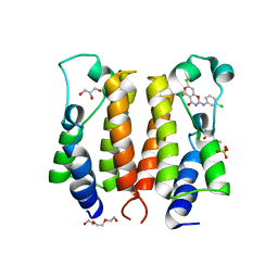
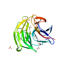 | | Crystal structure of Keap1 in complex with monoethyl fumarate (MEF) | | 分子名称: | (~{Z})-4-ethoxy-4-oxidanylidene-but-2-enoic acid, ACETATE ION, Kelch-like ECH-associated protein 1, ... | | 著者 | Padmanabhan, B, Unni, S, Deshmukh, P, Krishnappa, G. | | 登録日 | 2020-05-21 | | 公開日 | 2020-08-05 | | 最終更新日 | 2023-11-29 | | 実験手法 | X-RAY DIFFRACTION (1.95 Å) | | 主引用文献 | Structural insights into the multiple binding modes of Dimethyl Fumarate (DMF) and its analogs to the Kelch domain of Keap1.
Febs J., 288, 2021
|
|
7C5E
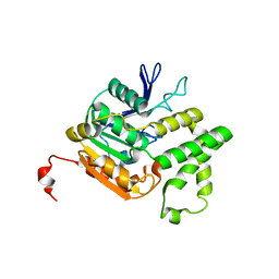
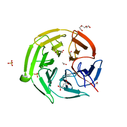 | | Crystal structure of Keap1 in complex with fumarate (FUM) | | 分子名称: | ACETATE ION, FUMARIC ACID, Kelch-like ECH-associated protein 1, ... | | 著者 | Padmanabhan, B, Unni, S, Deshmukh, P. | | 登録日 | 2020-05-19 | | 公開日 | 2020-08-05 | | 最終更新日 | 2023-11-29 | | 実験手法 | X-RAY DIFFRACTION (1.75 Å) | | 主引用文献 | Structural insights into the multiple binding modes of Dimethyl Fumarate (DMF) and its analogs to the Kelch domain of Keap1.
Febs J., 288, 2021
|
|
7EXI
 | |
2DWW
 | |
2DVV
 | |
2DWZ
 | |
2DX8
 | |
2E3K
 | |
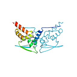
6JKE
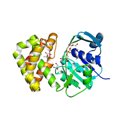
 | | Discovery and the crystal structure of NS5 in complex with the N-terminal bromodomain of BRD2. | | 分子名称: | 2,3-DIHYDROXY-1,4-DITHIOBUTANE, 7-chloranyl-2-[(3-chlorophenyl)amino]pyrano[3,4-e][1,3]oxazine-4,5-dione, Bromodomain-containing protein 2, ... | | 著者 | Padmanabhan, B, Mathur, S, Deshmukh, P. | | 登録日 | 2019-02-28 | | 公開日 | 2020-07-01 | | 最終更新日 | 2023-11-22 | | 実験手法 | X-RAY DIFFRACTION (1.5 Å) | | 主引用文献 | Novel pyrano 1,3 oxazine based ligand inhibits the epigenetic reader hBRD2 in glioblastoma.
Biochem.J., 477, 2020
|
|
4QLO
 | |
6K1R
 | |
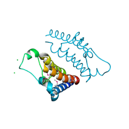
2K9Z
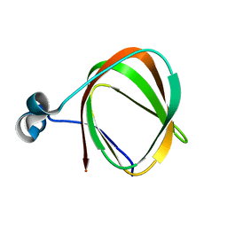 | | NMR structure of the protein TM1112 | | 分子名称: | uncharacterized protein TM1112 | | 著者 | Mohanty, B, Pedrini, B, Serrano, P, Geralt, M, Horst, R, Herrmann, T, Wilson, I.A, Wuthrich, K, Joint Center for Structural Genomics (JCSG) | | 登録日 | 2008-10-28 | | 公開日 | 2008-11-25 | | 最終更新日 | 2024-05-08 | | 実験手法 | SOLUTION NMR | | 主引用文献 | Comparison of NMR and crystal structures for the proteins TM1112 and TM1367.
Acta Crystallogr.,Sect.F, 66, 2010
|
|
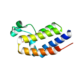
2KA0
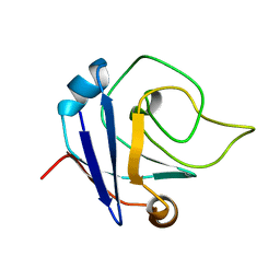 | | NMR structure of the protein TM1367 | | 分子名称: | uncharacterized protein TM1367 | | 著者 | Mohanty, B, Pedrini, B, Serrano, P, Geralt, M, Horst, R, Herrmann, T, Wilson, I.A, Wuthrich, K, Joint Center for Structural Genomics (JCSG) | | 登録日 | 2008-10-27 | | 公開日 | 2009-01-13 | | 最終更新日 | 2024-05-08 | | 実験手法 | SOLUTION NMR | | 主引用文献 | Comparison of NMR and crystal structures for the proteins TM1112 and TM1367.
Acta Crystallogr.,Sect.F, 66, 2010
|
|
2L6P
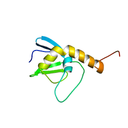 | | NMR solution structure of the protein NP_253742.1 | | 分子名称: | PhaC1, phaC2 and phaD genes | | 著者 | Mohanty, B, Serrano, P, Geralt, M, Horst, R, Wuthrich, K, Joint Center for Structural Genomics (JCSG) | | 登録日 | 2010-11-23 | | 公開日 | 2011-01-19 | | 最終更新日 | 2024-05-01 | | 実験手法 | SOLUTION NMR | | 主引用文献 | NMR solution structure of the protein NP_253742.1
To be Published
|
|
2N39
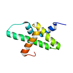 | |
2L9D
 | | Solution structure of the protein YP_546394.1, the first structural representative of the pfam family PF12112 | | 分子名称: | Uncharacterized protein | | 著者 | Mohanty, B, Serrano, P, Geralt, M, Horst, R, Wuthrich, K, Joint Center for Structural Genomics (JCSG) | | 登録日 | 2011-02-08 | | 公開日 | 2011-03-16 | | 最終更新日 | 2024-05-15 | | 実験手法 | SOLUTION NMR | | 主引用文献 | Solution structure of the protein YP_546394.1, the first structural representative of the pfam family PF12112
To be Published
|
|
2L6N
 | | NMR solution structure of the protein YP_001092504.1 | | 分子名称: | uncharacterized protein YP_001092504.1 | | 著者 | Mohanty, B, Serrano, P, Geralt, M, Horst, R, Wuthrich, K, Joint Center for Structural Genomics (JCSG) | | 登録日 | 2010-11-23 | | 公開日 | 2011-01-19 | | 最終更新日 | 2024-05-01 | | 実験手法 | SOLUTION NMR | | 主引用文献 | NMR solution structure of the protein YP_001092504.1
To be Published
|
|
2LA3
 | | The NMR structure of the protein NP_344798.1 reveals a CCA-adding enzyme head domain | | 分子名称: | Uncharacterized protein | | 著者 | Mohanty, B, Serrano, P, Geralt, M, Horst, R, Wuthrich, K, Joint Center for Structural Genomics (JCSG) | | 登録日 | 2011-03-01 | | 公開日 | 2011-03-30 | | 最終更新日 | 2024-05-15 | | 実験手法 | SOLUTION NMR | | 主引用文献 | Solution NMR structure of the protein NP_344798.1
To be Published
|
|
2KL4
 | | NMR structure of the protein NB7804A | | 分子名称: | BH2032 protein | | 著者 | Mohanty, B, Geralt, M, Augustyniak, W, Serrano, P, Horst, R, Wilson, I.A, Wuthrich, K, Joint Center for Structural Genomics (JCSG) | | 登録日 | 2009-06-30 | | 公開日 | 2009-07-14 | | 最終更新日 | 2024-05-22 | | 実験手法 | SOLUTION NMR | | 主引用文献 | Solution structure of the protein NB7804A from Bacillus Halodurans
To be Published
|
|
2KZC
 | | Solution NMR structure of the protein YP_510488.1 | | 分子名称: | Uncharacterized protein | | 著者 | Mohanty, B, Serrano, P, Geralt, M, Horst, R, Wilson, I.A, Wuthrich, K, Joint Center for Structural Genomics (JCSG) | | 登録日 | 2010-06-15 | | 公開日 | 2010-07-14 | | 最終更新日 | 2024-05-15 | | 実験手法 | SOLUTION NMR | | 主引用文献 | Solution NMR structure of the protein YP_510488.1
To be Published
|
|
2L1N
 | | Solution NMR structure of the protein YP_399305.1 | | 分子名称: | Uncharacterized protein | | 著者 | Mohanty, B, Serrano, P, Geralt, M, Horst, R, Wuthrich, K, Joint Center for Structural Genomics (JCSG) | | 登録日 | 2010-07-30 | | 公開日 | 2010-08-18 | | 最終更新日 | 2024-05-15 | | 実験手法 | SOLUTION NMR | | 主引用文献 | Solution NMR structure of the protein YP_399305.1
To be Published
|
|
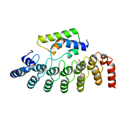
8XB9
 | | The Crystal Structure of polo-box domain of PLK1 from Biortus. | | 分子名称: | 1,2-ETHANEDIOL, Serine/threonine-protein kinase PLK1 | | 著者 | Wang, F, Cheng, W, Yuan, Z, Meng, Q, Zhang, B. | | 登録日 | 2023-12-06 | | 公開日 | 2023-12-27 | | 実験手法 | X-RAY DIFFRACTION (1.95 Å) | | 主引用文献 | The Crystal Structure of polo-box domain of PLK1 from Biortus
To Be Published
|
|
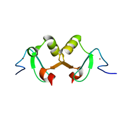
8XPG
 | | The Crystal Structure of polo box domain of Plk4 from Biortus. | | 分子名称: | SULFATE ION, Serine/threonine-protein kinase PLK4 | | 著者 | Wang, F, Cheng, W, Lv, Z, Meng, Q, Zhang, B. | | 登録日 | 2024-01-03 | | 公開日 | 2024-03-06 | | 実験手法 | X-RAY DIFFRACTION (2.7 Å) | | 主引用文献 | The Crystal Structure of polo box domain of Plk4 from Biortus.
To Be Published
|
|


