Loading
PDBj
MenuPDBj@FacebookPDBj@X(formerly Twitter)PDBj@BlueSkyPDBj@YouTubewwPDB FoundationwwPDB
RCSB PDBPDBeBMRBAdv. SearchSearch help
PDB: 687 results

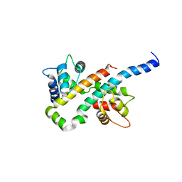
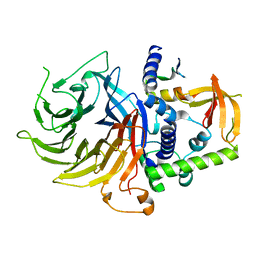
7N40
DownloadVisualize
BU of 7n40 by Molmil
Crystal structure of LIN9-RbAp48-LIN37, a MuvB subcomplex
Descriptor: Histone-binding protein RBBP4, Isoform 2 of Protein lin-9 homolog, Protein lin-37 homolog
Authors:Asthana, A, Ramanan, P, Tripathi, S.M, Rubin, S.M.
Deposit date:2021-06-02
Release date:2022-02-09
Last modified:2023-10-18
Method:X-RAY DIFFRACTION (2.55 Å)
Cite:The MuvB complex binds and stabilizes nucleosomes downstream of the transcription start site of cell-cycle dependent genes.
Nat Commun, 13, 2022
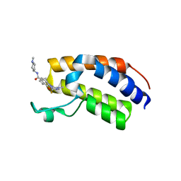
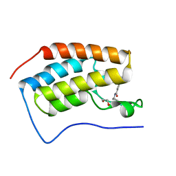
7BJY
DownloadVisualize
BU of 7bjy by Molmil
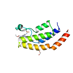
Crystal structure of the first bromodomain (BD1) of human BRDT bound to Ro3280
Descriptor: 1,2-ETHANEDIOL, 4-[(9-cyclopentyl-7,7-difluoro-5-methyl-6-oxo-6,7,8,9-tetrahydro-5H-pyrimido[4,5-b][1,4]diazepin-2-yl)amino]-3-methoxy-N-(1-methylpiperidin-4-yl)benzamide, Isoform 2 of Bromodomain testis-specific protein
Authors:Chan, A, Karim, M.R, Schonbrunn, E.
Deposit date:2021-01-14
Release date:2021-08-25
Last modified:2024-01-31
Method:X-RAY DIFFRACTION (2.22 Å)
Cite:Differential BET Bromodomain Inhibition by Dihydropteridinone and Pyrimidodiazepinone Kinase Inhibitors.
J.Med.Chem., 64, 2021
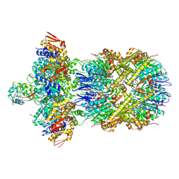
9C87
DownloadVisualize
BU of 9c87 by Molmil
Cryo-EM Structure of a Proteolytic ClpXP AAA+ Machine Poised to Unfold a Linear-Degron DHFR-ssrA Substrate Bound with MTX
Descriptor: ADENOSINE-5'-DIPHOSPHATE, ADENOSINE-5'-TRIPHOSPHATE, ATP-dependent Clp protease ATP-binding subunit ClpX, ...
Authors:Ghanbarpour, A, Sauer, R.T, Davis, J.H.
Deposit date:2024-06-12
Release date:2024-10-16
Method:ELECTRON MICROSCOPY (3.7 Å)
Cite:Cryo-EM Structure of a Proteolytic ClpXP AAA+ Machine Poised to Unfold a Linear-Degron DHFR-ssrA Substrate Bound with MTX
To Be Published
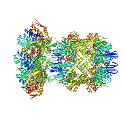
9C88
DownloadVisualize
BU of 9c88 by Molmil
Cryo-EM Structure of a Proteolytic ClpXP AAA+ Machine Translocating a Portion of a Branched-Degron DHFR Substrate
Descriptor: ADENOSINE-5'-DIPHOSPHATE, ADENOSINE-5'-TRIPHOSPHATE, ATP-dependent Clp protease ATP-binding subunit ClpX, ...
Authors:Ghanbarpour, A, Sauer, R.T, Davis, J.H.
Deposit date:2024-06-12
Release date:2024-10-16
Method:ELECTRON MICROSCOPY (2.6 Å)
Cite:Cryo-EM structure of a fully-engaged DHFR-ssrA substrate and the AAA+ ClpXP protease
To Be Published
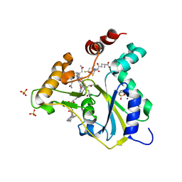
5UOS
DownloadVisualize
BU of 5uos by Molmil
Crystal Structure of CblC (MMACHC) (1-238), a human B12 processing enzyme, complexed with an Antivitamin B12
Descriptor: 1-ethynyl-2,4-difluorobenzene, 2-PHENYLAMINO-ETHANESULFONIC ACID, COBALAMIN, ...
Authors:Shanmuganathan, A, Karasik, A, Ruetz, M, Banerjee, R, Krautler, B, Koutmos, M.
Deposit date:2017-02-01
Release date:2017-06-07
Last modified:2023-10-04
Method:X-RAY DIFFRACTION (2.51 Å)
Cite:Antivitamin B12 Inhibition of the Human B12 -Processing Enzyme CblC: Crystal Structure of an Inactive Ternary Complex with Glutathione as the Cosubstrate.
Angew. Chem. Int. Ed. Engl., 56, 2017
1NJO
DownloadVisualize
BU of 1njo by Molmil
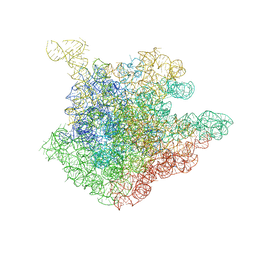
The crystal structure of the 50S Large ribosomal subunit from Deinococcus radiodurans complexed with a short substrate analog ACCPuromycin (ACCP)
Descriptor: 23S ribosomal RNA, RNA ACC(Puromycin)
Authors:Bashan, A, Agmon, I, Zarivatch, R, Schluenzen, F, Harms, J.M, Berisio, R, Bartels, H, Hansen, H.A, Yonath, A.
Deposit date:2003-01-02
Release date:2003-02-11
Last modified:2023-08-16
Method:X-RAY DIFFRACTION (3.7 Å)
Cite:Structural basis of the ribosomal machinery for Peptide bond formation, translocation, and nascent chain progression
Mol.Cell, 11, 2003
1NJM
DownloadVisualize
BU of 1njm by Molmil
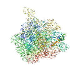
The crystal structure of the 50S Large ribosomal subunit from Deinococcus radiodurans complexed with a tRNA acceptor stem mimic (ASM) and the antibiotic sparsomycin
Descriptor: 23S ribosomal RNA, 50S ribosomal protein L16, GENERAL STRESS PROTEIN CTC, ...
Authors:Bashan, A, Agmon, I, Zarivatch, R, Schluenzen, F, Harms, J.M, Berisio, R, Bartels, H, Hansen, H.A, Yonath, A.
Deposit date:2003-01-02
Release date:2003-02-11
Last modified:2023-08-16
Method:X-RAY DIFFRACTION (3.6 Å)
Cite:Structural basis of the ribosomal machinery for Peptide bond formation, translocation, and nascent chain progression
Mol.Cell, 11, 2003
1NJP
DownloadVisualize
BU of 1njp by Molmil
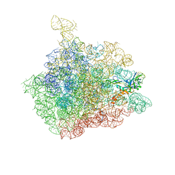
The crystal structure of the 50S Large ribosomal subunit from Deinococcus radiodurans complexed with a tRNA acceptor stem mimic (ASM)
Descriptor: 23S ribosomal RNA, 50S ribosomal protein L16, GENERAL STRESS PROTEIN CTC, ...
Authors:Bashan, A, Agmon, I, Zarivatch, R, Schluenzen, F, Harms, J.M, Berisio, R, Bartels, H, Hansen, H.A, Yonath, A.
Deposit date:2003-01-02
Release date:2003-02-11
Last modified:2023-08-16
Method:X-RAY DIFFRACTION (3.5 Å)
Cite:Structural basis of the ribosomal machinery for Peptide bond formation, translocation, and nascent chain progression
Mol.Cell, 11, 2003
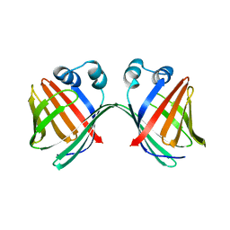
6E5E
DownloadVisualize
BU of 6e5e by Molmil
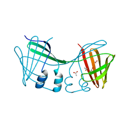
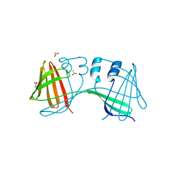
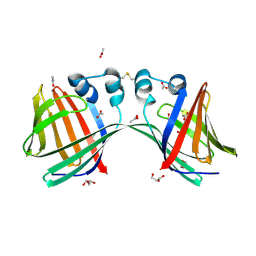
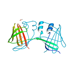
Crystal structure of the apo domain-swapped dimer Q108K:T51D mutant of human cellular retinol binding protein II
Descriptor: ACETATE ION, Retinol-binding protein 2
Authors:Ghanbarpour, A, Geiger, J.
Deposit date:2018-07-20
Release date:2019-10-16
Last modified:2023-10-11
Method:X-RAY DIFFRACTION (1.696 Å)
Cite:Engineering the hCRBPII Domain-Swapped Dimer into a New Class of Protein Switches.
J.Am.Chem.Soc., 141, 2019
6E5Q
DownloadVisualize
BU of 6e5q by Molmil
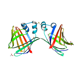
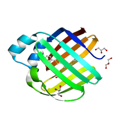
Crystal structure of the apo domain-swapped dimer Q108K:T51D:A28H mutant of human Cellular Retinol Binding Protein II
Descriptor: ACETATE ION, Retinol-binding protein 2
Authors:Ghanbarpour, A, Geiger, J.
Deposit date:2018-07-21
Release date:2019-10-16
Last modified:2023-10-11
Method:X-RAY DIFFRACTION (1.99 Å)
Cite:Engineering the hCRBPII Domain-Swapped Dimer into a New Class of Protein Switches.
J.Am.Chem.Soc., 141, 2019
6E51
DownloadVisualize
BU of 6e51 by Molmil
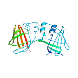
Crystal structure of the apo domain-swapped dimer Q108K:K40L:T51W mutant of human cellular retinol binding protein II
Descriptor: Retinol-binding protein 2
Authors:Ghanbarpour, A, Geiger, J.
Deposit date:2018-07-19
Release date:2019-10-16
Last modified:2023-10-11
Method:X-RAY DIFFRACTION (2.256 Å)
Cite:Engineering the hCRBPII Domain-Swapped Dimer into a New Class of Protein Switches.
J.Am.Chem.Soc., 141, 2019
6E50
DownloadVisualize
BU of 6e50 by Molmil
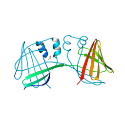
Crystal structure of the apo domain-swapped dimer Q108K:K40L:T51F mutant of human Cellular Retinol Binding Protein II
Descriptor: ACETATE ION, Retinol-binding protein 2
Authors:Ghanbarpour, A, Geiger, J.
Deposit date:2018-07-18
Release date:2019-10-16
Last modified:2023-10-11
Method:X-RAY DIFFRACTION (1.965 Å)
Cite:Engineering the hCRBPII Domain-Swapped Dimer into a New Class of Protein Switches.
J.Am.Chem.Soc., 141, 2019
6E5R
DownloadVisualize
BU of 6e5r by Molmil
Crystal structure of the apo domain-swapped dimer Q108K:T51D:A28C mutant of human Cellular Retinol Binding Protein II
Descriptor: ACETATE ION, GLYCEROL, Retinol-binding protein 2
Authors:Ghanbarpour, A, Geiger, J.
Deposit date:2018-07-22
Release date:2019-08-07
Last modified:2023-10-11
Method:X-RAY DIFFRACTION (2.592 Å)
Cite:Engineering the hCRBPII Domain-Swapped Dimer into a New Class of Protein Switches.
J.Am.Chem.Soc., 141, 2019
6E7M
DownloadVisualize
BU of 6e7m by Molmil
Crystal structure of the holo retinal-bound domain-swapped dimer Q108K:T51D:A28C mutant of human Cellular Retinol Binding Protein II
Descriptor: ACETATE ION, GLYCEROL, RETINAL, ...
Authors:Ghanbarpour, A, Geiger, J.
Deposit date:2018-07-26
Release date:2019-10-16
Last modified:2024-11-13
Method:X-RAY DIFFRACTION (2.7 Å)
Cite:Engineering the hCRBPII Domain-Swapped Dimer into a New Class of Protein Switches.
J.Am.Chem.Soc., 141, 2019
7MFY
DownloadVisualize
BU of 7mfy by Molmil
The Crystal Structure of Q108K:K40L:T51V:T53S:R58W:Y19W:A33W:L117E Mutant of HCRBPII Bound with LizFluor
Descriptor: 4-{5-[(2E)-but-2-en-2-yl]thiophen-2-yl}-N,N-dimethylaniline, ACETATE ION, GLYCEROL, ...
Authors:Ghanbarpour, A, Geiger, J.
Deposit date:2021-04-12
Release date:2022-01-12
Last modified:2023-10-18
Method:X-RAY DIFFRACTION (1.26 Å)
Cite:Design of Large Stokes Shift Fluorescent Proteins Based on Excited State Proton Transfer of an Engineered Photobase.
J.Am.Chem.Soc., 143, 2021
7MFZ
DownloadVisualize
BU of 7mfz by Molmil
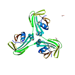
The Crystal Structure of Domain-Swapped Trimer Q108K:K40D:T53A:R58L:Q38F:Q4F Mutant of HCRBPII Bound with LizFluor3 Chromophore Showing Excited State Intermolecular Proton Transfer
Descriptor: 1-(4-{5-[(2E)-but-2-en-2-yl]thiophen-2-yl}phenyl)azetidine, ACETATE ION, GLYCEROL, ...
Authors:Ghanbarpour, A, Geiger, J.
Deposit date:2021-04-12
Release date:2022-01-12
Last modified:2024-10-09
Method:X-RAY DIFFRACTION (2.49 Å)
Cite:Design of Large Stokes Shift Fluorescent Proteins Based on Excited State Proton Transfer of an Engineered Photobase.
J.Am.Chem.Soc., 143, 2021
7MFX
DownloadVisualize
BU of 7mfx by Molmil
The Crystal Structure of Q108K:K40H:T53A:R58L:Q38F:Q4F Mutant of HCRBPII Bound with FR1 Chromophore Showing Excited State Intermolecular Proton Transfer
Descriptor: (4aP)-N,N-diethyl-9,9-dimethyl-7-[(1E)-prop-1-en-1-yl]-9H-fluoren-2-amine, GLYCEROL, Retinol-binding protein 2
Authors:Ghanbarpour, A, Geiger, J.
Deposit date:2021-04-11
Release date:2022-01-12
Last modified:2024-11-06
Method:X-RAY DIFFRACTION (1.59 Å)
Cite:Design of Large Stokes Shift Fluorescent Proteins Based on Excited State Proton Transfer of an Engineered Photobase.
J.Am.Chem.Soc., 143, 2021
6E6L
DownloadVisualize
BU of 6e6l by Molmil
Crystal structure of the holo retinal-bound domain-swapped dimer Q108K:K40L:T51F:Y60A mutant of human cellular retinol binding protein II
Descriptor: ACETATE ION, RETINAL, Retinol-binding protein 2
Authors:Ghanbarpour, A, Geiger, J.
Deposit date:2018-07-25
Release date:2019-10-16
Last modified:2024-11-06
Method:X-RAY DIFFRACTION (2.08 Å)
Cite:Engineering the hCRBPII Domain-Swapped Dimer into a New Class of Protein Switches.
J.Am.Chem.Soc., 141, 2019
6V1H
DownloadVisualize
BU of 6v1h by Molmil
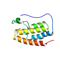
Crystal structure of the bromodomain of human BRD7 bound to bromosporine
Descriptor: 1,2-ETHANEDIOL, Bromodomain-containing protein 7, Bromosporine
Authors:Chan, A, Karim, M.R, Schonbrunn, E.
Deposit date:2019-11-20
Release date:2020-03-11
Last modified:2023-10-11
Method:X-RAY DIFFRACTION (1.93 Å)
Cite:Structural Basis of Inhibitor Selectivity in the BRD7/9 Subfamily of Bromodomains.
J.Med.Chem., 63, 2020
6V1L
DownloadVisualize
BU of 6v1l by Molmil
Crystal structure of the first bromodomain (BD1) of human BRD4 bound to BI9564
Descriptor: 4-[4-[(dimethylamino)methyl]-2,5-dimethoxy-phenyl]-2-methyl-2,7-naphthyridin-1-one, Bromodomain-containing protein 4
Authors:Chan, A, Karim, M.R, Schonbrunn, E.
Deposit date:2019-11-20
Release date:2020-03-11
Last modified:2023-10-11
Method:X-RAY DIFFRACTION (2.1 Å)
Cite:Structural Basis of Inhibitor Selectivity in the BRD7/9 Subfamily of Bromodomains.
J.Med.Chem., 63, 2020
6V1E
DownloadVisualize
BU of 6v1e by Molmil
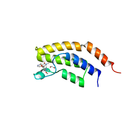
Crystal structure of the bromodomain of human BRD7 bound to BI7273
Descriptor: 1,2-ETHANEDIOL, 4-[4-[(dimethylamino)methyl]-3,5-dimethoxy-phenyl]-2-methyl-2,7-naphthyridin-1-one, Bromodomain-containing protein 7
Authors:Chan, A, Karim, M.R, Schonbrunn, E.
Deposit date:2019-11-20
Release date:2020-03-11
Last modified:2023-10-11
Method:X-RAY DIFFRACTION (2.3 Å)
Cite:Structural Basis of Inhibitor Selectivity in the BRD7/9 Subfamily of Bromodomains.
J.Med.Chem., 63, 2020
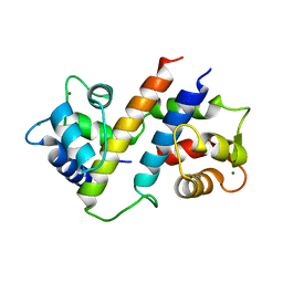
6B8M
DownloadVisualize
BU of 6b8m by Molmil
Crystal Structure of the Ca2+/CaM:Kv7.4 (KCNQ4) AB Domain Complex, 1 mM CaCl2 soak
Descriptor: CALCIUM ION, Calmodulin-1, Potassium voltage-gated channel subfamily KQT member 4, ...
Authors:Chang, A, Minor, D.L.
Deposit date:2017-10-09
Release date:2018-03-14
Last modified:2023-10-04
Method:X-RAY DIFFRACTION (2.3 Å)
Cite:A Calmodulin C-Lobe Ca
Neuron, 97, 2018
6MCV
DownloadVisualize
BU of 6mcv by Molmil
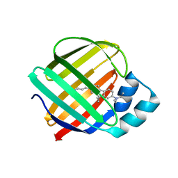
Crystal Structure of Holo Retinal-Bound Domain-Swapped Dimer of Wild Type Human Cellular Retinol Binding Protein II
Descriptor: RETINAL, Retinol-binding protein 2
Authors:Ghanbarpour, A, Geiger, J.
Deposit date:2018-09-02
Release date:2019-10-16
Last modified:2023-10-11
Method:X-RAY DIFFRACTION (3.3 Å)
Cite:Engineering the hCRBPII Domain-Swapped Dimer into a New Class of Protein Switches.
J.Am.Chem.Soc., 141, 2019
6V1U
DownloadVisualize
BU of 6v1u by Molmil
Crystal structure of the first bromodomain (BD1) of human BRD4 bound to TP-472
Descriptor: 1,2-ETHANEDIOL, 3-(6-acetylpyrrolo[1,2-a]pyrimidin-8-yl)-N-cyclopropyl-4-methylbenzamide, Bromodomain-containing protein 4
Authors:Chan, A, Karim, M.R, Schonbrunn, E.
Deposit date:2019-11-21
Release date:2020-03-11
Last modified:2023-10-11
Method:X-RAY DIFFRACTION (1.73 Å)
Cite:Structural Basis of Inhibitor Selectivity in the BRD7/9 Subfamily of Bromodomains.
J.Med.Chem., 63, 2020
6B8Q
DownloadVisualize
BU of 6b8q by Molmil
Crystal Structure of the Mg2+/CaM:Kv7.5 (KCNQ5) AB domain complex
Descriptor: Calmodulin-1, MAGNESIUM ION, Potassium voltage-gated channel subfamily KQT member 5
Authors:Chang, A, Abderemane-Ali, F, Minor, D.L.
Deposit date:2017-10-09
Release date:2018-03-14
Last modified:2023-10-04
Method:X-RAY DIFFRACTION (2.6 Å)
Cite:A Calmodulin C-Lobe Ca
Neuron, 97, 2018

227344

건을2024-11-13부터공개중

PDB statisticsPDBj update infoContact PDBjnumon