
Loading
PDBj
MenuPDBj@FacebookPDBj@TwitterPDBj@YouTubewwPDB FoundationwwPDB
RCSB PDBPDBeBMRBAdv. SearchSearch help
PDB: 52 results

4ML7
DownloadVisualize
BU of 4ml7 by Molmil
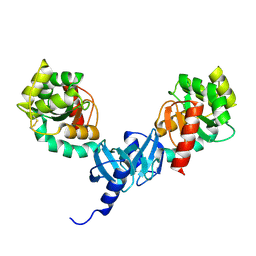
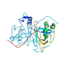
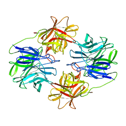
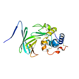
Crystal structure of Brucella abortus PliC in complex with human lysozyme
Descriptor: Humanlysozyme, Lysozyme C
Authors:Ha, N.C, Um, S.H, Kim, J.S.
Deposit date:2013-09-06
Release date:2014-07-23
Last modified:2017-11-08
Method:X-RAY DIFFRACTION (1.8 Å)
Cite:Structural Basis for the Inhibition of Human Lysozyme by PliC from Brucella abortus
Biochemistry, 52, 2013
4MIR
DownloadVisualize
BU of 4mir by Molmil
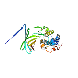
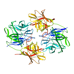
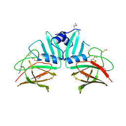
The structure of Brucella abortus PliC in the hexagonal crystal form
Descriptor: Putative uncharacterized protein
Authors:Ha, N.C, Um, S.H, Kim, J.S.
Deposit date:2013-09-02
Release date:2014-07-16
Last modified:2024-10-09
Method:X-RAY DIFFRACTION (2.4 Å)
Cite:Structural basis for the inhibition of human lysozyme by PliC from Brucella abortus
Biochemistry, 52, 2013
4MIS
DownloadVisualize
BU of 4mis by Molmil
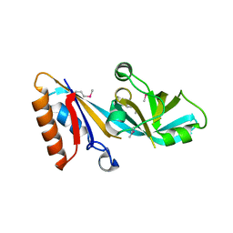
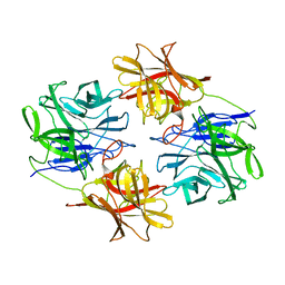
The structure of Brucella abortus PliC in the orthorombic crystal form
Descriptor: Putative uncharacterized protein
Authors:Ha, N.C, Um, S.H, Kim, J.S.
Deposit date:2013-09-02
Release date:2014-07-16
Method:X-RAY DIFFRACTION (2 Å)
Cite:Structural basis for the inhibition of human lysozyme by PliC from Brucella abortus
Biochemistry, 52, 2013
5C0Q
DownloadVisualize
BU of 5c0q by Molmil
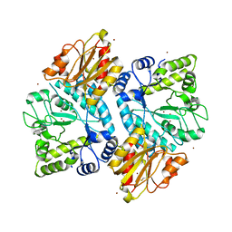
Crystal structure of Zn bound CbsA from Thermotoga neapolitana
Descriptor: Beta-N-acetylhexosaminidase, ZINC ION
Authors:Ha, N.C, Kim, J.S, Yoon, B.Y.
Deposit date:2015-06-12
Release date:2015-09-16
Last modified:2024-10-30
Method:X-RAY DIFFRACTION (2.499 Å)
Cite:Crystal structure of beta-N-acetylglucosaminidase CbsA from Thermotoga neapolitana
Biochem.Biophys.Res.Commun., 464, 2015
5BZA
DownloadVisualize
BU of 5bza by Molmil
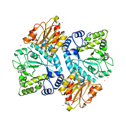
Crystal structure of CbsA from Thermotoga neapolitana
Descriptor: Beta-N-acetylhexosaminidase, CADMIUM ION
Authors:Ha, N.C, Kim, J.S, Yoon, B.Y.
Deposit date:2015-06-11
Release date:2015-09-16
Last modified:2020-02-19
Method:X-RAY DIFFRACTION (2.002 Å)
Cite:Crystal structure of beta-N-acetylglucosaminidase CbsA from Thermotoga neapolitana
Biochem.Biophys.Res.Commun., 464, 2015
2OLG
DownloadVisualize
BU of 2olg by Molmil
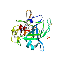
Crystal structure of the serine protease domain of prophenoloxidase activating factor-I in a zymogen form
Descriptor: 2-acetamido-2-deoxy-beta-D-glucopyranose, CALCIUM ION, Pro-phenoloxidase activating enzyme-I, ...
Authors:Ha, N.C, Piao, S.
Deposit date:2007-01-19
Release date:2007-02-06
Last modified:2023-10-25
Method:X-RAY DIFFRACTION (1.7 Å)
Cite:Crystal structure of the serine protease domain of prophenoloxidase activating factor-I
J.Biol.Chem., 282, 2007
4ILF
DownloadVisualize
BU of 4ilf by Molmil
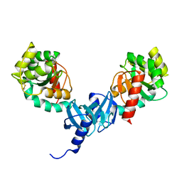
Crystal structure of DsbC R125A from Salmonella enterica serovar Typhimurium
Descriptor: Thiol:disulfide interchange protein DsbC
Authors:Ha, N.C, Li, J, Kim, J.S, Yoon, B.Y, Yeom, J.H, Lee, K.
Deposit date:2012-12-31
Release date:2013-10-16
Last modified:2017-11-15
Method:X-RAY DIFFRACTION (1.999 Å)
Cite:Crystal structure of the periplasmic disulfide-bond isomerase DsbC from Salmonella enterica serovar Typhimurium and the mechanistic implications.
J.Struct.Biol., 183, 2013
4GKL
DownloadVisualize
BU of 4gkl by Molmil
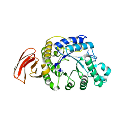
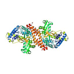
Crystal structure of a noncanonic maltogenic alpha-amylase AmyB from Thermotoga neapolitana
Descriptor: Alpha-amylase
Authors:Ha, N.C, Jun, S.Y, Kim, J.S.
Deposit date:2012-08-13
Release date:2013-02-27
Last modified:2024-03-20
Method:X-RAY DIFFRACTION (2.4 Å)
Cite:Structure of a novel alpha-amylase AmyB from Thermotoga neapolitana that produces maltose from the nonreducing end of polysaccharides
Acta Crystallogr.,Sect.D, 69, 2013
4QA8
DownloadVisualize
BU of 4qa8 by Molmil
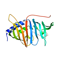
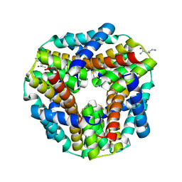
Crystal structure of LprF from Mycobacterium bovis
Descriptor: (2R)-2-(dodecanoyloxy)propyl (4E,6E,8E,10E,12E)-pentadeca-4,6,8,10,12-pentaenoate, Putative lipoprotein LprF
Authors:Ha, N.C, Jiao, L, Kim, J.S.
Deposit date:2014-05-02
Release date:2014-10-22
Last modified:2024-03-20
Method:X-RAY DIFFRACTION (1.1 Å)
Cite:Crystal structure and functional implications of LprF from Mycobacterium tuberculosis and M. bovis
Acta Crystallogr.,Sect.D, 70, 2014
4I5Q
DownloadVisualize
BU of 4i5q by Molmil
Crystal structure and catalytic mechanism for peroplasmic disulfide-bond isomerase DsbC from Salmonella enterica serovar Typhimurium
Descriptor: MAGNESIUM ION, Thiol:disulfide interchange protein DsbC
Authors:Ha, N.C, Li, J, Kim, J.S, Yoon, B.Y, Yeom, J.H, Lee, K.
Deposit date:2012-11-28
Release date:2013-10-16
Last modified:2017-11-15
Method:X-RAY DIFFRACTION (1.962 Å)
Cite:Crystal structure of the periplasmic disulfide-bond isomerase DsbC from Salmonella enterica serovar Typhimurium and the mechanistic implications.
J.Struct.Biol., 183, 2013
4YK9
DownloadVisualize
BU of 4yk9 by Molmil
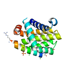
Complex structure of BCL-XL and mutated BIM BH3 domain
Descriptor: ACETATE ION, BH3BIM, Bcl-2-like protein 1, ...
Authors:Ha, N.C, Kim, J.S.
Deposit date:2015-03-04
Release date:2016-04-13
Method:X-RAY DIFFRACTION (1.7 Å)
Cite:Complex structure of BCL-XL and mutated BIM BH3 domain
To be published
4Z85
DownloadVisualize
BU of 4z85 by Molmil
Crystal structur of Pseudomonas fluorescens 2-nitrobenzoate 2-nitroreductase NbaA
Descriptor: 2-nitrobenzoate nitroreductase
Authors:Ha, N.C, Jiao, L, Kim, J.S.
Deposit date:2015-04-08
Release date:2016-01-20
Last modified:2023-11-08
Method:X-RAY DIFFRACTION (1.7 Å)
Cite:Structural and Mechanistic Insights into the Pseudomonas fluorescens 2-Nitrobenzoate 2-Nitroreductase NbaA
Appl.Environ.Microbiol., 81, 2015
3F6Z
DownloadVisualize
BU of 3f6z by Molmil
Crystal structure of Pseudomonas aeruginosa MliC in complex with hen egg white lysozyme
Descriptor: Lysozyme C, Putative uncharacterized protein
Authors:Ha, N.C, Yum, S.
Deposit date:2008-11-07
Release date:2008-12-23
Last modified:2024-10-30
Method:X-RAY DIFFRACTION (2.5 Å)
Cite:Structural basis for the recognition of lysozyme by MliC, a periplasmic lysozyme inhibitor in Gram-negative bacteria.
Biochem.Biophys.Res.Commun., 378, 2009
5C21
DownloadVisualize
BU of 5c21 by Molmil
Crystal structure of native HlyD from E. coli
Descriptor: Chromosomal hemolysin D
Authors:Ha, N.C, Kim, J.S, Yoon, B.Y.
Deposit date:2015-06-15
Release date:2016-02-17
Last modified:2024-03-20
Method:X-RAY DIFFRACTION (2.5 Å)
Cite:Crystal Structure of a Soluble Fragment of the Membrane Fusion Protein HlyD in a Type I Secretion System of Gram-Negative Bacteria
Structure, 24, 2016
5C59
DownloadVisualize
BU of 5c59 by Molmil
Crystal structure of the periplasmic region of MacB from E. coli
Descriptor: Macrolide export ATP-binding/permease protein MacB
Authors:Ha, N.C, Kim, J.S.
Deposit date:2015-06-19
Release date:2016-06-22
Last modified:2020-02-19
Method:X-RAY DIFFRACTION (3 Å)
Cite:Crystal structure of the periplasmic region of MacB from E. coli
To Be Published
5C22
DownloadVisualize
BU of 5c22 by Molmil
Crystal structure of Zn-bound HlyD from E. coli
Descriptor: Chromosomal hemolysin D, ZINC ION
Authors:Ha, N.C, Kim, J.S.
Deposit date:2015-06-15
Release date:2016-02-17
Last modified:2024-03-20
Method:X-RAY DIFFRACTION (2.302 Å)
Cite:Crystal Structure of a Soluble Fragment of the Membrane Fusion Protein HlyD in a Type I Secretion System of Gram-Negative Bacteria
Structure, 24, 2016
5ZQS
DownloadVisualize
BU of 5zqs by Molmil
Crystal structure of beta-xylosidase mutant (E186Q/F503Y) from Bacillus pumilus
Descriptor: Beta-xylosidase, beta-D-xylopyranose, beta-D-xylopyranose-(1-4)-beta-D-xylopyranose
Authors:Ha, N.C, Hong, S, Jo, I.
Deposit date:2018-04-20
Release date:2018-05-30
Last modified:2023-11-22
Method:X-RAY DIFFRACTION (1.782 Å)
Cite:Structure-based protein engineering of bacterial beta-xylosidase to increase the production yield of xylobiose from xylose
Biochem. Biophys. Res. Commun., 501, 2018
5ZQJ
DownloadVisualize
BU of 5zqj by Molmil
Crystal structure of beta-xylosidase from Bacillus pumilus
Descriptor: Beta-xylosidase, GLYCEROL
Authors:Ha, N.C, Hong, S, Jo, I.
Deposit date:2018-04-19
Release date:2018-05-30
Last modified:2023-11-22
Method:X-RAY DIFFRACTION (1.73 Å)
Cite:Structure-based protein engineering of bacterial beta-xylosidase to increase the production yield of xylobiose from xylose
Biochem. Biophys. Res. Commun., 501, 2018
5ZQX
DownloadVisualize
BU of 5zqx by Molmil
Crystal structure of beta-xylosidase mutant (E186Q) from Bacillus pumilus
Descriptor: Beta-xylosidase, beta-D-xylopyranose-(1-4)-beta-D-xylopyranose
Authors:Ha, N.C, Hong, S, Jo, I.
Deposit date:2018-04-20
Release date:2018-05-30
Last modified:2023-11-22
Method:X-RAY DIFFRACTION (2 Å)
Cite:Structure-based protein engineering of bacterial beta-xylosidase to increase the production yield of xylobiose from xylose
Biochem. Biophys. Res. Commun., 501, 2018
3U95
DownloadVisualize
BU of 3u95 by Molmil
Crystal structure of a putative alpha-glucosidase from Thermotoga neapolitana
Descriptor: Glycoside hydrolase, family 4, MANGANESE (II) ION
Authors:Ha, N.C, Jun, S.Y, Yun, B.Y, Yoon, B.Y, Piao, S.
Deposit date:2011-10-17
Release date:2012-09-26
Last modified:2024-03-20
Method:X-RAY DIFFRACTION (1.998 Å)
Cite:Crystal structure and thermostability of a putative alpha-glucosidase from Thermotoga neapolitana
Biochem.Biophys.Res.Commun., 416, 2011
4GQZ
DownloadVisualize
BU of 4gqz by Molmil
Crystal Structure of S.CueP
Descriptor: CHLORIDE ION, Putative periplasmic or exported protein
Authors:Ha, N.C, Yoon, B.Y.
Deposit date:2012-08-24
Release date:2013-08-14
Last modified:2013-10-23
Method:X-RAY DIFFRACTION (1.799 Å)
Cite:Structure of the periplasmic copper-binding protein CueP from Salmonella enterica serovar Typhimurium
Acta Crystallogr.,Sect.D, 69, 2013
7Y4R
DownloadVisualize
BU of 7y4r by Molmil
Structure of RclX
Descriptor: CMD domain-containing protein, HYDROGEN PEROXIDE, LYSINE
Authors:Ki, N, Ha, N.C.
Deposit date:2022-06-16
Release date:2023-07-05
Last modified:2024-10-30
Method:X-RAY DIFFRACTION (2.51 Å)
Cite:Crystal structure of the putative HOCl and HOSCN-responsive peroxiredoxin RclX from Pseudomonas aeruginosa
To Be Published
8OHM
DownloadVisualize
BU of 8ohm by Molmil
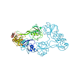
CRYSTAL STRUCTURE OF RNA HELICASE FROM GENOTYPE 1B HEPATITIS C VIRUS: MECHANISM OF UNWINDING DUPLEX RNA
Descriptor: RNA HELICASE
Authors:Cho, H.S, Ha, N.C, Kang, L.W, Oh, B.H.
Deposit date:1998-03-13
Release date:1999-04-20
Last modified:2024-02-14
Method:X-RAY DIFFRACTION (2.3 Å)
Cite:Crystal structure of RNA helicase from genotype 1b hepatitis C virus. A feasible mechanism of unwinding duplex RNA.
J.Biol.Chem., 273, 1998
8I33
DownloadVisualize
BU of 8i33 by Molmil
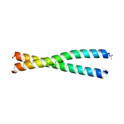
Coil 1a of lamin A (residue 25-65)
Descriptor: Prelamin-A/C
Authors:Jeong, S, Ha, N.C.
Deposit date:2023-01-16
Release date:2024-01-24
Method:X-RAY DIFFRACTION (1.62 Å)
Cite:Coil 1a of lamin A (residue 25-65)
To Be Published
6JLB
DownloadVisualize
BU of 6jlb by Molmil
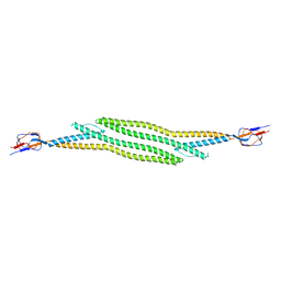
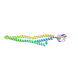
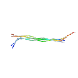
Crystal structure of lamin A/C fragment and assembly mechanisms of intermediate filaments
Descriptor: Lamin A/C
Authors:Ahn, J, Jo, I, Ha, N.C.
Deposit date:2019-03-04
Release date:2019-09-11
Last modified:2024-10-23
Method:X-RAY DIFFRACTION (3.205 Å)
Cite:Structural basis for lamin assembly at the molecular level.
Nat Commun, 10, 2019


123>

226707

PDB entries from 2024-10-30

PDB statisticsPDBj update infoContact PDBjnumon