Loading
PDBj
MenuPDBj@FacebookPDBj@TwitterPDBj@YouTubewwPDB FoundationwwPDB
RCSB PDBPDBeBMRBAdv. SearchSearch help
PDB: 74 results

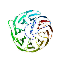
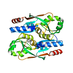
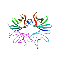
6IY0
DownloadVisualize
BU of 6iy0 by Molmil
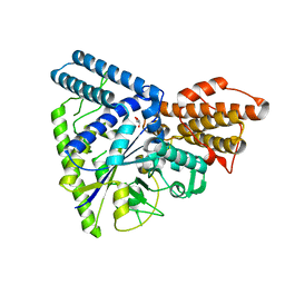
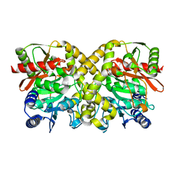
Crystal structure of conserved hypothetical protein SAV0927 from Staphylococcus aureus subsp. aureus Mu50
Descriptor: CHLORIDE ION, SAV0927
Authors:Jeong, S, Ha, N.-C.
Deposit date:2018-12-12
Release date:2019-12-18
Last modified:2020-07-01
Method:X-RAY DIFFRACTION (2.5 Å)
Cite:Crystal Structure of SAV0927 and Its Functional Implications.
J Microbiol Biotechnol., 29, 2019
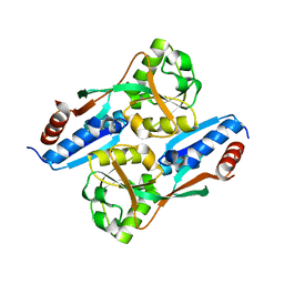
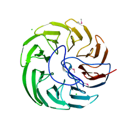
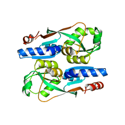
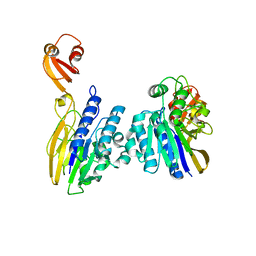
5X15
DownloadVisualize
BU of 5x15 by Molmil
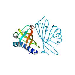
Crystal structure of Streptomyces coelicolor RraAS2, an unusual member of the RNase ES inhibitor RraA protein family
Descriptor: Putative transferase
Authors:Park, N, Jo, I, Ha, N.-C.
Deposit date:2017-01-24
Release date:2017-05-17
Last modified:2024-03-20
Method:X-RAY DIFFRACTION (3.094 Å)
Cite:Crystal structure of Streptomyces coelicolor RraAS2, an unusual member of the RNase E inhibitor RraA protein family
J. Microbiol., 55, 2017
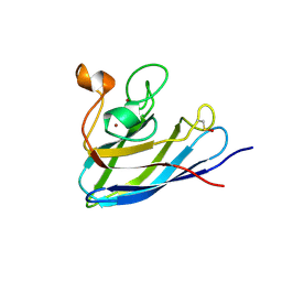
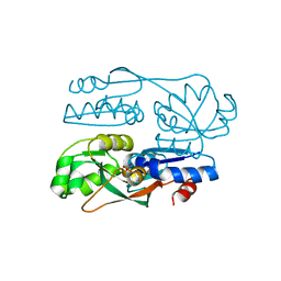
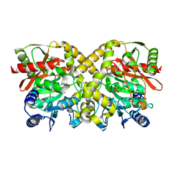
5WU7
DownloadVisualize
BU of 5wu7 by Molmil
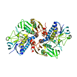
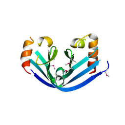
Crystal structure of GH57-type branching enzyme from hyperthermophilic archaeon Pyrococcus horikoshii
Descriptor: GLYCEROL, Uncharacterized protein
Authors:Na, S, Jo, I, Ha, N.-C.
Deposit date:2016-12-16
Release date:2017-03-08
Last modified:2023-11-08
Method:X-RAY DIFFRACTION (2.31 Å)
Cite:Structural basis for the transglycosylase activity of a GH57-type glycogen branching enzyme from Pyrococcus horikoshii.
Biochem. Biophys. Res. Commun., 484, 2017
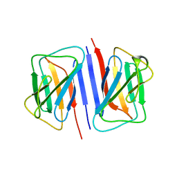
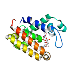
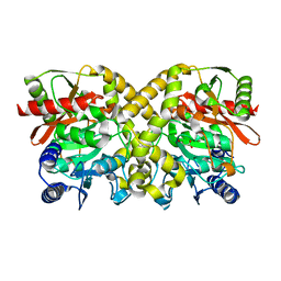
6K22
DownloadVisualize
BU of 6k22 by Molmil
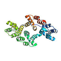
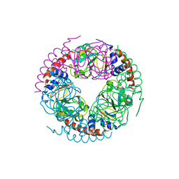
Crystal structure of Ca-bound human Annexin A5 in low salt condition
Descriptor: Annexin A5, CALCIUM ION
Authors:Hong, S, Ha, N.-C.
Deposit date:2019-05-13
Release date:2020-03-25
Last modified:2023-11-22
Method:X-RAY DIFFRACTION (2.747 Å)
Cite:High-resolution structures of annexin A5 in a two-dimensional array.
J.Struct.Biol., 209, 2020
6KGY
DownloadVisualize
BU of 6kgy by Molmil
HOCl-induced flavoprotein disulfide reductase RclA from Escherichia coli
Descriptor: CHLORIDE ION, FLAVIN-ADENINE DINUCLEOTIDE, Pyridine nucleotide-disulphide oxidoreductase dimerisation region
Authors:Baek, Y, Ha, N.-C.
Deposit date:2019-07-12
Release date:2020-02-05
Last modified:2024-04-03
Method:X-RAY DIFFRACTION (2.9 Å)
Cite:Structure and function of the hypochlorous acid-induced flavoprotein RclA fromEscherichia coli.
J.Biol.Chem., 295, 2020
1OPY
DownloadVisualize
BU of 1opy by Molmil
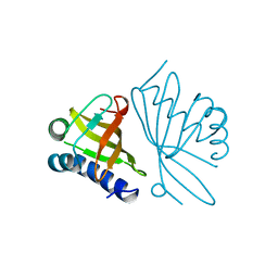
KSI
Descriptor: DELTA5-3-KETOSTEROID IOSMERASE
Authors:Kim, S.-W, Cha, S.-S, Cho, H.-S, Kim, J.-S, Ha, N.-C, Cho, M.-J, Choi, K.-Y, Oh, B.-H.
Deposit date:1997-05-23
Release date:1998-04-08
Last modified:2024-02-14
Method:X-RAY DIFFRACTION (1.9 Å)
Cite:High-resolution crystal structures of delta5-3-ketosteroid isomerase with and without a reaction intermediate analogue.
Biochemistry, 36, 1997
1CVM
DownloadVisualize
BU of 1cvm by Molmil
CADMIUM INHIBITED CRYSTAL STRUCTURE OF PHYTASE FROM BACILLUS AMYLOLIQUEFACIENS
Descriptor: CADMIUM ION, CALCIUM ION, PHYTASE
Authors:Shin, S, Ha, N.-C, Oh, B.-H.
Deposit date:1999-08-24
Release date:2000-02-24
Last modified:2024-02-07
Method:X-RAY DIFFRACTION (2.4 Å)
Cite:Crystal structures of a novel, thermostable phytase in partially and fully calcium-loaded states.
Nat.Struct.Biol., 7, 2000
7V5V
DownloadVisualize
BU of 7v5v by Molmil
LysR family transcriptional regulator RipR from Salmonella Typhimurium
Descriptor: RipR
Authors:Ki, N, Ha, N.-C.
Deposit date:2021-08-18
Release date:2022-08-24
Last modified:2024-04-03
Method:X-RAY DIFFRACTION (2.37 Å)
Cite:The activation mechanism of the itaconic acid-responsive LTTR RipR in the pathogenesis of the foodborne-pathogen Salmonella enterica
To Be Published
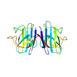
7FB9
DownloadVisualize
BU of 7fb9 by Molmil
Crystal Structure of Human Cu, Zn Superoxide Dismutase (SOD1)
Descriptor: COPPER (II) ION, Superoxide dismutase [Cu-Zn], ZINC ION
Authors:Baek, Y, Ha, N.-C.
Deposit date:2021-07-08
Release date:2022-10-12
Last modified:2023-11-29
Method:X-RAY DIFFRACTION (2.7 Å)
Cite:Structural analysis of the overoxidized Cu/Zn-superoxide dismutase in ROS-induced ALS filament formation.
Commun Biol, 5, 2022
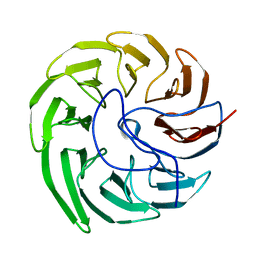
7EVF
DownloadVisualize
BU of 7evf by Molmil
Plant growth-promoting factor YxaL mutant from Bacillus velezensis - T175W/S213G/W215A
Descriptor: PQQ_3 domain-containing protein
Authors:Kim, J, Ha, N.-C.
Deposit date:2021-05-21
Release date:2022-01-12
Last modified:2023-11-29
Method:X-RAY DIFFRACTION (1.5 Å)
Cite:Structure of the plant growth-promoting factor YxaL from the rhizobacterium Bacillus velezensis and its application to protein engineering.
Acta Crystallogr D Struct Biol, 78, 2022
7EQ5
DownloadVisualize
BU of 7eq5 by Molmil
Plant growth-promoting factor YxaL mutant from Bacillus velezensis - T175W/W215G
Descriptor: Membrane associated protein kinase with beta-propeller domain, pyrrolo-quinoline quinone beta-propeller repeat
Authors:Kim, J, Ha, N.-C.
Deposit date:2021-04-30
Release date:2022-01-12
Last modified:2023-11-29
Method:X-RAY DIFFRACTION (2.6 Å)
Cite:Structure of the plant growth-promoting factor YxaL from the rhizobacterium Bacillus velezensis and its application to protein engineering.
Acta Crystallogr D Struct Biol, 78, 2022
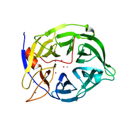
7DXN
DownloadVisualize
BU of 7dxn by Molmil
Plant growth-promoting factor YxaL from Bacillus velezensis
Descriptor: CHLORIDE ION, Membrane associated protein kinase with beta-propeller domain, pyrrolo-quinoline quinone beta-propeller repeat
Authors:Baek, Y, Ha, N.-C.
Deposit date:2021-01-19
Release date:2022-01-19
Method:X-RAY DIFFRACTION (1.8 Å)
Cite:Structure of the plant growth-promoting factor YxaL from the rhizobacterium Bacillus velezensis and its application to protein engineering.
Acta Crystallogr D Struct Biol, 78, 2022
7FB6
DownloadVisualize
BU of 7fb6 by Molmil
C57D/C146D mutant of Human Cu, Zn Superoxide Dismutase (SOD1)
Descriptor: CHLORIDE ION, COPPER (II) ION, Superoxide dismutase [Cu-Zn], ...
Authors:Baek, Y, Ha, N.-C.
Deposit date:2021-07-08
Release date:2022-10-12
Last modified:2023-11-29
Method:X-RAY DIFFRACTION (1.8 Å)
Cite:Structural analysis of the overoxidized Cu/Zn-superoxide dismutase in ROS-induced ALS filament formation.
Commun Biol, 5, 2022
7DTG
DownloadVisualize
BU of 7dtg by Molmil
Crystal structure of lamin B1 Ig-like domain from human
Descriptor: Lamin-B1
Authors:Ahn, J, Lee, J, Ha, N.-C.
Deposit date:2021-01-05
Release date:2021-11-03
Last modified:2023-11-29
Method:X-RAY DIFFRACTION (3.6 Å)
Cite:Beta-strand-mediated dimeric formation of the Ig-like domains of human lamin A/C and B1.
Biochem.Biophys.Res.Commun., 550, 2021
7ERQ
DownloadVisualize
BU of 7erq by Molmil
The regulatory domain of YeiE, a sulfite sensing LysR-type transcriptional regulator from Cronobacter sakazakii (ligand-free form)
Descriptor: LysR family transcriptional regulator
Authors:Hong, S, Ha, N.-C.
Deposit date:2021-05-06
Release date:2022-03-23
Last modified:2023-11-29
Method:X-RAY DIFFRACTION (1.95 Å)
Cite:Crystal structures of YeiE from Cronobacter sakazakii and the role of sulfite tolerance in gram-negative bacteria.
Proc.Natl.Acad.Sci.USA, 119, 2022
7ERP
DownloadVisualize
BU of 7erp by Molmil
The regulatory domain of YeiE, a sulfite sensing LysR-type transcriptional regulator from Cronobacter sakazakii (sulfite-bound form)
Descriptor: LysR family transcriptional regulator, SULFITE ION
Authors:Hong, S, Ha, N.-C.
Deposit date:2021-05-06
Release date:2022-03-23
Last modified:2024-04-03
Method:X-RAY DIFFRACTION (2.03 Å)
Cite:Crystal structures of YeiE from Cronobacter sakazakii and the role of sulfite tolerance in gram-negative bacteria.
Proc.Natl.Acad.Sci.USA, 119, 2022
7FDF
DownloadVisualize
BU of 7fdf by Molmil
The E145S mutant of the regulatory domain of YeiE, a sulfite sensing LysR-type transcriptional regulator from Cronobacter sakazakii (sulfate-bound form)
Descriptor: LysR family transcriptional regulator, SULFATE ION
Authors:Hong, S, Ha, N.-C.
Deposit date:2021-07-16
Release date:2022-03-23
Last modified:2023-11-29
Method:X-RAY DIFFRACTION (2.05 Å)
Cite:Crystal structures of YeiE from Cronobacter sakazakii and the role of sulfite tolerance in gram-negative bacteria.
Proc.Natl.Acad.Sci.USA, 119, 2022
7DNP
DownloadVisualize
BU of 7dnp by Molmil
Structure of Brucella abortus SagA
Descriptor: (2R)-2-[[(2S)-2-[[(2R)-2-[(2R,3S,4R,5R,6S)-5-acetamido-3-[(2S,3R,4R,5S,6R)-3-acetamido-6-(hydroxymethyl)-4,5-bis(oxidanyl)oxan-2-yl]oxy-2-(hydroxymethyl)-6-oxidanyl-oxan-4-yl]oxypropanoyl]amino]propanoyl]amino]pentanedioic acid, Secretion activator protein, hypothetical
Authors:Hyun, Y, Ha, N.-C.
Deposit date:2020-12-10
Release date:2021-06-23
Last modified:2024-05-29
Method:X-RAY DIFFRACTION (2 Å)
Cite:Structure and Function of the Autolysin SagA in the Type IV Secretion System of Brucella abortus .
Mol.Cells, 44, 2021
7DPY
DownloadVisualize
BU of 7dpy by Molmil
Structure of Brucella abortus PhiA
Descriptor: Brucella Abortus PhiA
Authors:Hyun, Y, Ha, N.-C.
Deposit date:2020-12-22
Release date:2021-06-23
Last modified:2023-11-29
Method:X-RAY DIFFRACTION (1.8 Å)
Cite:Structure and Function of the Autolysin SagA in the Type IV Secretion System of Brucella abortus .
Mol.Cells, 44, 2021
7DOG
DownloadVisualize
BU of 7dog by Molmil
Crystal structure of a nuclease and capping domain of SbcD from Staphylococcus aureus
Descriptor: MANGANESE (II) ION, Nuclease SbcCD subunit D
Authors:Lee, J, Ha, N.-C.
Deposit date:2020-12-14
Release date:2021-05-05
Last modified:2023-11-29
Method:X-RAY DIFFRACTION (2.91 Å)
Cite:Crystal structure of the nuclease and capping domain of SbcD from Staphylococcus aureus.
J.Microbiol, 59, 2021
1OCM
DownloadVisualize
BU of 1ocm by Molmil
THE CRYSTAL STRUCTURE OF MALONAMIDASE E2 COVALENTLY COMPLEXED WITH PYROPHOSPHATE FROM BRADYRHIZOBIUM JAPONICUM
Descriptor: MALONAMIDASE E2, PYROPHOSPHATE 2-
Authors:Shin, S, Ha, N.-C, Lee, T.-H, Oh, B.-H.
Deposit date:2003-02-08
Release date:2003-02-25
Last modified:2023-12-13
Method:X-RAY DIFFRACTION (1.9 Å)
Cite:Characterization of a Novel Ser-Cisser-Lys Catalytic Triad in Comparison with the Classical Ser-His-Asp Triad.
J.Biol.Chem., 278, 2003
1OCL
DownloadVisualize
BU of 1ocl by Molmil
THE CRYSTAL STRUCTURE OF MALONAMIDASE E2 COMPLEXED WITH MALONATE FROM BRADYRHIZOBIUM JAPONICUM
Descriptor: MALONAMIDASE E2, MALONIC ACID
Authors:Shin, S, Ha, N.-C, Lee, T.-H, Oh, B.-H.
Deposit date:2003-02-08
Release date:2003-02-25
Last modified:2023-12-13
Method:X-RAY DIFFRACTION (2 Å)
Cite:Characterization of a Novel Ser-Cisser-Lys Catalytic Triad in Comparison with the Classical Ser-His-Asp Triad.
J.Biol.Chem., 278, 2003
1OCK
DownloadVisualize
BU of 1ock by Molmil
THE CRYSTAL STRUCTURE OF MALONAMIDASE E2 FROM BRADYRHIZOBIUM JAPONICUM
Descriptor: MALONAMIDASE E2
Authors:Shin, S, Ha, N.-C, Lee, T.-H, Oh, B.-H.
Deposit date:2003-02-08
Release date:2003-03-03
Last modified:2024-05-08
Method:X-RAY DIFFRACTION (1.8 Å)
Cite:Characterization of a Novel Ser-Cisser-Lys Catalytic Triad in Comparison with the Classical Ser-His-Asp Triad.
J.Biol.Chem., 278, 2003
1GS3
DownloadVisualize
BU of 1gs3 by Molmil
High resolution crystal structure of PI delta-5-3-Ketosteroid Isomerase mutants Y30F/Y55F/Y115F/D38N (Y32F/Y57F/Y119F/D40N, PI numbering)complexed with equilenin at 2.1 A resolution
Descriptor: EQUILENIN, STEROID DELTA-ISOMERASE
Authors:Shin, S, Ha, N.-C, Oh, B.-H.
Deposit date:2001-12-27
Release date:2003-01-23
Last modified:2023-12-13
Method:X-RAY DIFFRACTION (2.1 Å)
Cite:Contribution of a Low-Barrier Hydrogen Bond to Catalysis by Delta-5-3-Ketosteroid Isomerase is not Extremely High Compared to that of an Ordinary Hydrogen Bond. Low-Barrier Hydrogen Bond of Pi Ksi
To be Published
<123


224004

数据于2024-08-21公开中

PDB statisticsPDBj update infoContact PDBjnumon